 Fatima Adam

Vision (Physiology 2)
Vision - Dr Penny Murphy Lecture Outline Vision is the best-developed sense in man, and for the majority of people it is the most important source of information about the external world. Loss of vision, especially late in life, can severely restrict mobility, remove the ability to read and enjoy many forms of entertainment, and make even the most basic tasks difficult and potentially dangerous. This lecture covers the structure and function of the eye and early visual pathways, focusing on the capture of the light image and the conversion of that image into information that the brain can use to generate a percept. Desired learning outcomes By the end of this lecture unit, students should be able to: Explain how the eye captures the pattern of light reflected off the world around Explain how the retina converts that pattern into neural signals and extracts the salient features Describe the pathway that carries information from the retina to the brain Explain how and why central vision is special Describe how visual information is encoded at different levels in the primary visual pathway and cortex Session description This is an on-line asynchronous session that is divided into 4 sections. The lecture draws on previous learning, which you will be encouraged to identify and review before starting some sections. Please don't move on if you aren't confident in the relevant background knowledge. Each section has a PowerPoint presentation and video recording. NB to see the powerpoint slides properly you'll need to use the slide show view to activate the animations. Each PowerPoint includes an outline of the contents and a check-list of things you should be able to do before moving on. Session Recording and Powerpoints Part a: The eye as a camera This segment introduces the components of the eye that are essential for capturing the visual image and transmitting the information gathered by the eye to the brain. It covers how the eye focuses an image of the visual scene onto the light-sensitive photoreceptors, and the neural pathway from the retina via the dorsal lateral geniculate nucleus of the thalamus to the primary visual cortex. Preparation - before going further: review previous learning around the overall structure of the brain and the sub-divisions of the cerebral cortex identify the "Cranial Nerves" that carry signals to and from the eye. Once you've done that, go ahead with the lecture. Part a powerpoint slidesDownload Part a powerpoint slides Panopto Part b: Photoreceptors The optical apparatus of the eye focuses the visual image onto a single layer of photoreceptors that line the back of the main eye chamber. You probably know that there are two types of photoreceptor - rods and cones. They share the same basic structure and biochemical processes, but rods are far more sensitive than cones and are useful only for night (scotopic) vision. It is the cones that support our colourful, far more detailed and essential "photopic" vision. This segment gives a brief description of a cone photoreceptor, and of the intracellular cascade of biochemical reactions that link the action of a photon of light to a change in receptor potential. Rather intriguingly, the response to an increase in light is hyperpolarisation of the receptor - the cone acts as though a decrease in brightness is the stimulus. Preparation - before going further: review what you have previously learned about the ionic basis of membrane potentials and depolarising / hyperpolarising electrical activity (receptor potentials have similar properties to synaptic potentials). Once you've done that, go ahead with the lecture. Part b powerpoint slidesDownload Part b powerpoint slides Part c: Retinal processing This segment will concentrate on the structure of the retina, and the connections that gather signals from the photoreceptors and pass then through the retinal circuitry to the retinal afferents. The afferents are retinal ganglion cells (RGCs), the first set of cells in this chain to have axons. It is their axons that transmit the information through the first part of the visual pathway. The exact point in the visual image that you are looking directly at is focused onto the "centre" of the retina (NB this is the functional centre, rather than the physical centre). This location is highly specialised, providing a small window of very high resolution vision. It allows us to see fine detail, but at the expense of constantly moving our eyes to "scan" the image. The differences between central and peripheral retina will be described, and their consequences explained. Part c powerpoint slidesDownload Part c powerpoint slides Part d: Visual Receptive Fields Cells in the retina and the visual areas of the brain have "receptive fields". This concept encapsulates all of the elements that must be present in the visual image to activate that particular cell. They include: the location within the person's visual field that the cell will respond to ( ~ equivalent to the location of the photoreceptors providing the cell's input) the extent of that region ( ~ equivalent to the size of the patch of photoreceptors) the pattern of light within that location (the cells don't just respond to the presence of light at their receptive field location - each cell receives a mixture of excitatory and inhibitory inputs, which ensure that only very specific patterns will activate the cell). This segment will explain the concept of the visual field, and illustrate how the retinal circuitry generates a number of different classes of retinal ganglion cell, each with a characteristic set of visual properties that suit them to particular roles. It will briefly introduce the primary and higher visual cortical areas, and the way in which different higher cortical areas are specialised for different tasks. Preparation - before going further: review what you have previously learned about fast excitatory and inhibitory synapses (the type that use ionotropic receptors, generally glutamatergic and GABAergic) and in particular how a single nerve cell integrates the many synaptic inputs that it receives. Once you've done that, go ahead with the lecture. Part d powerpoint slidesDownload Part d powerpoint slides Well done, you've reached the end of the session If you want to test test yourself, either now or during exam revision, this link will take you to a quiz that uses exam-style questions and provides feedback on incorrect answers. Vision Follow up activities This is where the real learning takes place. Discuss the material in this lecture with others You need to practice using the information, and to do so in different contexts. Answering quiz questions is one way to do that, but any quiz will cover only a small part of the material. Active discussion is more wide-ranging and very effective. Each PowerPoint presentation includes a check-list of things you should be able to do before moving on to the next part. If you are struggling with any of the concepts and the reading material hasn't helped, then post questions on the "Vision Discussion Forum". Use the forum to discuss your questions with other students and to offer suggestions to others who have posted questions of their own. I will monitor the forum and add explanations where needed. Some suggested reading material: You need to read around to consolidate your knowledge and clear up any difficulties in understanding the lecture material. NB - use the reading to back up and better understand the information given in the lecture, but don't get bogged down in the extra information given or the additional fine detail: "Neuroscience: Exploring the Brain" Bear, Connors and Paradiso, 3rd ed or higher; Chapters 9 & 10. Some important terms: Retina – nerve cells and glia forming a thin, layered structure that lines the back of the eye, including: Photoreceptors that are light-sensitive sensory cells (rods for low light levels, cones for daylight vision) Interneurons forming a circuit that extracts salient information from the pattern of photoreceptor responses Retinal ganglion cells that are the retinal afferents Fovea – a tiny region in the centre of the retina, where the point in visual space that you are directly looking at is focussed, that is structurally adapted for fine detailed vision. Often used as synonymous with “macula”, a more extensive region distinguished by yellow pigmentation. Photopigment – the molecule that captures a photon of light and triggers a change in a photoreceptor’s membrane potential. Consists of a protein, “opsin”, and a light-sensitive chromophore, “11-cis retinal”. Receptive field – region of the visual image that directly increases or decreases the action potential firing rate of a given cell. The term also encapsulates the pattern of light that is needed in that region to generate this response. Binocular cell – a cell that receives input from both eyes, and hence has matching receptive fields in the two eye fields. These cells contribute to depth perception. Dorsal lateral geniculate nucleus (dLGN) – the thalamic nucleus that relays visual information from the retina to the primary visual cortex, in the pathway that serves perception. Primary visual cortex – area 17 of the cortex, receives input from the retina via the dLGN and distributes it to many “higher” visual areas for further and more specific processing. Higher visual cortical areas - there are at least 20 identified regions in the primate cortex that contribute to vision, and it has been estimated that up to half of all the cortex may be involved in one way or another. The main cortical areas can be roughly divided between two pathways. Infero-temporal pathway - supports our ability to see objects in detail and identify them. Parietal pathway - supports our ability to understand the location and relationships of multiple objects within our visual field, how they relate to us and therefore how we can interact with them, how they are moving and hence how these relationships are changing.
-
What are the key components of the anatomy of the eye that provide rigidity and stability for the visual image? (2)
Sclera: The tough, outer layer of the eye that maintains its shape and protects internal components.
Vitreous humor: The clear, gel-like substance inside the eye that helps maintain its shape and optical properties.
-
How does the optical apparatus of the eye achieve good focus of the image? (3)
Cornea: Provides most of the refractive power of the eye.
Lens: Adjusts its shape (accommodation) to focus light onto the retina for clear vision.
Pupil: Regulates the amount of light entering the eye to enhance focus.
-
Describe the organisation of the primary visual pathway that carries information from the retina to the visual cortex. (4)
Retina: Converts light into electrical signals via photoreceptors (rods and cones).
Optic nerve: Transmits signals from the retina to the brain.
Optic chiasm: The point where some nerve fibers from each eye cross to the opposite side of the brain.
Visual cortex: Processes visual information for perception in the occipital lobe.
-
Identify the regions of the retina based on orientation and provide an example of their directional labels. (4)
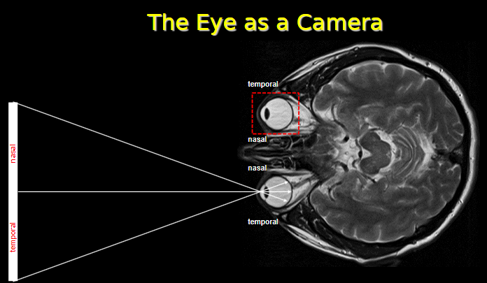
Temporal retina: Located on the side closer to the temples.
Nasal retina: Located on the side closer to the nose.
Temporal-nasal orientation: Refers to the arrangement of visual field regions.
Example: Light from the right visual field falls on the nasal retina of the right eye and temporal retina of the left eye.
-
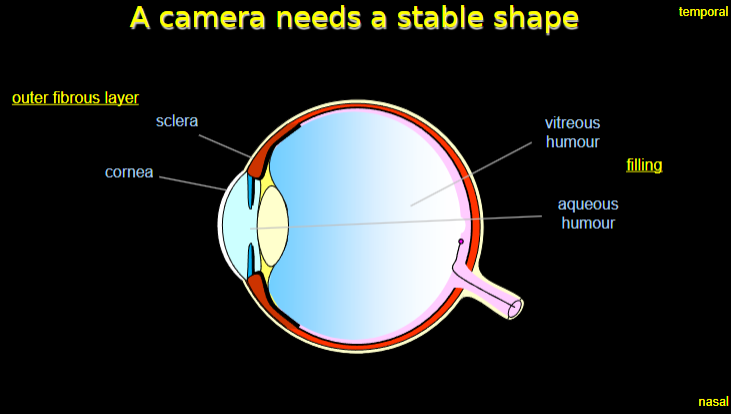
What components of the eye contribute to its stable shape, similar to a camera? (4)

Sclera: The tough outer fibrous layer that maintains the shape of the eye.
Cornea: Part of the outer fibrous layer that contributes to the rigidity and optical properties.
Aqueous humor: A clear fluid in the anterior chamber that helps maintain intraocular pressure.
Vitreous humor: A gel-like substance that fills the eye’s interior, supporting its shape.
-
What are the regions of the retina based on orientation, and why are they important? (4)
Nasal retina: Detects light from the outer part of the visual field.
Temporal retina: Detects light from the inner part of the visual field.
Importance: These regions work together to create a complete visual field.
Example: Light from the left visual field hits the nasal retina of the left eye and the temporal retina of the right eye.
-
What makes up the outer fibrous layer of the eye? (2)

Sclera: Provides structure and protection.
Cornea: Transparent and allows light to enter the eye, aiding in focus.
-
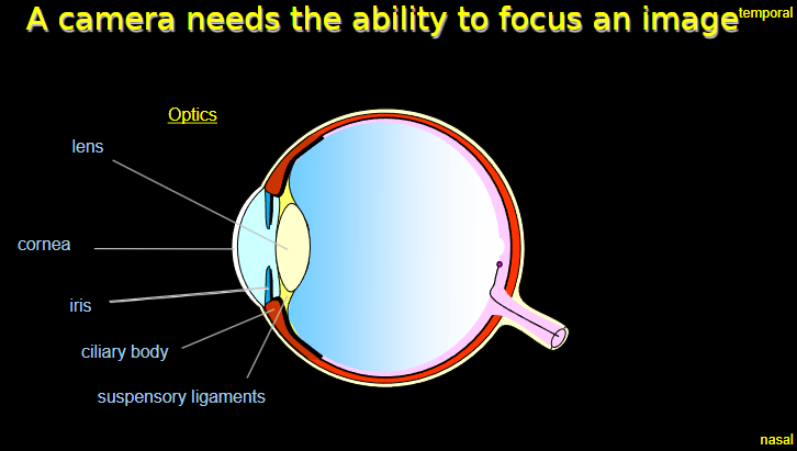
What components of the eye contribute to its ability to focus an image, similar to a camera? (6)

Lens: Changes shape (accommodation) to focus light onto the retina.
Cornea: Provides the majority of the eye's refractive power.
Iris: Controls the amount of light entering the eye by adjusting the size of the pupil.
Ciliary body: Adjusts the lens shape by contracting or relaxing.
Suspensory ligaments: Connect the ciliary body to the lens, facilitating its movement.
Optics: The combined action of these structures ensures a focused image on the retina.
-
How does the orientation of the nasal and temporal retina contribute to focusing an image? (2)
Nasal retina: Detects light from the outer parts of the visual field.
Temporal retina: Detects light from the inner parts of the visual field, aiding in image stability and focus.
-
What structures in the eye contribute to its ability to focus an image? (4)
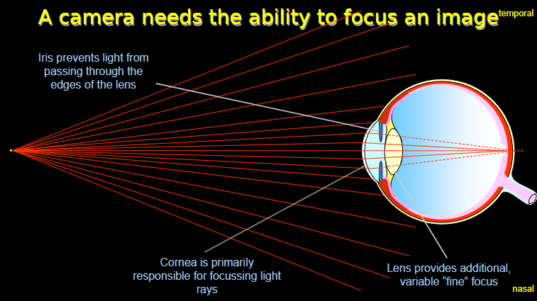
Lens: Provides additional, variable "fine" focus by changing its shape.
Cornea: Primarily responsible for focusing light rays onto the retina.
Iris: Prevents light from passing through the edges of the lens, improving image clarity.
Retina (nasal and temporal): Processes light from different parts of the visual field to create a complete image.
-
How does the cornea and lens work together to focus light? (2)

Cornea: Handles most of the focusing by bending incoming light rays.
Lens: Adjusts its curvature to fine-tune focus for near or distant objects.
-
What components of the eye are involved in focusing an image? (6)

Cornea: Primarily responsible for bending light rays to focus them onto the retina.
Lens: Provides fine focus by adjusting its curvature for near and far objects.
Iris: Regulates the amount of light entering the eye, preventing distortion.
Temporal retina: Processes light from the inner visual field.
Nasal retina: Processes light from the outer visual field.
Retina: Captures the focused image and transmits visual information to the brain.
-
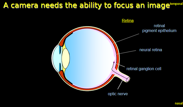
What are the layers of the retina, and what are their roles in vision? (4)

Retinal pigment epithelium: Supports photoreceptor cells by providing nutrients and absorbing excess light.
Neural retina: Contains photoreceptor cells (rods and cones) that detect light and convert it into electrical signals.
Retinal ganglion cells: Receive signals from photoreceptors and transmit them to the brain via the optic nerve.
Optic nerve: Carries visual information from the retina to the visual cortex in the brain.
-
How do the temporal and nasal regions of the retina contribute to vision? (2)
Temporal retina: Detects light from the nasal (inner) part of the visual field.
Nasal retina: Detects light from the temporal (outer) part of the visual field.
-
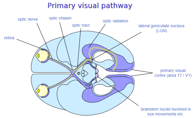
What is the primary visual pathway, and what structures are involved? (7)

Retina: Converts light into electrical signals.
Optic nerve: Transmits signals from the retina to the brain.
Optic chiasm: The point where some fibers cross to the opposite side of the brain for binocular vision.
Optic tract: Carries visual information from the optic chiasm to the brain.
Lateral geniculate nucleus (LGN): Relays visual information to the visual cortex.
Optic radiation: Transmits signals from the LGN to the primary visual cortex.
Primary visual cortex (area 17 / V1): Processes visual information for interpretation.
-
What is the function of the lateral geniculate nucleus (LGN) in the primary visual pathway? (2)
Acts as a relay station between the optic tract and the visual cortex.
Organizes and processes visual information before sending it to area 17 (V1).
-
What brainstem nuclei are involved in eye movements and other visual processing tasks? (3)

Superior colliculus: Coordinates eye movements and visual reflexes.
Pretectal nuclei: Involved in the pupillary light reflex.
Oculomotor nuclei: Controls eye muscle movements for tracking and focus.
-
What is the structure of a cone photoreceptor? (4)
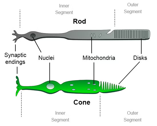
Outer segment: Contains photopigments (e.g., opsins) that capture light.
Inner segment: Houses cellular organelles and produces energy to sustain phototransduction.
Cell body: Contains the nucleus of the photoreceptor cell.
Synaptic terminal: Transmits signals to bipolar cells in the retina.
-
What is the phototransduction cascade, and how does it convert light energy into a receptor potential? (5)
Light absorption: Photopigments in the outer segment absorb photons, activating the opsin protein.
Activation of transducin: Light-induced opsin changes activate the G-protein transducin.
Phosphodiesterase activation: Transducin activates phosphodiesterase (PDE), which breaks down cyclic GMP (cGMP).
Closing of ion channels: Reduced cGMP levels cause sodium channels to close, leading to hyperpolarization of the photoreceptor.
Receptor potential: The hyperpolarization alters neurotransmitter release to bipolar cells.
-
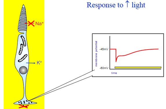
How does a photoreceptor respond to increases in illumination? (3)
Increased light decreases cGMP levels.
Sodium channels close, leading to hyperpolarization.
Reduced neurotransmitter release occurs at the synaptic terminal.
-
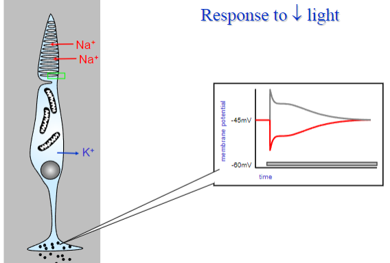
How does a photoreceptor respond to decreases in illumination? (3)
Decreased light increases cGMP levels.
Sodium channels open, leading to depolarization.
Increased neurotransmitter release occurs at the synaptic terminal.
-
What are the main layers visible in a histological section through the retina? (5)
Pigment epithelium: The outermost layer that provides support and absorbs excess light.
Photoreceptors: Rods and cones that detect light and initiate visual signals.
Interneurons: Bipolar, horizontal, and amacrine cells that process and relay signals.
Ganglion cells: The innermost layer of neurons that transmit signals to the optic nerve.
Vitreous humour: The gel-like substance adjacent to the retina that provides structural support.
-
What is the function of the retinal pigment epithelium (RPE)? (3)
Absorbs excess light to prevent scattering.
Provides nutrients to photoreceptors.
Phagocytoses shed photoreceptor outer segments.
-
What is the role of interneurons in the retina? (3)
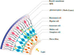
Bipolar cells: Connect photoreceptors to ganglion cells.
Horizontal cells: Integrate signals across photoreceptors for contrast.
Amacrine cells: Modulate signals in the ganglion and bipolar layers.
-
What is the role of ganglion cells in the retina? (2)
Collect visual information from interneurons.
Transmit visual signals to the brain via the optic nerve.
-
What are the main structural components of a cone photoreceptor? (6)
Membrane discs: Found in the outer segment; contain photopigments (opsins) that absorb light.
Outer segment: Houses the membrane discs; responsible for capturing light.
Inner segment: Contains organelles like mitochondria for energy production.
Nucleus: Located in the cell body; controls cellular activities.
“Axon”: A thin extension that connects the cell body to the synaptic terminal.
Synaptic terminal: Releases neurotransmitters to communicate with bipolar cells.
-
What is the function of the outer and inner segments in a cone photoreceptor? (4)
Outer segment: Captures light using photopigments in the membrane discs.
Inner segment: Produces energy and supports phototransduction processes.
-
How does the synaptic terminal of a cone photoreceptor function? (2)
Converts electrical signals into neurotransmitter release.
Transmits visual information to bipolar cells for further processing
-
Picture demonstrating a photoreceptor responding to increased light:
-
Picture demonstrating a photoreceptor responding to decreased light:
-
What are the key components involved in the initiation of the light response in photoreceptors? (7)
cGMP: A secondary messenger that maintains the opening of ion channels in the dark.
Opsin: A protein found in photopigments that binds to retinal, enabling the photoreceptor to absorb light.
Retinal: A light-sensitive molecule that binds to opsin to form the photopigment.
Membrane disc in outer segment: Contains photopigments and is where light is absorbed to initiate the transduction process.
Cell membrane: The boundary of the photoreceptor that houses ion channels and receptors.
Na+: Sodium ions that are involved in the dark current by entering the photoreceptor.
Photopigment: The combination of opsin and retinal that absorbs light and begins the phototransduction cascade.
-
What role does cGMP play in the dark current of photoreceptors? (2)

cGMP: Maintains the opening of sodium channels in the dark, allowing Na+ to enter the photoreceptor.
This results in a depolarized state of the photoreceptor in the absence of light.
-
How does the photopigment initiate the light response? (2)
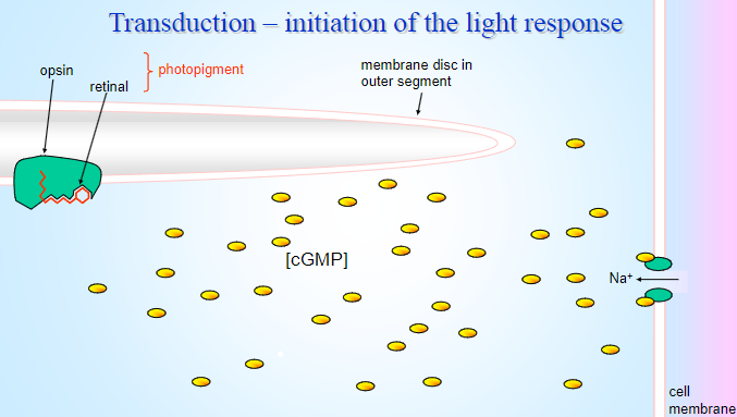
Photopigment (opsin + retinal): Absorbs light, causing a conformational change in retinal.
This change activates opsin, triggering a biochemical cascade that ultimately leads to a decrease in cGMP levels, closing sodium channels and hyperpolarizing the photoreceptor.
-
What steps are involved in the amplification of the biochemical cascade during phototransduction? (7)
![Opsin: The light-activated protein that initiates the biochemical cascade by changing its conformation.Membrane disc in outer segment: The location where photopigments are found, and where light is absorbed to activate opsin.Cell membrane: Houses the opsin and ion channels, which are essential for the transduction process.Trans retinal: The product formed when retinal undergoes a conformational change upon light absorption, activating opsin.Activated photopigment: The opsin-retinal complex that, when activated by light, initiates the cascade.Fall in [cGMP]: Light activation causes a reduction in cyclic GMP levels, leading to ion channel closure.G-protein (G): The G-protein activated by the photopigment that triggers further signaling events, including the activation of phosphodiesterase.](/flashcards/cardimage2/a2105009/804/8804258_back.png)
Opsin: The light-activated protein that initiates the biochemical cascade by changing its conformation.
Membrane disc in outer segment: The location where photopigments are found, and where light is absorbed to activate opsin.
Cell membrane: Houses the opsin and ion channels, which are essential for the transduction process.
Trans retinal: The product formed when retinal undergoes a conformational change upon light absorption, activating opsin.
Activated photopigment: The opsin-retinal complex that, when activated by light, initiates the cascade.
Fall in [cGMP]: Light activation causes a reduction in cyclic GMP levels, leading to ion channel closure.
G-protein (G): The G-protein activated by the photopigment that triggers further signaling events, including the activation of phosphodiesterase.
-
How does the fall in [cGMP] affect the photoreceptor cell? (2)
![Fall in [cGMP]: Causes the closing of sodium (Na+) channels.This leads to hyperpolarization of the photoreceptor cell, changing its electrical potential and reducing neurotransmitter release.](/flashcards/cardimage2/a2105009/804/8804259_back.png)
Fall in [cGMP]: Causes the closing of sodium (Na+) channels.
This leads to hyperpolarization of the photoreceptor cell, changing its electrical potential and reducing neurotransmitter release.
-
What is the role of G-proteins in amplifying the light signal? (3)
G-protein (G): Is activated by the photopigment (opsin-retinal complex) upon light absorption.
It activates phosphodiesterase, which reduces cGMP levels.
The cascade of events amplifies the signal, allowing a single photon to cause a significant change in the photoreceptor's response.
-
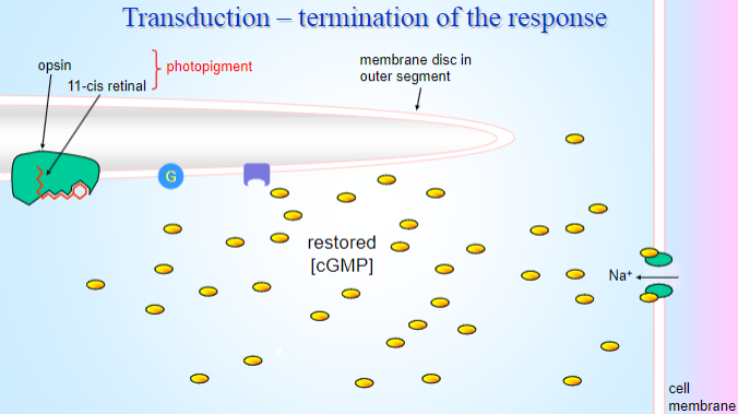
What is the role of 11-cis retinal in terminating the response? (2)

11-cis retinal: After phototransduction, retinal is converted back from the all-trans form to 11-cis form, which can rebind to opsin.
This restores the photopigment to its original state, ready to absorb light again.
-
How is the photopigment regenerated during the termination phase? (3)

11-cis retinal: Is synthesized from trans retinal in the retinal pigment epithelium.
Photopigment regeneration: The 11-cis retinal binds to opsin, regenerating the functional photopigment.
This process allows the photoreceptor to be ready for the next light stimulus.
-
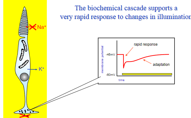
How does the biochemical cascade allow photoreceptors to respond to both increases and decreases in illumination? (3)

Decreased illumination:
cGMP levels increase.
Sodium channels reopen, leading to depolarization of the photoreceptor cell.
This results in a weaker signal being sent to the bipolar cells, signaling reduced light.
Increased illumination:
Light activates rhodopsin, which stimulates a G-protein (transducin).
This leads to a cascade that decreases cGMP levels.
Sodium channels close, causing hyperpolarization and a signal indicating increased light intensity.
Rapid adaptation:
Photoreceptors adjust their sensitivity through calcium ion regulation, enabling them to respond to both bright and dim light with minimal delay.
-
Why is amplification essential for rapid visual responses? (2)
Signal amplification: A single photon can cause a large change in the electrical state of the photoreceptor, allowing for the detection of even faint light.
Fast adaptation: This amplification ensures that photoreceptors can quickly adapt to changes in light intensity, supporting rapid visual processing.
-
What are the key components of retinal structure and circuitry? (5)
Pigment epithelium: A layer of cells that nourishes the photoreceptors and absorbs excess light.
Ganglion cells: The final output cells that send visual information to the brain via the optic nerve.
Photoreceptors: Rods and cones that detect light and initiate the transduction process.
Interneurons: Cells like horizontal, amacrine, and bipolar cells that process visual information between photoreceptors and ganglion cells.
Bipolar cells: Connect photoreceptors to ganglion cells, transmitting the visual signals.
-
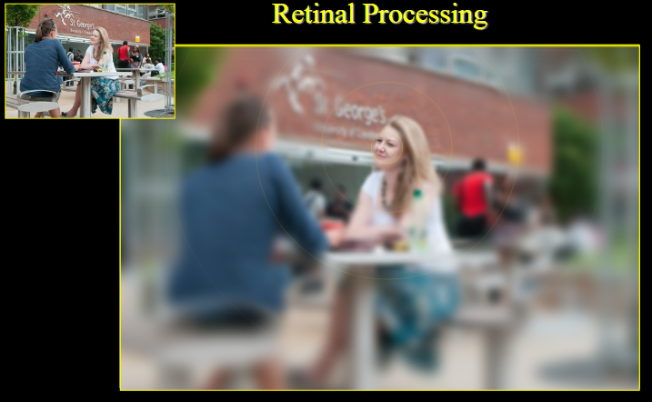
How does the retinal structure differ between the central and peripheral regions? (4)
Central retina (fovea): Contains a high density of cones, with minimal convergence, allowing for sharp, detailed vision.
Peripheral retina: Contains more rods, with significant convergence, leading to less detailed but more sensitive vision in low light.
Central retina: Features tightly packed photoreceptors, resulting in a higher resolution image.
Peripheral retina: Features large gaps in the sampling array, causing a decrease in visual resolution.
-
How does retinal structure affect visual resolution in central vs. peripheral vision? (3)
Central vision (fovea): The high density of cones and lack of convergence allow for high-resolution, detailed vision.
Peripheral vision: Greater convergence of rods leads to lower resolution but better sensitivity to movement and low-light conditions.
Difference in resolution: Central vision is specialized for tasks like reading and recognizing faces, while peripheral vision detects motion and provides broader visual awareness.
-
What is the importance of central vision? (2)
Central vision (foveal vision): Allows for detailed tasks such as reading, recognizing faces, and distinguishing fine details.
High resolution: It provides the sharpest and clearest visual information due to the high density of cones.
-
What causes the loss of peripheral vision, and which conditions are associated with it? (3)
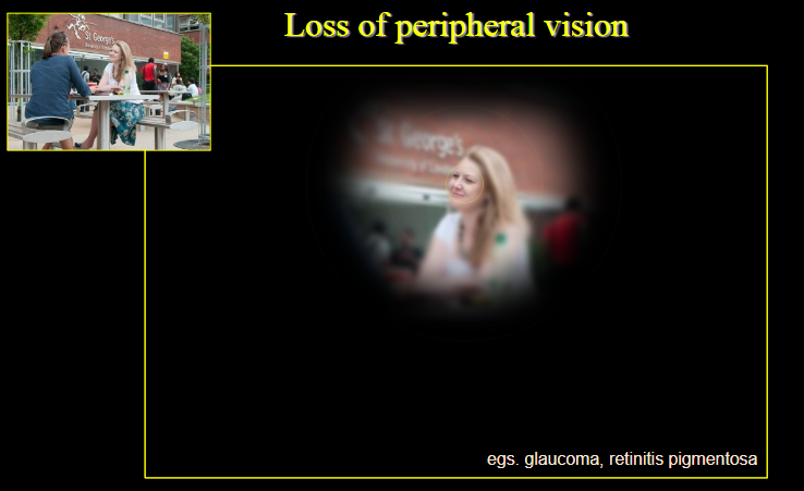
Loss of peripheral vision: Results from conditions such as glaucoma or retinitis pigmentosa.
Glaucoma: Damages the optic nerve, leading to gradual peripheral vision loss.
Retinitis pigmentosa: Degenerates photoreceptors in the peripheral retina, causing night blindness and loss of peripheral vision.
-
What causes the loss of central vision, and which condition is commonly associated with it? (2)
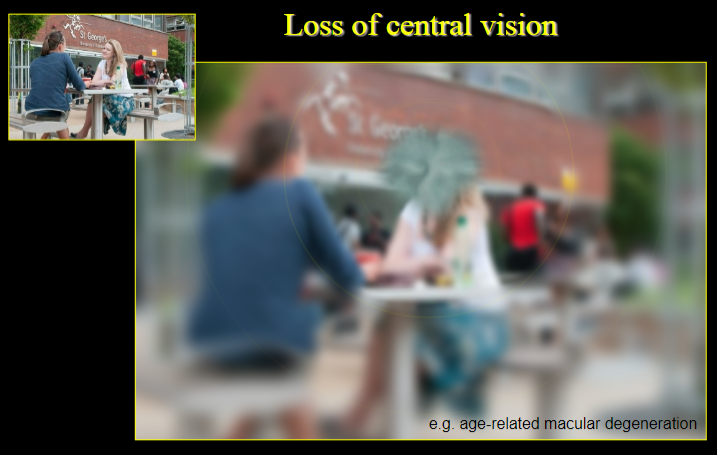
Loss of central vision: Occurs when the macula or fovea is damaged, resulting in loss of detailed vision.
Age-related macular degeneration (AMD): A common condition causing degeneration of the macula, leading to a loss of central vision.
-
How does the convergence of photoreceptors affect visual quality in the peripheral retina? (3)
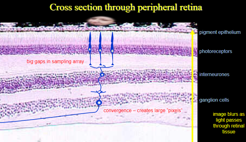
Convergence: Multiple photoreceptors (rods) send their signals to a single ganglion cell, creating large “pixels.”
Lower resolution: This convergence results in a decrease in visual sharpness, making peripheral vision less detailed.
Image blurring: As light passes through the retinal tissue, the image becomes blurred due to the large sampling area and reduced resolution.
-
How does the retina map onto the primary visual pathway, and what is the retinotopic map? (3)

Retina mapping: The retinal image is projected onto the primary visual cortex via the optic nerve, optic chiasm, and optic tract.
Retinotopic map: The organization of the visual field in the brain corresponds to the spatial arrangement of the retina.
Distorted scale: The central part of the retina (fovea) occupies a disproportionally large area in the visual cortex, creating a distorted scale.
-
What is central vision, and why is it special? (3)
Central vision (foveal vision): It provides the sharpest and most detailed visual information.
High resolution: This is due to the dense concentration of cones and minimal convergence in the central retina.
Specialized tasks: It enables precise activities like reading, recognizing faces, and distinguishing fine details.
-
What are the key components of the central retina? (3)
Fovea centralis: The center of the retina, where visual acuity is highest due to a high density of cones and minimal convergence.
Macula lutea: The area surrounding the fovea, contributing to sharp central vision.
Retinal blood vessels: Supply oxygen and nutrients to the retina, ensuring proper functioning for detailed vision.
-
What is the fovea centralis, and what is its role in central vision? (2
Fovea centralis: A small depression in the retina that contains a high concentration of cones.
Role in central vision: It is responsible for the sharpest visual acuity and is critical for tasks requiring high-detail focus, such as reading.
-
What is the macula lutea, and how does it contribute to vision? (2)
Macula lutea: The region surrounding the fovea centralis, containing fewer photoreceptors.
Role in vision: It enhances visual acuity by assisting in central vision and focusing on detailed objects, though it has a slightly lower resolution than the fovea.
-
How do retinal blood vessels affect central vision? (2)
Retinal blood vessels: Supply oxygen and nutrients to the retina, ensuring its proper functioning.
Impact on vision: Well-functioning blood vessels are essential for maintaining the health of the retina and supporting detailed vision.
-
Why does peripheral vision represent the majority of the visual field? (2)
Wider field: Peripheral vision covers a larger area of the visual field, allowing us to detect movement and changes in the environment outside the direct line of sight.
Larger coverage: It is responsible for detecting peripheral stimuli and situational awareness rather than detailed focus.
-
Why is peripheral vision considered coarse? (5)
Optical blur: The visual image in peripheral vision is more optically blurred compared to central vision.
Large, widely spaced cones: Cone photoreceptors in the periphery are larger and more spaced out.
More rods present: The periphery contains a higher number of rods, which are specialized for detecting movement and low light rather than detailed color vision.
Convergence onto ganglion cells: Multiple cone signals converge onto a single ganglion cell, reducing spatial resolution.
Increasing convergence with distance from the fovea: As you move further from the fovea, the convergence of signals from cones onto ganglion cells increases, further lowering the resolution of peripheral vision.
-
How does the structure of peripheral photoreceptors affect vision quality? (3)
Larger cones: In peripheral vision, the cones are larger and more sparsely distributed, which compromises the ability to resolve fine details.
Higher rod concentration: Rods are more numerous in the periphery, supporting better night vision and motion detection but contributing to lower resolution for color and detail.
Signal convergence: The convergence of multiple cones onto a single ganglion cell reduces the resolution, making peripheral vision less sharp.
-
How does resolution change with distance from the fovea? (2)
Decreases with distance: As you move away from the fovea, visual resolution decreases due to increased convergence of signals and a higher rod-to-cone ratio.
Effect on vision: The further from the fovea, the blurrier and coarser the visual image becomes, particularly for fine detail and color discrimination.
-
What is the significance of foveal vision in terms of visual field coverage? (2)
Small area: Foveal vision represents a tiny portion of the visual field, specifically the central part where visual acuity is highest.
Specialized function: It is specialized for tasks requiring high detail, like reading, recognizing faces, and distinguishing fine details.
-
How is the visual image in foveal vision well-focused? (2)
Minimal obstruction: The obstructing layers of the retina are pushed to one side in the fovea, allowing light to directly reach the photoreceptors.
High focus: This arrangement ensures that the visual image is sharply focused and free from distortion or scattering.
-
How do the photoreceptors in the fovea contribute to high resolution? (3)
Red and green cones: The fovea primarily contains red and green photoreceptors, which are essential for color vision and sharp focus.
Thin and tightly packed: These cones are smaller and packed tightly together, maximizing light reception and clarity.
No convergence: Signals from individual cones are kept separate with no convergence onto a single ganglion cell, ensuring high spatial resolution.
-
How does the lack of convergence in the fovea enhance vision? (2)
Individual signal transmission: In the fovea, cones transmit signals directly to separate ganglion cells, allowing for high precision.
Tiny receptive fields: The lack of convergence means each ganglion cell receives input from just one cone, which creates very small receptive fields and maximizes resolution.
-
What does it mean for the primary visual pathway to be retinotopic? (2)
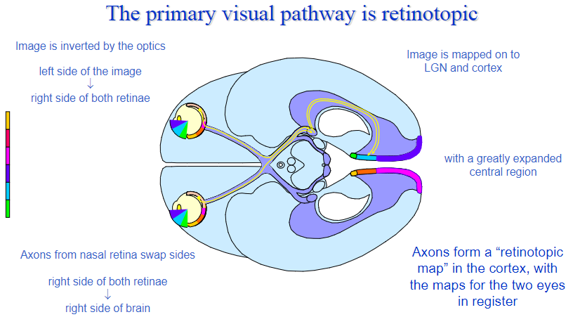
Mapped by location: The visual pathway maintains a spatial map of the retina, meaning that adjacent regions of the retina correspond to adjacent areas of the visual cortex.
Image processing: This mapping allows the brain to process visual information based on its location on the retina, preserving spatial relationships.
-
How is the image inverted by the optics of the eye? (2)

Inversion of the image: The lens and cornea of the eye invert the image as it enters the eye.
Effect on retina: The top of the image is mapped to the bottom of the retina, and the left side is mapped to the right side of both retinae.
-
How does the visual information from the retina get mapped onto the brain? (3)

Image inversion maintained: The left side of the image is processed by the right side of the retina, and the right side by the left side of the retina.
Mapping to the brain: This inverted image is transmitted to the lateral geniculate nucleus (LGN) and the visual cortex.
Expanded central region: The central region of the visual field, particularly from the fovea, has an expanded representation in the brain, allowing for detailed processing.
-
What happens to the axons from the nasal retina? (2)

Axon crossing: Axons from the nasal retina cross over to the opposite side of the brain at the optic chiasm.
Retinotopic map: This crossing ensures that the left visual field from both eyes is processed by the right hemisphere and vice versa, maintaining a retinotopic map of the visual field in the brain.
-
How are the maps for the two eyes aligned in the cortex? (2)

In-register maps: The retinotopic maps for the left and right eyes are aligned, or "in register," in the visual cortex.
Shared representation: This alignment allows for binocular vision and depth perception, as the brain can integrate information from both eyes.
-
How are visual cells designed to respond to changes in the visual image? (2)
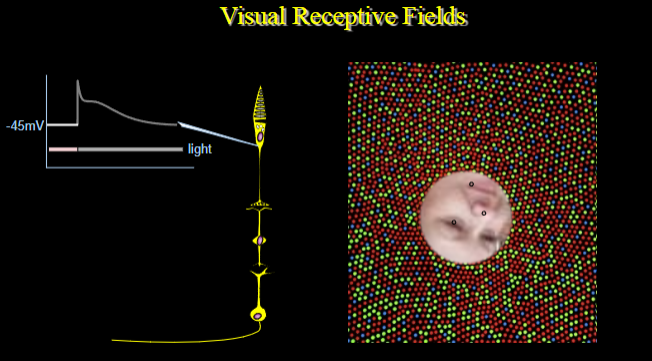
Sensitivity to change: Visual cells, particularly those in the retina and visual cortex, are designed to respond to changes in the visual scene, such as movement, brightness, or contrast.
Receptive fields: These cells have receptive fields that are sensitive to specific areas of the visual field and react to variations in stimuli within these areas.
-
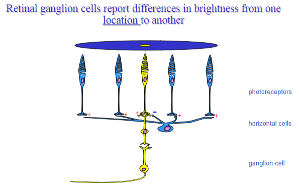
What are centre-surround receptive fields and where are they found? (2)
Centre-surround organization: Receptive fields in retinal ganglion cells (RGCs) and lateral geniculate nucleus (LGN) cells are structured with a central area that responds to light and a surrounding area that inhibits or counteracts this response.
Functional role: This arrangement enhances the contrast between light and dark areas of the image, improving edge detection and spatial resolution.
-
What are the main types of retinal ganglion cells (RGCs) in humans and their role in vision? (3)
Types of RGCs: There are two main types of retinal ganglion cells: M cells (magnocellular) and P cells (parvocellular).
M cells: These cells are large, have a fast response, and are involved in detecting movement and coarse visual detail.
P cells: These smaller cells are involved in detecting color and fine detail, contributing to high-resolution vision.
-
How are the receptive field properties of RGCs generated by retinal circuitry? (3)

Retinal network: The receptive field properties of RGCs are shaped by the circuitry in the retina, where bipolar cells and amacrine cells influence the response of ganglion cells to specific stimuli.
Centre-surround arrangement: The interaction between excitatory and inhibitory inputs from photoreceptors, bipolar cells, and horizontal cells creates the centre-surround receptive fields.
Role of convergence: Multiple photoreceptors converge onto a single RGC, influencing the size and sensitivity of its receptive field.
-
What is the significance of the increased stimulus selectivity seen in the visual cortex? (2)
Higher processing: The visual cortex is capable of more selective responses to stimuli, allowing it to detect specific features such as orientation, movement, and color.
Feature detectors: Neurons in the cortex are tuned to respond to specific features in the visual field, such as lines, edges, or motion, enhancing the brain’s ability to analyze complex visual information.
-
Which higher cortical areas are specialized for different aspects of vision? (3)
V1 (Primary visual cortex): This area is responsible for processing basic visual features like orientation, contrast, and spatial frequency.
V2-V4: These areas contribute to more complex visual processing, including color perception (V4) and shape recognition.
MT (Middle temporal area): Specialized for motion detection and processing of dynamic visual stimuli.
-
Why do visual cells respond to changes rather than absolutes? (2)
Adaptation: Photoreceptors and ganglion cells are designed to detect changes in illumination, which allows for better adaptation to varying light conditions.
Efficiency: The system prioritizes detecting contrasts or differences rather than absolute brightness, which is more useful for interpreting dynamic visual scenes.
-
What are the centre-surround receptive fields of retinal ganglion and lateral geniculate nucleus cells, and what do they encode? (3)
Centre-surround structure: These receptive fields have a central region that responds positively to light and a surrounding region that has an inhibitory response.
Encoding contrast: This structure enhances edge detection by highlighting differences in illumination between adjacent regions.
Retinal ganglion cells: Encode spatial differences in light intensity across the visual field, facilitating the detection of objects and their movement.
-
What are the main types of retinal ganglion cells (RGCs) in humans, and how do their receptive field properties relate to their role in vision? (4)
Parvocellular (P) cells:
Small receptive fields for fine detail and high-resolution vision.
Specialised in detecting color and providing information about stationary objects.
Require a stable image for accurate processing.
Magnocellular (M) cells:
Large receptive fields for coarse resolution and detecting fast movement.
Involved in processing dynamic visual information, such as motion.
Receptive field properties: These are influenced by the convergence of photoreceptor signals and the properties of the retinal circuitry.
Role in vision: P cells are important for color perception and detailed vision, while M cells excel at detecting movement and changes in the environment.
-
How do retinal ganglion cells compare the outputs from different types of cones? (2)
Comparison of cones: Some retinal ganglion cells receive inputs from different cone types, comparing their outputs to detect specific color contrasts.
Color opponency:
Parvocellular cells: Compare red and green cone signals.
Blue-yellow ganglion cells: Compare blue and yellow cone signals.
-
How do the receptive fields of lateral geniculate nucleus (LGN) cells compare to those of retinal ganglion cells? (2)
LGN as a faithful relay: The receptive fields of LGN cells closely resemble those of retinal ganglion cells.
Role in processing: Both retinal ganglion cells and LGN cells play a role in encoding spatial and contrast information, but the LGN is a relay station, sending this information to the visual cortex for further processing.
-
How do cortical receptive fields differ from retinal and LGN receptive fields? (3)
Cortical receptive fields: These are more complex than those in the retina and LGN. They are capable of responding to more specific visual features, such as orientation, direction of movement, and spatial frequency.
Higher selectivity: Visual cells in the cortex show greater selectivity for features of visual stimuli (e.g., edges, shapes).
Integration of information: Cortical cells integrate information from multiple sources, allowing for the processing of more abstract visual features.
-
What is the difference between the higher visual cortical areas in the temporal and parietal cortex, and how does this relate to the input they receive? (4)
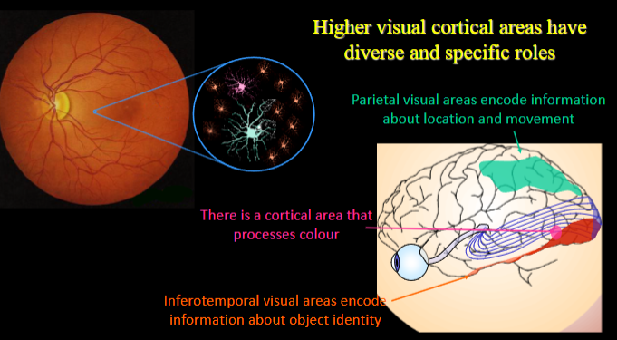
Temporal cortex:
Specializes in object recognition and encoding information about object identity.
Receives input from the ventral visual stream, processing detailed features of objects.
Parietal cortex:
Specializes in encoding information about the location and movement of objects.
Receives input from the dorsal visual stream, focusing on motion and spatial awareness.
Differentiated roles: The temporal cortex is crucial for “what” we see, while the parietal cortex is vital for “where” and “how” we see objects.
Input sources: The two cortical areas receive distinct input from different parts of the visual pathway: the ventral stream feeds into the temporal lobe, and the dorsal stream feeds into the parietal lobe.
-
What is the retina and what types of cells does it consist of? (3)
Retina: A thin, layered structure that lines the back of the eye and is responsible for visual processing.
Photoreceptors: Light-sensitive sensory cells; rods for low light levels and cones for daylight vision.
Interneurons and Retinal Ganglion Cells: Interneurons form circuits that process photoreceptor responses, while retinal ganglion cells serve as afferents, transmitting visual information to the brain.
-
What is the fovea and how is it adapted for fine detailed vision? (2)
Fovea: A tiny region at the center of the retina, responsible for focusing the point in visual space directly in front of us.
Structural Adaptation: The fovea has a high density of cones, allowing for detailed, sharp vision. It is often used interchangeably with the macula, a larger region distinguished by yellow pigmentation.
-
What is a photopigment and its role in vision? (2)
Photopigment: A molecule that captures photons of light and triggers changes in a photoreceptor's membrane potential.
Composition: Consists of a protein called opsin and a light-sensitive chromophore, 11-cis retinal.
-
What is a receptive field and what does it encode? (2)
Receptive Field: The region of the visual image that affects the action potential firing rate of a given cell.
Encoding: It encompasses the specific pattern of light needed in that region to elicit a response from the cell.
-
What is a binocular cell and its function? (2)
Binocular Cell: A cell that receives input from both eyes.
Function: These cells contribute to depth perception by matching receptive fields in the two eye fields.
-
What is the Dorsal Lateral Geniculate Nucleus (dLGN) and what is its role in vision? (2)
dLGN: The thalamic nucleus that relays visual information from the retina to the primary visual cortex.
Role: It serves as part of the perceptual pathway for visual processing.
-
What is the primary visual cortex and what role does it play in visual processing? (2)
Primary Visual Cortex: Also known as Area 17, it receives input from the retina via the dLGN.
Role: It is responsible for initial processing of visual information, which is then distributed to higher visual areas for more specific processing.
-
What are higher visual cortical areas, and how are they divided between the two main pathways? (3)
Higher Visual Cortical Areas: At least 20 regions in the primate cortex contribute to vision, with some estimates suggesting up to half of the cortex is involved.
Infero-Temporal Pathway: Supports object identification and detailed vision.
Parietal Pathway: Supports understanding the location, relationships, and movement of objects, helping us interact with the environment.

