Erika Beaudin
Unit 15 - digestive system pt 1
Overview, digestive system (oral cavity, pharynx, and esophagus to anus), GI tract histology (esophagus to rectum), and peritoneum
-
What does the digestive system include?
1) Gastrointestinal (GI) tract
- tube from mouth to anus
2) Accessory organs
- teeth, tongue, salivary glands
- pancreas
- liver, gall bladder
-
What are the digestive system processes?
- Ingestion (food into oral cavity)
- Digestion (mechanical and chemical (enzymes and acid secretions))
- absorption (end products of digestion enter blood or lymph)
- defecation(elimination of undigested material)
-
What is the oral cavity?
lined by a mucosa (mucous membrane) made of a stratified squamous epithelium and lamina propria
includes:
a) lips
b) cheeks
c) palate (hard and soft)
d) tongue
-
What are the palates in the oral cavity?
- hard palate (2 maxillae and 2 palatine bones)
-soft palate (posterior to hard, skeletal muscle, posterior projection is uvula to close nasopharynx when swallowing)
-
What are the details of the tonge?
attached to hyoid bone
skeletal muscle
projections of mucosa = papillae (taste buds)
-
What are the salivary glands?
3 pairs:
a) parotid
inferior and anterior to ears
mumps = inflammation of 1 or both parotids
b) submandibular
floor of mouth
c) sublingual
below tongue on floor of mouth
-
What is the make up of saliva?
o 99.5% water
o 0.5% solutes (e.g. enzymes)
-
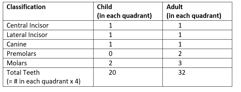
Where are dentition (teeth), what are the two types?
- in maxillae and mandible
- child dentition are primary dentition – deciduous (“baby”) teeth
- adult dentition are secondary dentition – permanent teeth
-
What are the classifications of teeth? How many do children and adults have of each?
-
What makes up a tooths structure?
crown, root, neck, periodontal ligaments, root canal extends to pulp cavity
-
What is a crown and root (in tooth structure)?
above the gum
dentin forms the majority of tooth
enamel overlay is acellular, highly calcified – hard!
root: dentin with cementum overlay
note: dentin, enamel, and cementum are similar to bone but avascular
-
What are the neck and periodontal ligaments (in tooth structure)?
a) neck
enamel and cementum boundary (gums)
b) periodontal ligaments
attach root to bones
-
What does the pulp cavity that the root canal extends into contain?
contains connective tissue, blood/lymph vessels, and nerves
-
What are the oropharynx and laryngopharynx made of?
only muscularis externa (skeletal muscle) and stratified squamous epithelium
-
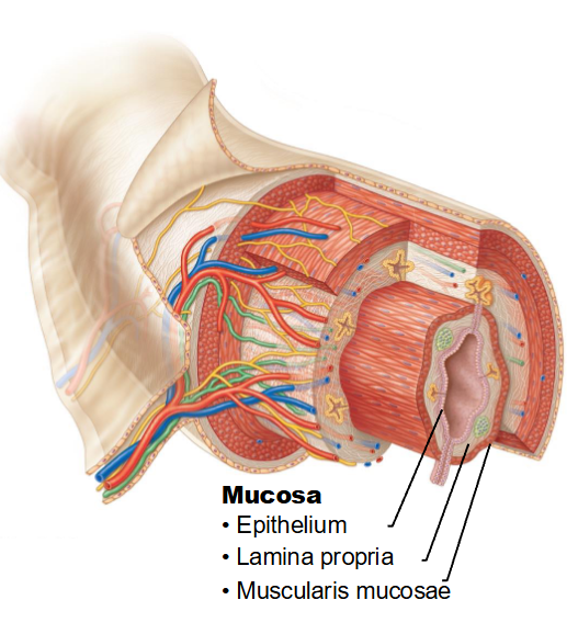
What are the 4 basic layers of the Gastrointestinal Tract (esophagus to rectum)?
mucosa, submucosa, muscularis externa, and serosa or adventitia (double walled membrane)
-
What are the 3 layers of mucosa in the Gastrointestinal Tract?
a) epithelium with numerous goblet cells
stratified squamous: esophagus, anal canal
simple columnar: stomach, small and large intestines, rectum
b) lamina propria (areolar connective tissue)
contains blood, lymph vessels, lymph nodules/tissues (immune)
c) muscularis mucosa
smooth muscle - allows movement of mucosa
-
What are the 3 layers of mucosa in the Gastrointestinal Tract (in order, visualize)?
-
What does the submucosa of the Gastrointestinal Tract contain? what's it made of?
areolar CT
contains: blood, lymphatic vessels, and a network of nerve cells
-
What are the two layers of the muscularis externa of the GI track?
- two layers of smooth muscle separated by a second network of nerve cells
o inner circular layer (contraction constricts the lumen)
o outer longitudinal layer (contraction shortens gut length)
- contractions controlled by the nerve network cause motility (mixing and movement)
-
What is peritoneum and what are its specializations?
serous membrane that lines the abdominopelvic cavity and most of the abdominal organs
specializations: omenta and mesentry
-
What are the Retroperitoneal organs (exceptions to being completly lined?
located posterior to the parietal peritoneum
peritoneum lines only one side of the organ
e.g. pancreas, duodenum
o anterior surface is covered by parietal peritoneum
o posterior surface is covered by adventitia that connects the organ to the body wall
-
What is the structure of serosa?
1) visceral peritoneum (against organ wall)
2) parietal peritoneum (against abdominal cavity wall)
3) peritoneal cavity (space between parietal and visceral peritoneum)
- filled with serous fluid to lessen friction between parts digestive tract during motility
-
What is omenta?
folds of serosa between organs made of a sheet of 2 fused layers of visceral peritoneum
contains blood/lymph vessels and nerves
examples:
- greater omentum (“fatty apron”) (connects stomach to transverse colon, forms large fold that hangs down over transverse colon and small intestine)
- lesser omentum (connects liver to stomach)
-
What is mesentry?
fold of serosa between the posterior abdominal cavity wall and the small/large intestine made of a sheet of 2 fused layers of parietal peritoneum
entry and exit point for blood vessels, nerves, and lymphatic vessels supplying digestive organs
-
What is the organ route of the Digestive System: Esophagus to Anus?
Esophagus, stomach, small intestine, large intestine,
-
What is the esophagus?
posterior to trachea
passes through the diaphragm entering into the abdominal cavity
-
What is the histology of the esophagus? (transition from superior to inferior)
a) muscularis externa
upper 1/3 = skeletal muscle
middle 1/3 = skeletal and smooth muscle
lower 1/3 = smooth muscle
b) the outer layer is adventitia within thoracic cavity and serosa within the abdominal cavity
-
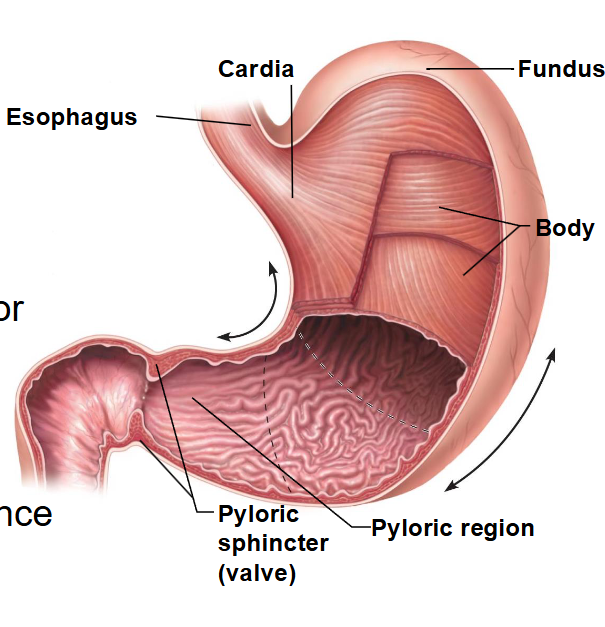
What does the stomach do and what are its 4 regions?
Stores, partially digests, and regulates emptying of chyme (food and gastric juices) into the small intestine
4 regions: cardiac region (cardia), fundus, body, pyloric region (pylorus)
-
What do each of the 4 regions of the stomach do?
- cardiac region (cardia): attached to inferior esophagus
- fundus: superior to esophageal entrance
- body: middle portion
- pyloric region (pylorus): inferior portion of the stomach
the pyloric sphincter regulates release of stomach contents into small intestine
-
What are the unique histological features of the stomach?
the mucosal surface is:
- formed exclusively by mucous cells
- has millions of gastric pits (invaginations of the epithelium) that are connected to the underlying gastric glands
-
What are gastric glands?
- exocrine glands that secrete gastric juice into the gastric pits where it then enters the lumen of the stomach
-
What cell types do gastric glands contain?
- goblet cells
- chief cells (secrete enzymes for protein and fat digestion)
- parietal cells (secrete hydrochloric acid (HCl) which lowers the pH of the stomach)
- G cells (enteroendocrine cells) (secrete a hormone into the blood that regulates activity of parietal cells and other digestive processes.)
-
What are rugae?
- are folds of the mucosa and submucosa due to contraction of muscularis mucosa
- visible when stomach is empty
- allows expansion of stomach without tearing
-
What is muscularis externa?
o function = churning chyme
o 3 layers instead of 2:
i. inner oblique
ii. middle circular
iii. outer longitudinal
-
What is the small intestine?
- pyloric sphincter to ileocaecal valve
- where most food digestion/absorption occurs
- 3 segments: duodenum, jejunum (middle section), ileum
-
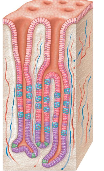
What is the histology of the small intestine?
simple columnar epithelium 3 main cell types
1. enterocytes: simple columnar cells (absorptive cells that form most of the mucosa)
2. goblet cells: secrete mucus
3. enteroendocrine cells: located within intestinal glands secrete hormones into the blood that help regulate digestive processes
-
What is the duodenum?
first fold (short) – is retroperitoneal
extra glands here secrete alkaline mucous to protect against stomach acid
ducts of accessory organs (liver, gall bladder, pancreas) enter the digestive system here
-
What is the ileum?
- last portion of small intestine before large intestine
- attached to caecum (part of large intestine)
- has groups of lymph nodules called Peyer’s patches (prevent infection of small intestine and prevent bacteria from entering blood)
-
What are the ways the segments increase absorption surface area?
- plicae circulares: submucosa thrown into large folds
- villi: projections of mucosa into lumen of small intestine (contains blood capillaries, lacteals (are lymphatic capillaries that absorb fats))
- microvilli: projections of the enterocyte cell membranes that extend into the lumen of the small intestine, forming a fuzzy “brush border” on the surface of the mucosa
-
What is the Large Intestine?
- from ileocaecal valve to anus
- basic functions: absorption of water, electrolytes, vitamins and formation and temporary storage of feces
- contains: caecum, appendix, colon, rectum, and anal canal
-
What is the histology of the large intestine?
mucosa has a smooth surface with no folds or villi
-
Where is the caecum?
connected to ileum by ileocaecal valve
-
What is the anal canal?
- last segment of large intestine, but external to the abdominopelvic cavity
- opening and closing of the inferior anal canal during defecation is controlled by two anal sphincters: internal anal sphincter (smooth muscle), and external anal sphincter (skeletal muscle – voluntary control)
- opening at inferior end of anal canal is the anus
-
What is the histology of the anal canal?
- histology: mucosa transitions to stratified squamous epithelium
-
What is the colon?
longitudinal layer of muscularis externa is reduced to three bands of muscle called teniae coli (contraction of the teniae coli forms pouches called haustra)
-
What are the parts of the colon?
- ascending colon (right side of abdominal cavity)
- hepatic flexure
- transverse colon
- splenic flexure
- descending colon (left side of abdominal cavity)
- sigmoid colon
-
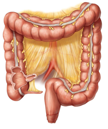
What is this?
Large intestine
-
What are the 4 regions of the stomach (visualize)?