-

PATHOGENESIS: Primary tuberculosis
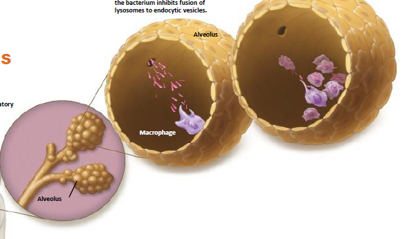
1. Mycobacterium typically infects the respiratory tract via inhalation of respiratory droplets from infected individuals.
-
PATHOGENESIS: Primary tuberculosis
2. Macrophages in alveoli
phagocytize mycobacteria but are unable to digest them, in part because the bacterium inhibits the fusion of lysosomes to endocytic vesicles.
-
PATHOGENESIS: Primary tuberculosis
3. Instead, bacteria replicate freely within macrophages, gradually killing the phagocytes. Bacteria released from dead macrophages are phagocytized by other macrophages, beginning the cycle anew.
-

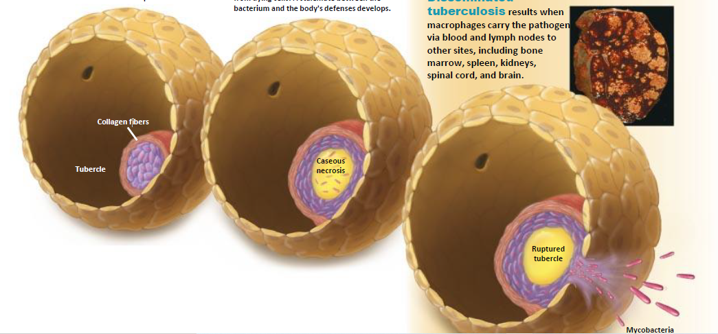
4. Tubercle and Collagen fibers
Infected macrophages present
antigen to T lymphocytes, which
produce lymphokines that attract and
activate more macrophages and
trigger inflammation. Tightly packed
macrophages surround the site of
infection, forming a tubercle over a
two- to three-month period.
-
5. Caseous necrosis
Other cells deposit collagen fibers,
enclosing infected macrophages and lung
cells within the tubercle. Infected cells in the
center die, releasing M. tuberculosis and
producing caseous necrosis—the death of
tissue that takes on a cheese-like consistency due to protein and fat released from dying cells. A stalemate between the bacterium and the body's defenses develops.
-
Secondary/reactivated tuberculosis
Results, when M. tuberculosis breaks the stalemate, ruptures the tubercle, and reestablishes an active infection. Reactivation occurs in about 10% of patients, especially those whose immune systems are weakened by disease, poor nutrition, drug or alcohol abuse, or other factors.
-
6. Disseminated Ruptured tubercle
Mycobacteria
Tuberculosis results when
macrophages carry the pathogen
via blood and lymph nodes to
other sites, including bone
marrow, spleen, kidneys,
the spinal cord, and brain.

