 Fatima Adam
The Respiratory System (Anatomy)
Introduction: This lecture serves as an introduction to the respiratory system. We will offer a brief overview of the skeletal structure and muscles within the thorax. Furthermore, we will delve into the mechanics of respiratory movements. Additionally, we will explore both the upper and lower respiratory organs, as well as the pleura and pleural cavity A pre-recorded lecture is provided for you to introduce the learning objectives and direct you through. Learning objectives: At the end of this session, you will be able to: Relate skeletal and soft tissue structures in the thorax to the mechanism of breathing Identify the major structures of the respiratory tract Locate and identify the paranasal sinuses Identify and recall the divisions of the pharynx (nasopharynx, oropharynx and laryngopharynx) Locate and describe the functional anatomy of the larynx Identify and describe the gross anatomy of the lungs (include hilum structures, borders, lobes, fissures, and surfaces)
-
What is the thorax?
The thorax is the region between the neck (above) and the abdomen (below).
-
What are the functions of the thoracic wall?
🍮To protect the heart and lungs.
🍮To facilitate the movements of breathing.
🍮To accommodate breast tissue for lactation.
-
What does the thoracic cavity contain?

🍮The thoracic cavity is located within the chest walls.
🍮It contains vital organs, also known as viscera.
🍮Major vessels and nerves are also found within the thoracic cavity.
🍮The thoracic cavity consists of the mediastinum and the right and left pleural cavities.
-
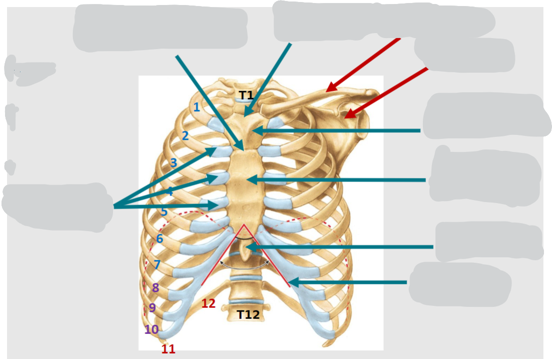
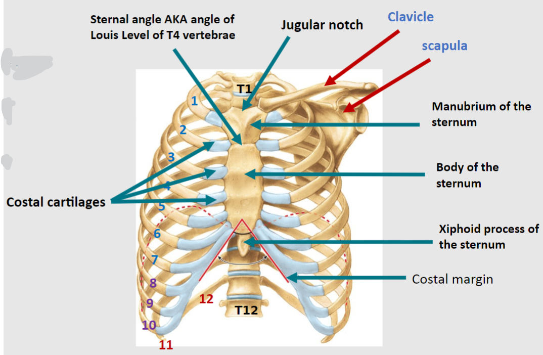
How many pairs of ribs are present in the skeleton of the thoracic or chest wall?
12 pairs of ribs.
-
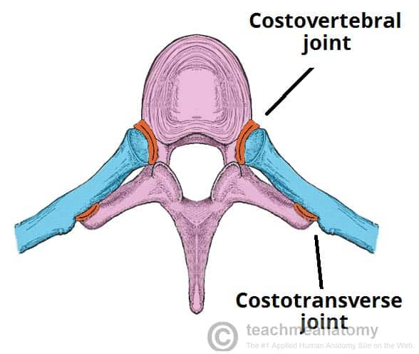
What are the three categories of ribs based on their attachment to the sternum?

True ribs (1-7): Attach via their costal cartilage directly to the sternum.
False ribs (8-10): Attach via their costal cartilage below to the sternum.
Floating ribs (11 & 12): Have no attachment to the sternum.
-
What are intercostal spaces?

Intercostal spaces are the spaces between adjacent ribs.
-
What is the costal margin?

The costal margin refers to the lower edge of the chest formed by the bottom edges of the rib cage.
-
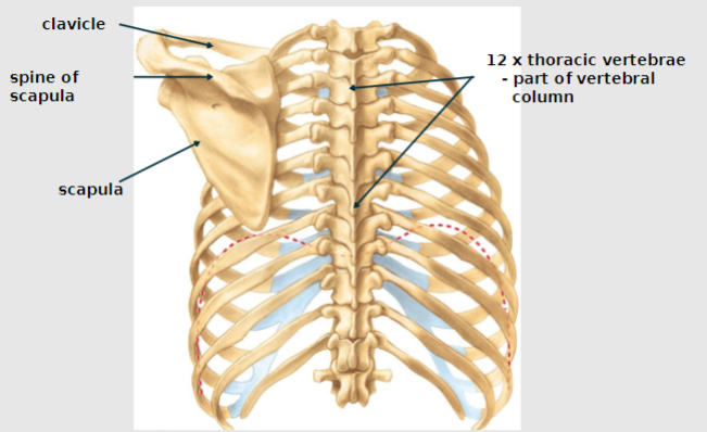
Besides ribs, what other bones are part of the skeleton of the thoracic or chest wall?

🍮12 thoracic vertebrae
🍮Clavicle and scapula
🍮Sternum, consisting of the manubrium, body, xiphoid process, and sternal angle.
-
Name the parts of The Skeleton of the Thoracic/ Chest Wall ANTERIOR:
-
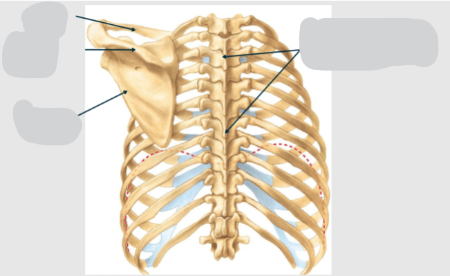
Name the parts of The Skeleton of the Thoracic/ Chest Wall POSTERIOR:
-
What is the intercostal space?
The intercostal space refers to the space between adjacent ribs, which is numbered according to the rib superior to it.
-
What structures are found within each intercostal space, and what is their function?
Each intercostal space contains intercostal muscles.
These muscles help move the ribs during breathing and change the volume within the thoracic cavity during respiration.
-
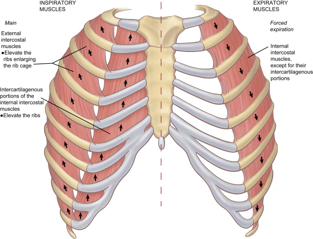
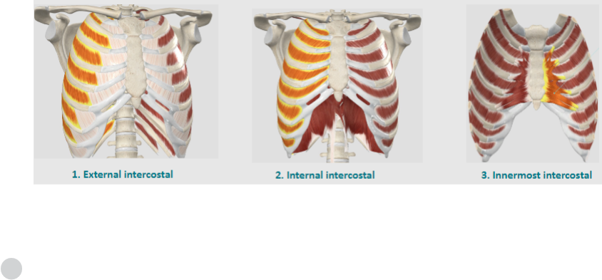
What are the three types of intercostal muscles, and what are their roles?
External intercostal muscles: Assist in elevating the ribs during inhalation.
Internal intercostal muscles: Aid in depressing the ribs during forced exhalation.
Innermost intercostal muscles: Assist in stabilizing the rib cage during breathing movements.
-
What is the function of the external intercostal muscles?

The external intercostal muscles function to elevate the ribs during inhalation.
-
What is the function of the internal intercostal muscles?
The internal intercostal muscles only function in forced exhalation.
-
What is the function of the innermost intercostal muscles?

The innermost intercostal muscles, the deepest layer, assist in stabilizing the rib cage during breathing movements.
They are separated from the other two layers of muscles by the neurovascular bundle.
-
What is the arrangement of the neurovascular bundle supplying each intercostal space?
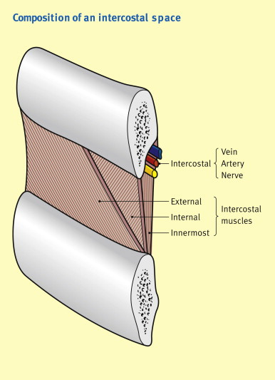
The neurovascular bundle consists of the intercostal vein, intercostal artery, and intercostal nerve, and it runs along the inferior aspect of the rib in the costal groove.
-
Describe the path of the neurovascular bundle from the spinal column to the sternum.
It runs along the inferior aspect of each rib within the intercostal space. The neurovascular bundle typically consists of the intercostal vein, artery, and nerve, and it travels laterally along the ribcage, supplying each intercostal space with blood vessels and nerves.
-

What is the consistent arrangement of structures within the neurovascular bundle from superior to inferior?
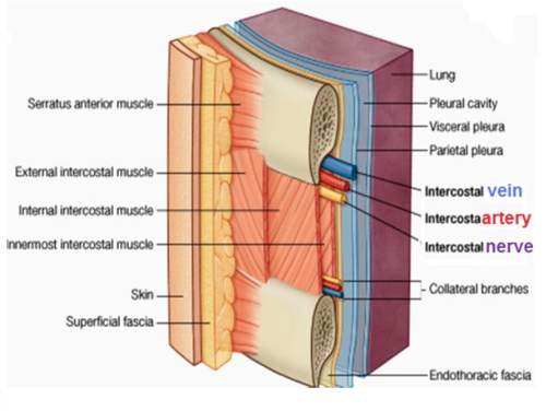
🍮Intercostal Vein
🍮Intercostal Artery
🍮Intercostal Nerve
REMEMBER V-A-N
-
What is the role of the diaphragm in the body?
The diaphragm forms the floor of the chest cavity and the roof of the abdominal cavity.
-
How does the diaphragm permit structures to pass between the chest and abdominal cavities?
The diaphragm has openings that allow structures, such as blood vessels and nerves, to pass between the two cavities.
-
Describe the structure of the diaphragm.
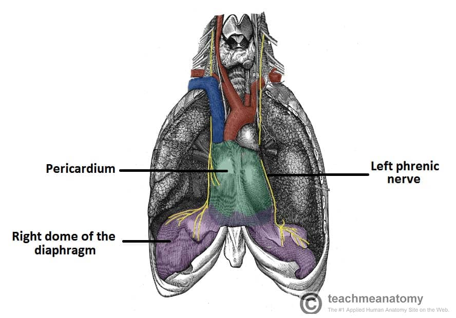
The diaphragm is a skeletal muscle with a central tendon.
It is anatomically arranged as right and left "domes," with the right dome typically being more superior due to the presence of the liver inferiorly.
-
What are the attachments of the muscular part of the diaphragm?
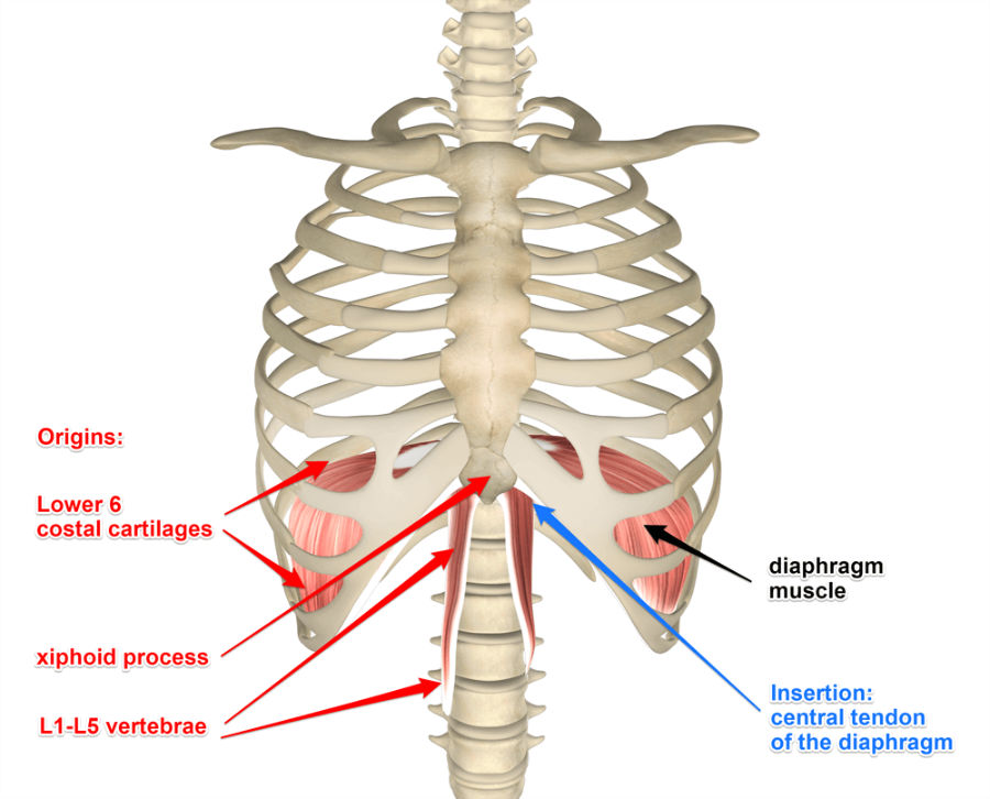
The muscular part of the diaphragm attaches peripherally to the sternum, the lower 6 ribs and their costal cartilages, and the L1-L3 vertebral bodies.
-
How is the muscular part of the diaphragm supplied neurologically?
The muscular part of the diaphragm is supplied by the phrenic nerve, arising from the anterior rami of spinal nerves C3, C4, and C5.
-
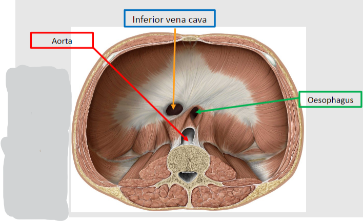
How many major openings are there in the diaphragm, and what structures do they allow to pass from the thoracic to the abdominal cavity?

There are three major openings in the diaphragm.
They allow the following structures to pass from the thoracic to the abdominal cavity:
🍮Inferior vena cava (IVC) passes through at the T8 level.
🍮Esophagus passes through at the T10 level.
🍮Aorta passes through at the T12 level.
-
How is the anteroposterior increase in thoracic volume illustrated during inspiration?

Anteroposterior increase is illustrated with a pump handle movement.
-
How is the transverse increase in thoracic volume illustrated during inspiration?

Transverse increase is illustrated using a bucket with two handles. Lifting handles is like raising right and left ribs at the costovertebral and costosternal joints.
-
How is the vertical dimension increased during inspiration?

The vertical dimension is increased by the downward pull of the diaphragm.
-
Which muscle is primarily responsible for inspiration?

The diaphragm is the primary muscle in inspiration.
-
Describe the process of relaxed inhalation.
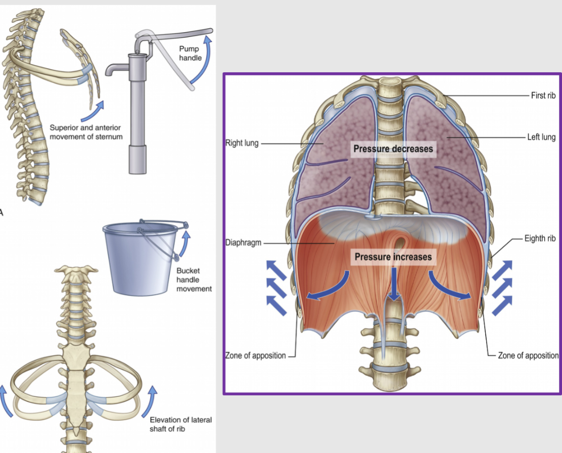
During relaxed inhalation, the diaphragm contracts, and the external intercostal muscles elevate the thoracic cavity, allowing air to enter the lungs.
-
Explain the process of relaxed exhalation.

During relaxed exhalation, the muscles above relax, allowing the lungs to recoil. This recoil pushes air out of the lungs.
-
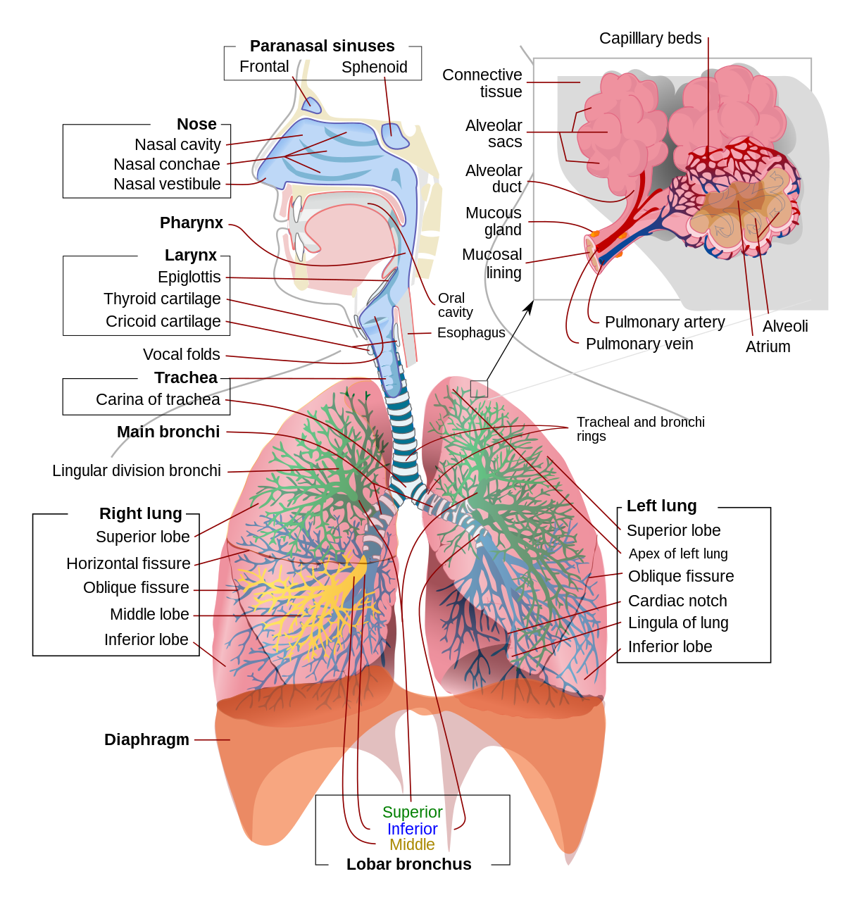
What structures are included in the upper respiratory tract?

The upper respiratory tract includes the nose, nasal passages, paranasal sinuses, pharynx, and the portion of the larynx above the vocal cords.
-
What structures are included in the lower respiratory tract?
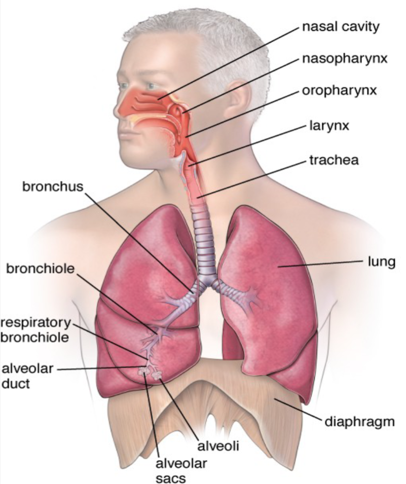
The lower respiratory tract includes the larynx below the vocal cords, trachea, bronchi, bronchioles, and lungs.
-
How does air enter the nasal cavity?

Air enters the nasal cavity.
-
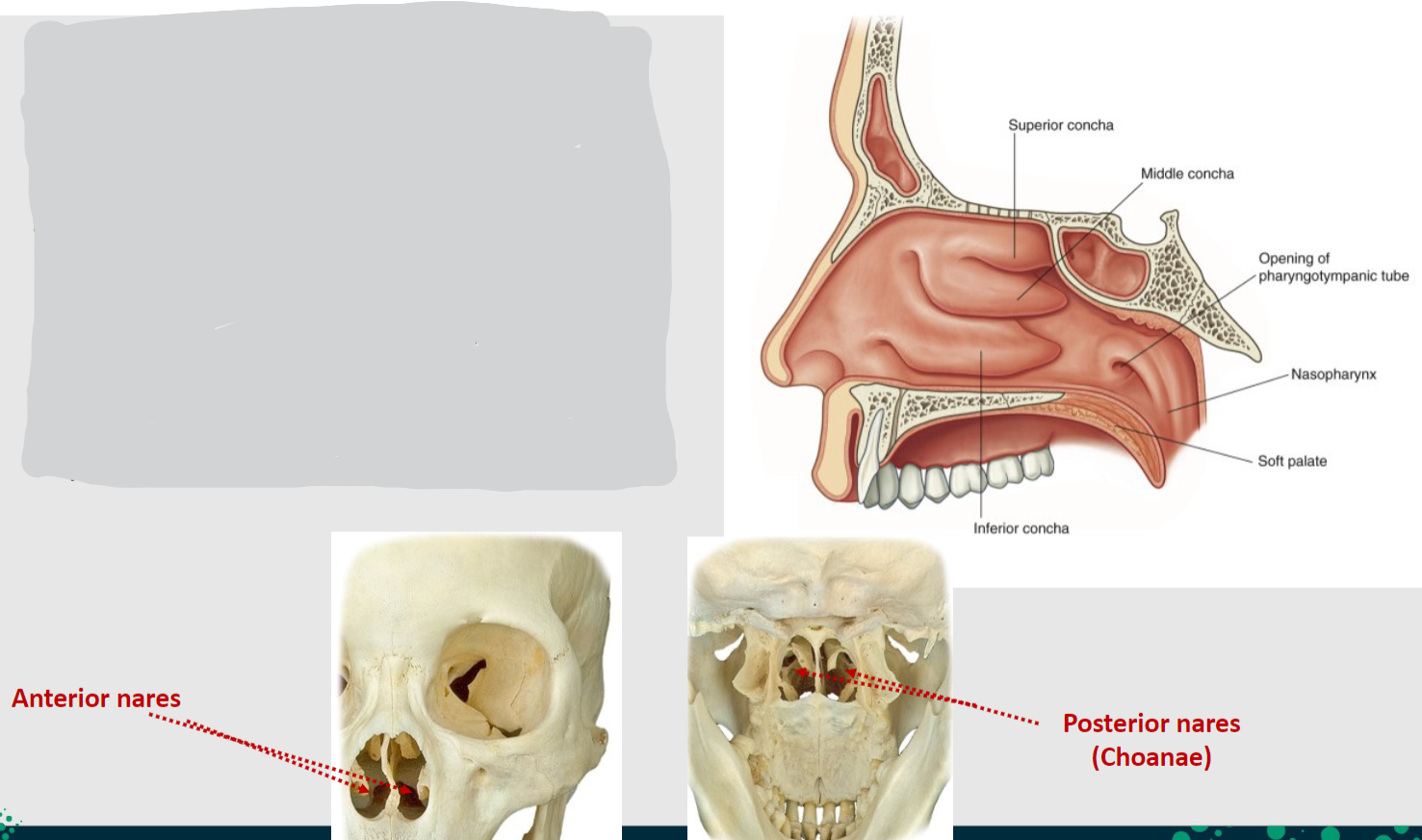
What structures serve as entry points into the nasal cavity?

The nasal cavity is entered anteriorly through the anterior nares (nostrils).
-
How does the nasal cavity communicate with the nasopharynx?

The nasal cavity opens posteriorly into the nasopharynx through the choanae (posterior nares).
-
What lines the nasal cavity, and what is the exception?
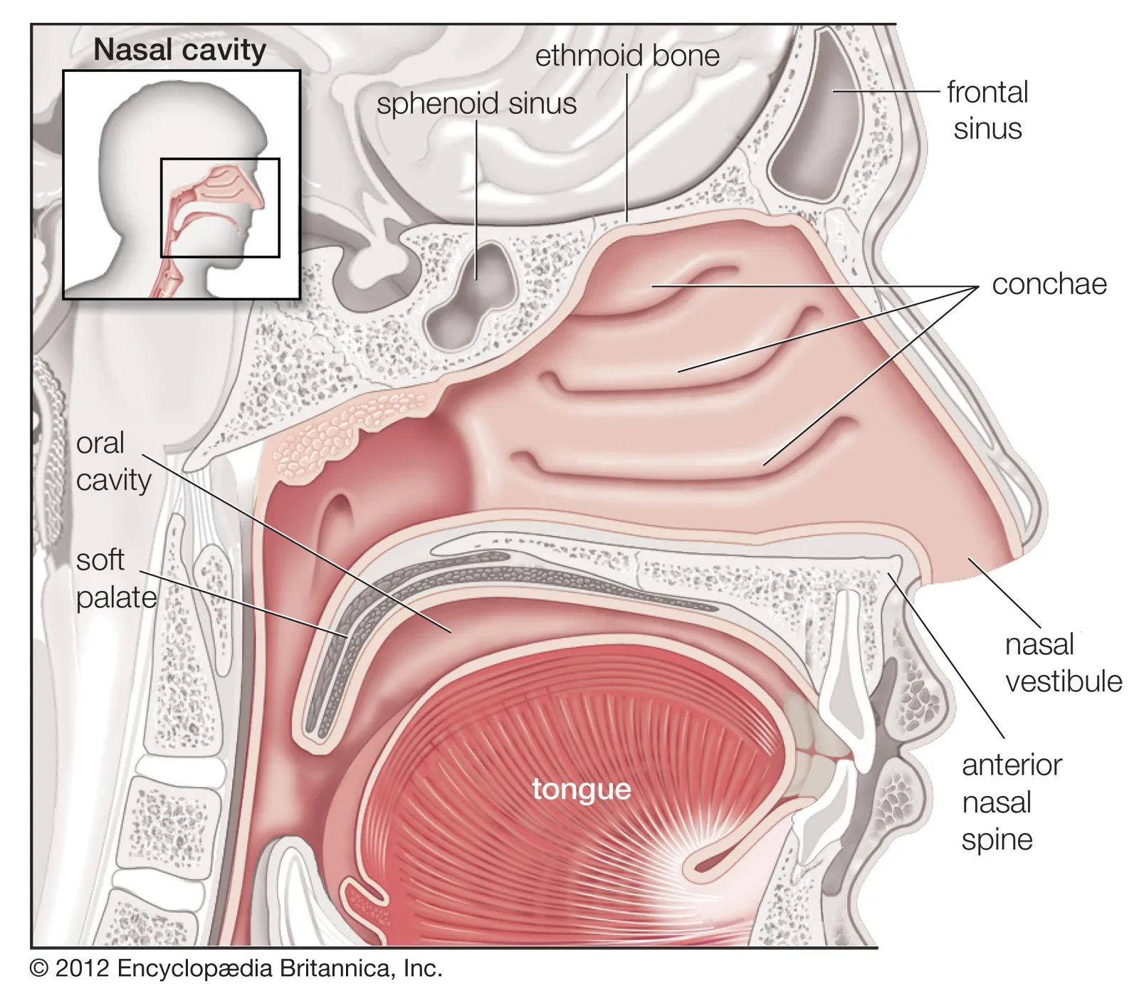
The nasal cavity is lined with mucosa, except for the nasal vestibule, which is lined with skin.
-
What type of epithelium lines the majority of the nasal cavity?
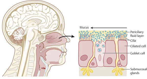
The majority of the nasal cavity is lined by respiratory epithelium.
-
What type of epithelium lines the nasal vestibule?
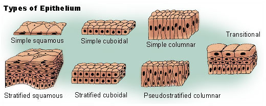
The nasal vestibule is lined by stratified squamous epithelium.
-
Describe the characteristics of respiratory epithelium.
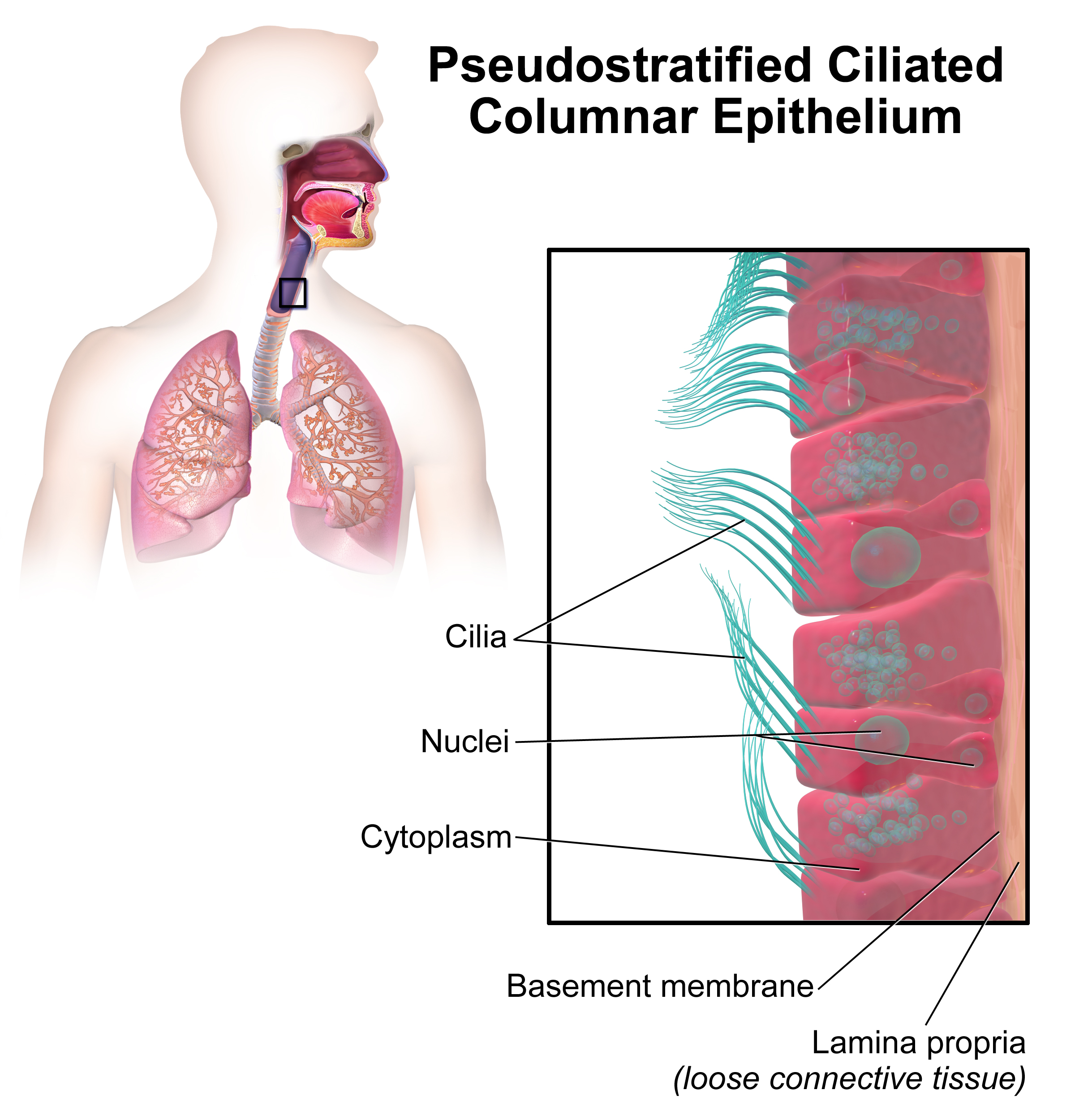
Respiratory epithelium is pseudostratified columnar ciliated epithelium with goblet cells.
-
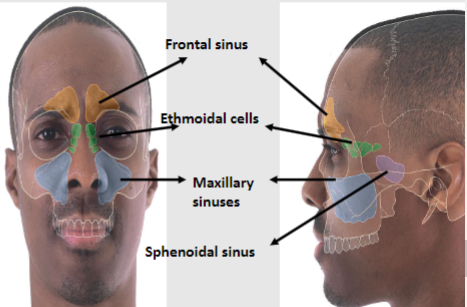
What are paranasal sinuses?
Paranasal sinuses are cavities within the skull filled with air.
-
What type of epithelial cells line the paranasal sinuses?
The paranasal sinuses are lined by ciliated pseudostratified columnar epithelial cells.
-
How are the paranasal sinuses connected to the nasal cavity?
The paranasal sinuses are connected to the lateral walls of the nasal cavity.
-
List three functions of the paranasal sinuses. (3)
🍮Lighten the skull.
🍮Increase resonance of voice.
🍮Humidify inspired air.
-
What innervates the paranasal sinuses, and what is their blood supply?
The paranasal sinuses are innervated by the trigeminal nerve (CN V) and supplied by branches of the external carotid artery.
-
Besides the nasal cavity, what other function does the oral cavity serve?
The oral cavity also acts as an air inlet.
-
What are the boundaries of the oral cavity? (5)
Anteriorly: Oral fissure.
Posteriorly: Oropharynx.
Laterally: Cheeks.
Superiorly: Palate (hard and soft).
Inferiorly: Muscular floor and the tongue.
-
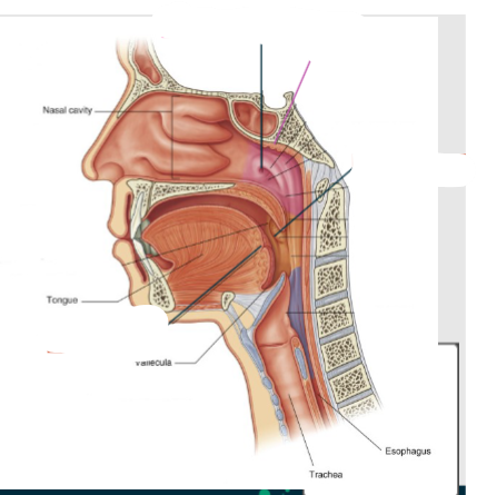
How many parts can the pharynx be divided into, and what are they?
The pharynx can be divided into three parts: Nasopharynx, oropharynx, and laryngopharynx.
-
Where is the nasopharynx located?
The nasopharynx is located posterior to the nasal cavities and above the soft palate.
-
Describe the location of the oropharynx.
The oropharynx is located posterior to the oral cavity, inferior to the level of the soft palate, and superior to the upper margin of the epiglottis.
-
Where does the laryngopharynx extend from and to?
The laryngopharynx extends from the superior margin of the epiglottis to the top of the esophagus.
-
What pathways do food and air take from the pharynx?
Food travels down the esophagus to the stomach, while air travels down the trachea to the lungs.
-
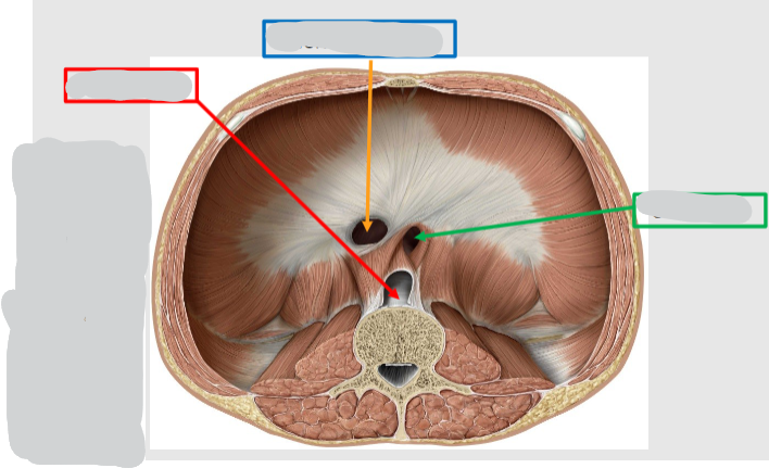
Name these openings in the diaphragm:
-
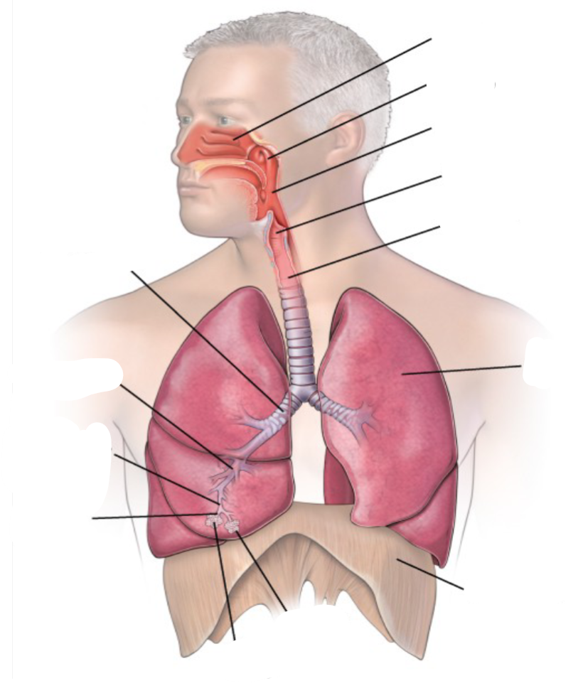
Name these parts of the lower and upper respiratory tract:

-
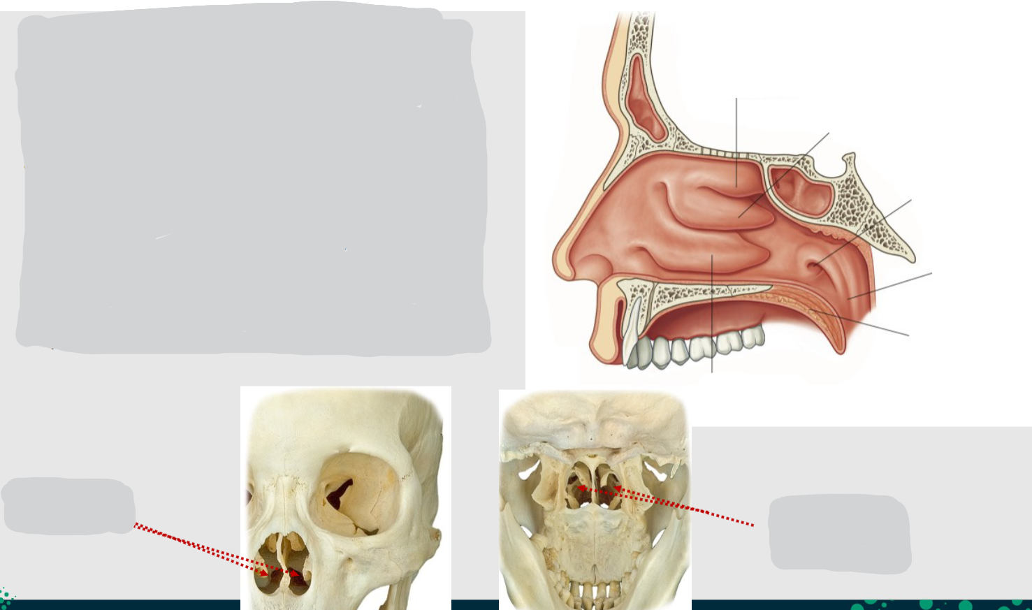
Name these parts of the nasal cavity:
-
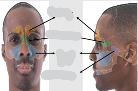
Name these sinuses:
-
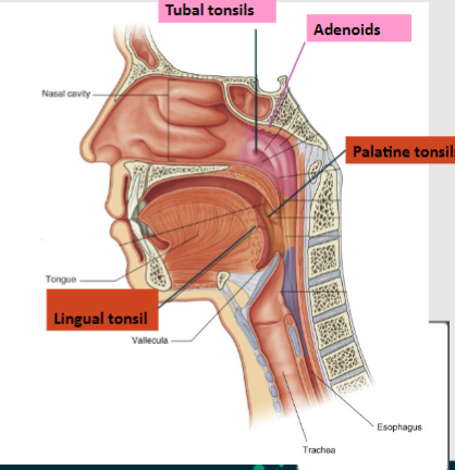
Name these parts of the tonsillar region:
-
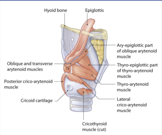
Picture demonstrating the parts of the larynx:
-
What is the larynx commonly known as?
The larynx is commonly known as the voice box.
-
How is sound created in the larynx?
Sound is created by forcing air through the vocal folds of the larynx, which vibrate to make noise. This process is how we talk and sing.
-
What is the function of the larynx in relation to air passage?
The larynx serves as a passage for air to travel into the trachea.
-
What structures surround the larynx, keeping it patent and protected?
The larynx is surrounded by several cartilage structures that maintain its patency and provide protection.
-
What is another name for the trachea?

The trachea is also known as the windpipe.
-
Describe the path of air through the trachea.
Air travels through the trachea, which is the tube transporting air to the lungs, after passing through the larynx.
-
What are the anatomical boundaries of the trachea?

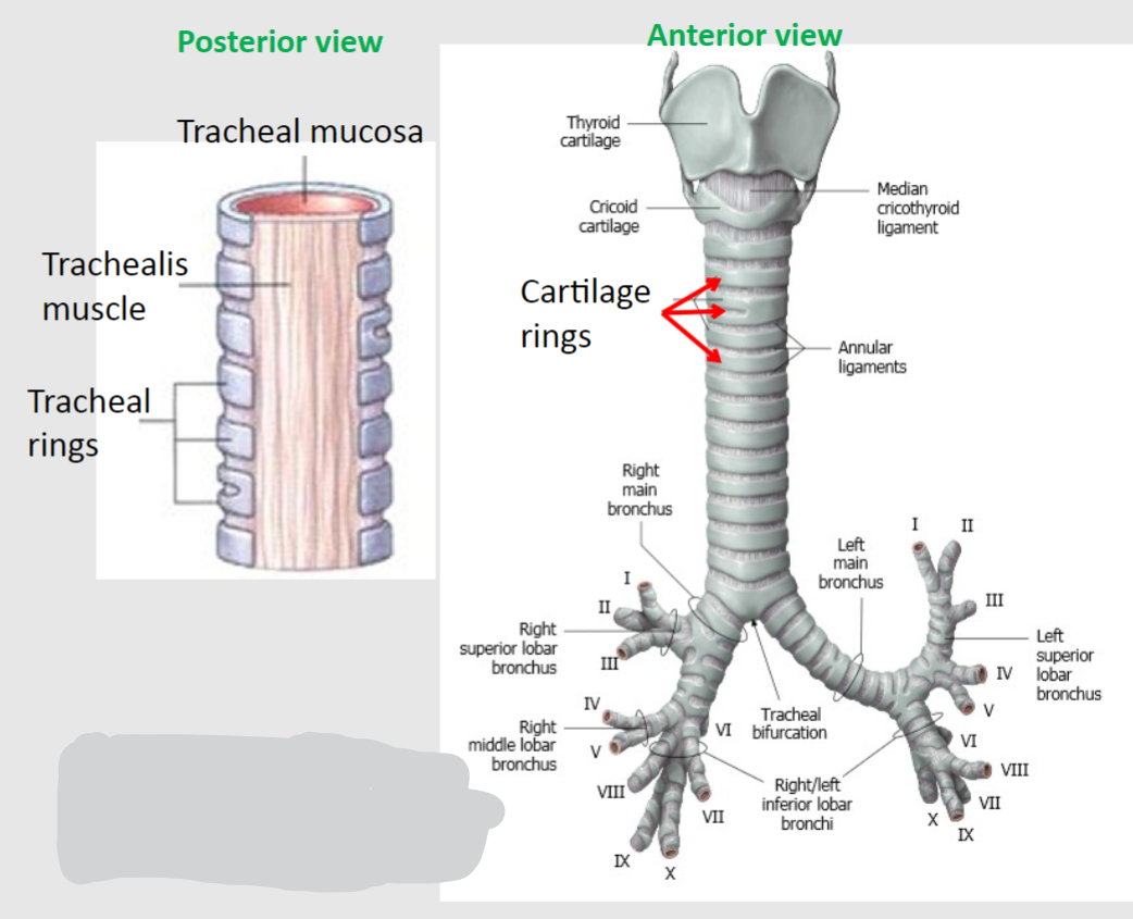
The trachea runs from the level of the lower border of the cricoid cartilage (C6/C7) to the T4/T5 vertebral level.
-
What happens at the bifurcation of the trachea?

At the T4/T5 vertebral level, the trachea bifurcates into the left and right primary (main) bronchi, leading to the left and right lungs.
-
Describe the composition of the trachea.

🍮The trachea is formed of "horse shoe" or "C-shaped" rings of hyaline cartilage held together by dense connective tissue.
🍮Posteriorly, at the junction where the trachea is in contact with the esophagus, there is a membrane void of cartilage covered in smooth muscle, known as the trachealis muscle.
-
Why are the tracheal rings C-shaped instead of being full circles?

🍮The tracheal rings are C-shaped rather than full circles to allow flexibility and movement during breathing and swallowing.
🍮This design prevents collapse of the trachea while also accommodating the movement of the adjacent esophagus during swallowing.
-
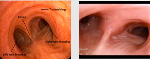
What is the carina in the context of the trachea?
The carina is a ridge located at the bifurcation of the trachea, which can be observed during bronchoscopy.
-
Why is the carina considered a key landmark?
The carina is a key landmark because it plays a crucial role in determining various pathologies. Widening or distortion of the carina can often indicate cancer in the lymph nodes located just inferior to the carina.
-
What are the lymph nodes found just inferior to the carina called?
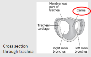
The lymph nodes located just inferior to the carina are called the inferior tracheobronchial lymph nodes.
-
What is the process of bronchoscopy?

Bronchoscopy involves the insertion of a bronchoscope down the trachea to enter a main bronchus.
-
What obstacle does the bronchoscope encounter during the procedure?
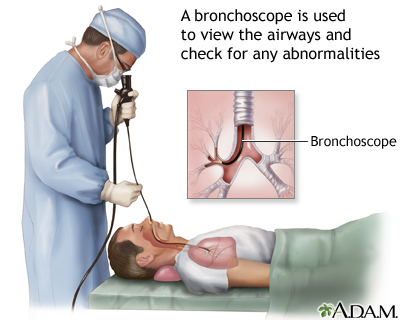
The bronchoscope must maneuver around the carina to reach the bronchus.
-
How can enlargement of the inferior tracheobronchial lymph nodes affect the carina during bronchoscopy?
Enlargement of the inferior tracheobronchial lymph nodes can blunt the carina, which can indicate pathology.
-
What are the characteristics of the bronchi in terms of cartilage support?
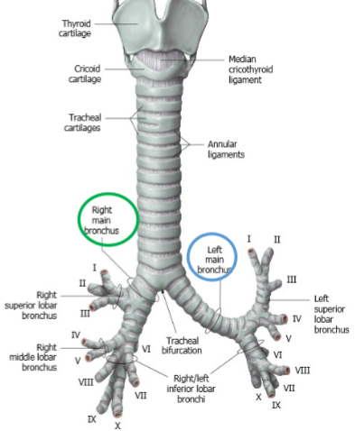
The bronchi have characteristic hyaline cartilage rings supporting them, similar to those found in the trachea.
-
How do the positions of the right and left bronchi differ?

The right bronchus is wider, shorter, and lies at a steeper vertical angle, while the left bronchus is narrower and more horizontal.
-
Why is the difference in position between the right and left bronchi important to know?

Inhaled foreign objects are more likely to get lodged in the right side bronchus due to its wider and more vertical orientation compared to the left bronchus.
-
From where does the trachea bifurcate into the left and right primary bronchi?

The trachea bifurcates into the left and right primary bronchi.
-
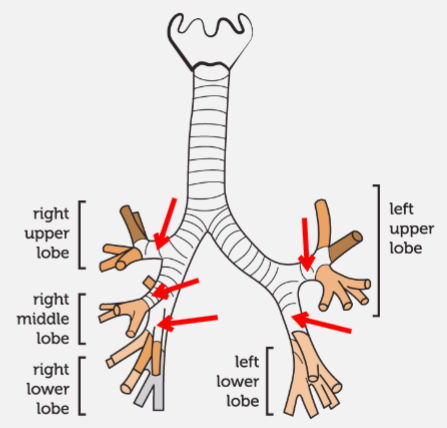
How do the left and right primary (main) bronchi further divide?

The left and right primary bronchi divide further into secondary (lobar) bronchi; there are 2 on the left and 3 on the right.
-
What is the relationship between the divisions of the bronchi and the lobes of the lungs?

The divisions of the bronchi correspond to the number of lobes in each lung.
-
What is the next level of bronchial division after the secondary bronchi?
The secondary bronchi further divide to give tertiary (segmental) bronchi; usually 10 for each lung.
-
What differentiates terminal bronchioles from earlier bronchial structures?
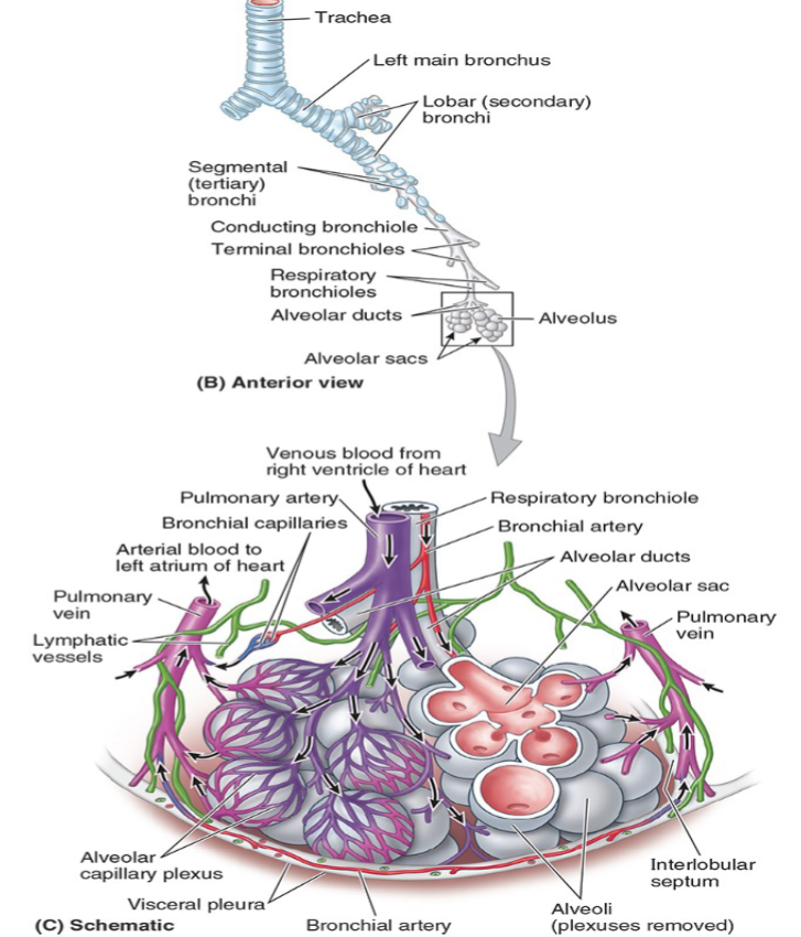
Terminal bronchioles no longer have hyaline cartilage in their walls.
-
What structures branch from terminal bronchioles?

Respiratory bronchioles branch from terminal bronchioles.
-
What is the structure at the end of each respiratory bronchiole?

Each respiratory bronchiole ends in an acinus of clustered alveoli.
-
What are the lungs primarily responsible for?

The lungs are the functional organs of respiration, primarily responsible for oxygenating blood.
-
How are the lungs positioned in relation to the mediastinum and ribcage?

The lungs are located on either side of the mediastinum and are protected by the ribcage.
-
What anatomical structure separates the thoracic cavity from the abdominal cavity?

The diaphragm lies inferior to the lungs and separates the thoracic cavity from the abdominal cavity.
-
How do the lungs achieve oxygenation of blood?

The lungs achieve oxygenation of blood by bringing inhaled air into close contact with oxygen-poor blood in the pulmonary capillaries.
-
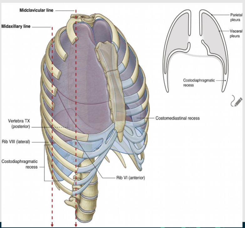
What are the boundaries of lung expansion in the body?

The lungs can expand down to the lower border of the ribs, known as the costal margin, and extend as far up as the root of the neck, above the 1st rib.
-
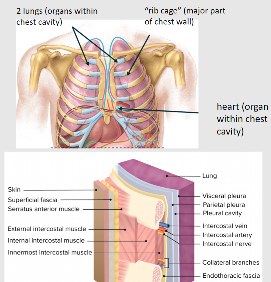
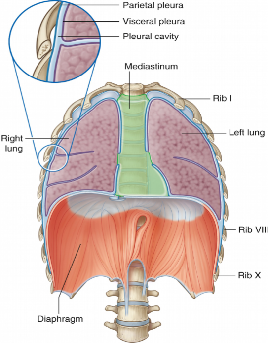
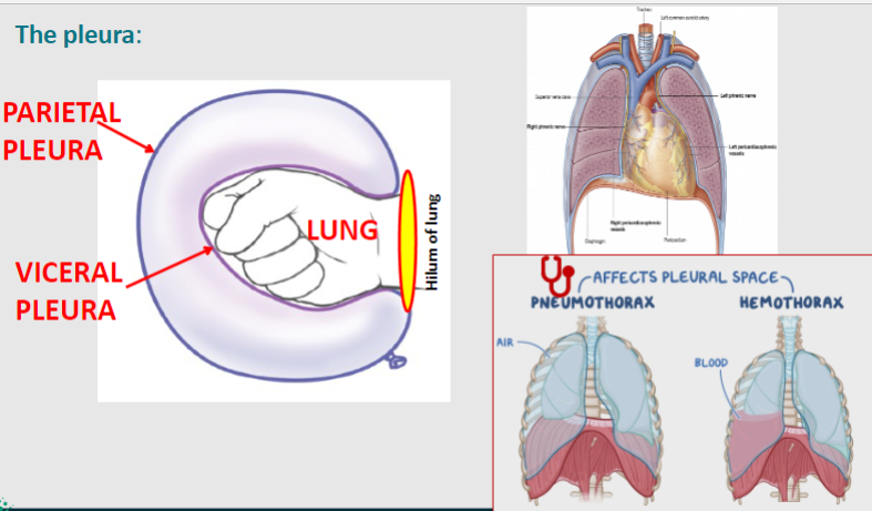
What is the pleura?

The pleura is a serous membrane that contains each lung within a pleural sac, flanking both sides of the heart and occupying most of the thoracic cavity.
-
What are the two serous layers composing each pleural sac?
Each pleural sac is composed of two serous layers: the parietal pleura and visceral pleura
-
What is the space between the parietal and visceral pleura called?

The space between the parietal and visceral pleura is called the pleural cavity.
-
What is the function of the pleural fluid?

The pleural fluid, found within the pleural cavity, provides lubrication between the two layers of pleura as the lungs expand, helping to prevent friction.
-
Picture demonstrating the pleura:
-
What is the parietal pleura, and what does it line?

The parietal pleura lines the internal surface of the thoracic cavity.
-
How is the parietal pleura named, depending on the area it lines?

Diaphragmatic parietal pleura: lines the superior surface of the diaphragm.
Mediastinal parietal pleura: lines the lateral surface of the mediastinum, which contains the heart.
Costal parietal pleura: lines the internal surface of the ribs.
Cervical parietal pleura: extends above rib 1 to the root of the neck.
-
What does the visceral pleura surround, and how is it attached?
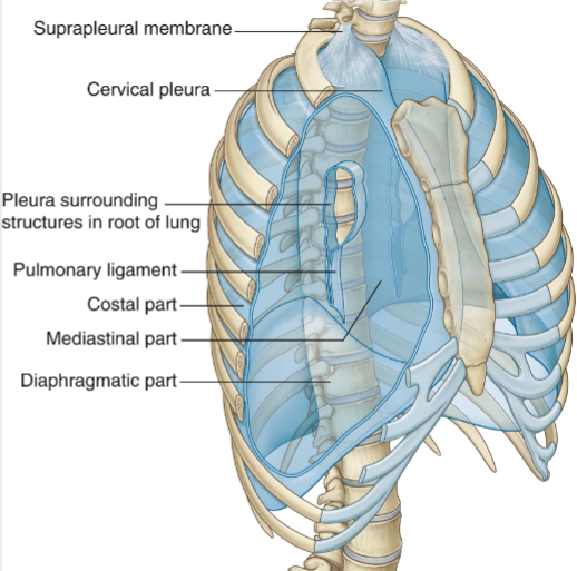
The visceral pleura surrounds and is intimately attached to each lung, following the contour of the lobes.
-
Where is the visceral pleura contiguous with the parietal pleura?

The visceral pleura is contiguous with the parietal pleura at the hilum of each lung.
-
How many lobes are there in the left lung, and how are they divided?

The left lung has 2 lobes: superior and inferior, which are divided by the oblique fissure.
-
How many lobes are there in the right lung, and how are they divided?
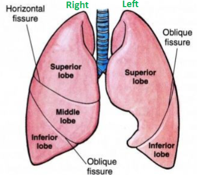
The right lung has 3 lobes: superior, middle, and inferior, which are divided by the oblique fissure and horizontal fissure.

