 Fatima Adam

The Nervous System (Anatomy 2)
Lecture: The Nervous System Session Introduction Dear Students, This session has been broken down into smaller chunks based on the learning objectives, for you to understand different concepts easily. Please remember to take breaks between the talks and use the additional resources given to support your learning. Learning outcomes At the end of this session, you will be able to: Define, in general terms, the central and peripheral nervous systems. Describe and identify the basic anatomy of the brain and brain stem. Briefly describe the names and functions of the twelve cranial nerves. Describe and identify the basic anatomy of the spinal cord, spinal nerves and peripheral nerves. Describe the meningeal coverings of brain and spinal cord.
-
What is the nervous system, and what are its main functions? (6)
Control center of the body
Controls and coordinates the activities of the body's systems
Collects information about the state of the body and its surroundings
Transmits information to the relevant area of the nervous system
Analyzes information
Initiates appropriate responses
-
What is a neuron? (1)
A cell forming the elements of the nervous system
-
How is the nervous system anatomically divided? (2)
Central Nervous System (CNS): Brain and spinal cord
Peripheral Nervous System (PNS): Spinal nerves (Peripheral nerves) and cranial nerves
-
How is the nervous system classified functionally? Provide an example of each type. (2)
Autonomic (subconscious): e.g., Control of heart rate
Somatic (conscious/voluntary): e.g., Picking up a pencil
-
What are ganglia and nuclei? (2)
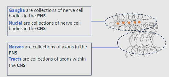
Ganglia: Collections of nerve cell bodies in the PNS
Nuclei: Collections of nerve cell bodies in the CNS
-
What are nerves and tracts? (2)

Nerves: Collections of axons in the PNS
Tracts: Collections of axons in the CNS
-
What are the three embryological regions of the brain? (3)
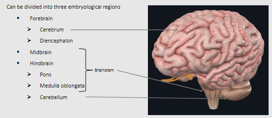
Forebrain: Cerebrum and diencephalon
Midbrain
Hindbrain: Pons, medulla oblongata, and cerebellum
-
What are the main features and functions of the cerebrum? (5)

Largest area of the brain, divided into two cerebral hemispheres (longitudinal fissure)
Conscious thought processes and intellectual function
Memory storage, processing, and retrieval
Regulation of skeletal muscle contraction (conscious and subconscious)
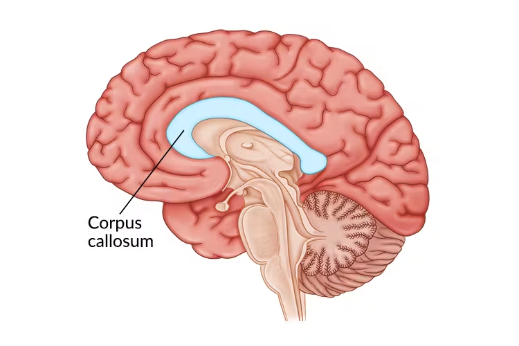
Connected by the corpus callosum
-
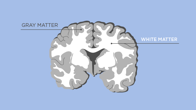
What are the layers of the cerebrum? (3)

Surface layer: Cortex (gray matter - neuronal cell bodies)
Inner layers: White matter (neuronal axons)
Innermost: Deep nuclei (gray matter - diencephalon)
-
What are the functions of the cerebellum? (3)
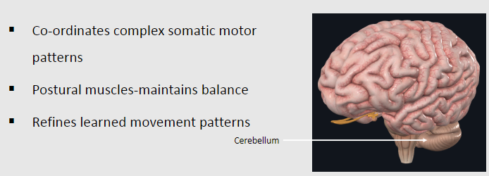
Coordinates complex somatic motor patterns
Maintains balance via postural muscles
Refines learned movement patterns
-
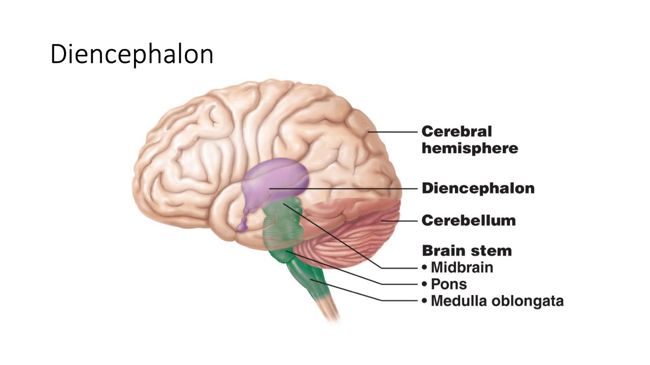
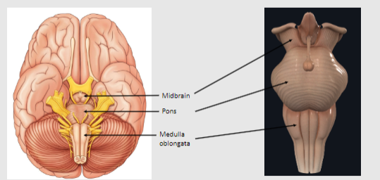
What are the main components of the brainstem? (3)

Midbrain
Pons
Medulla oblongata
-
What are the functions of the brainstem? (4)
Processes and relays information between the cerebrum and cerebellum
Contains reflex centres for respiratory and cardiovascular functions
Houses cranial nerve nuclei
Continuation into the spinal cord
-
What are the components of the diencephalon and their functions? (2)
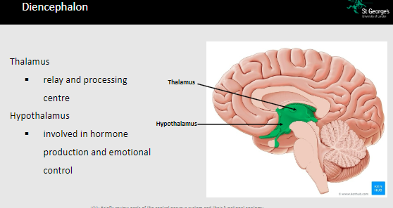
Thalamus: Relay and processing centre
Hypothalamus: Involved in hormone production and emotional control
-
What are the features of the spinal cord? (3)
Continuation of the brainstem into the vertebral column
Contains outer white matter and inner gray matter
Transmits information to and from the brain, connecting to the PNS
-
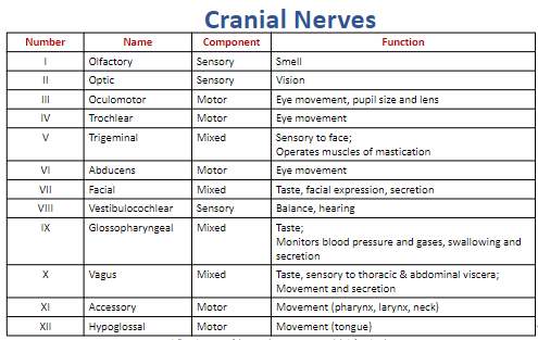
What is the function and classification of the olfactory nerve (Cranial Nerve I)? (2)
Classification: Sensory
Function: Smell
-
What is the function and classification of the optic nerve (Cranial Nerve II)? (2)
Classification: Sensory
Function: Vision
-
What is the function and classification of the oculomotor nerve (Cranial Nerve III)? (4)
Classification: Motor
Function:
Eye movement
Pupil size regulation
Lens accommodation
-
What is the function and classification of the trochlear nerve (Cranial Nerve IV)? (2)
Classification: Motor
Function: Eye movement
-
What is the function and classification of the trigeminal nerve (Cranial Nerve V)? (3)
Classification: Mixed
Function:
Sensory: Face sensation
Motor: Operates muscles of mastication
-
What is the function and classification of the abducens nerve (Cranial Nerve VI)? (2)
Classification: Motor
Function: Eye movement
-
What is the function and classification of the facial nerve (Cranial Nerve VII)? (4)
Classification: Mixed
Function:
Sensory: Taste
Motor: Facial expression
Secretion (e.g., salivary and tear glands)
-
What is the function and classification of the vestibulocochlear nerve (Cranial Nerve VIII)? (2)
Classification: Sensory
Function:
Balance
Hearing
-
What is the function and classification of the glossopharyngeal nerve (Cranial Nerve IX)? (4)
Classification: Mixed
Function:
Sensory: Taste, monitors blood pressure and gases
Motor: Swallowing
Secretion (e.g., saliva)
-
What is the function and classification of the vagus nerve (Cranial Nerve X)? (4)
Classification: Mixed
Function:
Sensory: Taste, sensation to thoracic and abdominal viscera
Motor: Movement and secretion
-
What is the function and classification of the accessory nerve (Cranial Nerve XI)? (2)
Classification: Motor
Function: Movement of the pharynx, larynx, and neck
-
What is the function and classification of the hypoglossal nerve (Cranial Nerve XII)? (2)
Classification: Motor
Function: Tongue movement
-
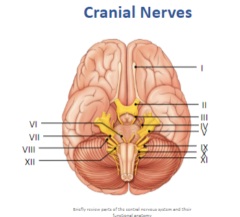
Label these cranial nerves:
-
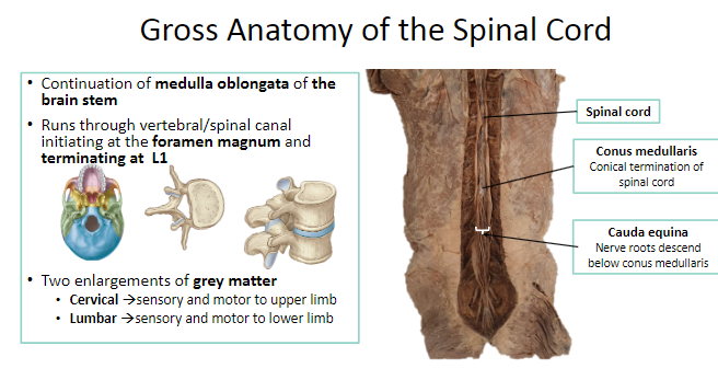
Describe the anatomy of the spinal cord and its coverings. (4)
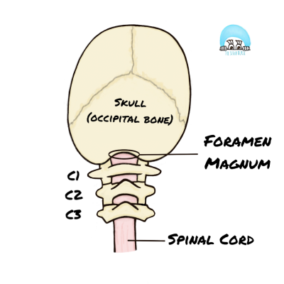
Continuation of the brainstem into the vertebral column through the foramen magnum.
Runs through the vertebral/spinal canal.
Initiates at the foramen magnum and terminates at L1.
Covered by meninges (dura mater, arachnoid mater, and pia mater).
-
What are the two enlargements of the spinal cord, and what are their functions? (2)
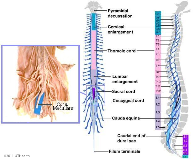
Cervical enlargement: Provides sensory and motor innervation to the upper limbs.
Lumbar enlargement: Provides sensory and motor innervation to the lower limbs.
-
What is the cauda equina? (1)
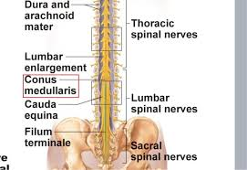
A collection of nerve roots descending below the conus medullaris.
-
What is the conus medullaris? (1)
The conical termination of the spinal cord.
-
Identify the major landmarks of the spinal cord within the vertebral canal. (3)
Foramen magnum: Entry point of the spinal cord into the vertebral column.
Termination at L1: End of the spinal cord.
Cauda equina: Nerve roots below the conus medullaris.
-
Describe the anatomical relationships of the meninges to the spinal cord. (4)
Dura mater: Tough outer layer protecting the spinal cord.
Arachnoid mater: Middle layer containing cerebrospinal fluid (CSF).
Pia mater: Inner layer adhering to the spinal cord.
Dorsal and ventral nerve roots pass through the meninges, with potential for compression in conditions like herniated discs.
-
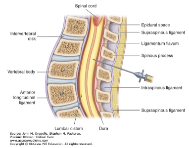
What is the anatomy relevant to performing a lumbar puncture? (4)

Performed below L1 to avoid damaging the spinal cord.
Inserted into the subarachnoid space to collect cerebrospinal fluid (CSF).
Needle passes through skin, subcutaneous tissue, supraspinous ligament, interspinous ligament, and ligamentum flavum.
Avoids the conus medullaris; targets the cauda equina.
-
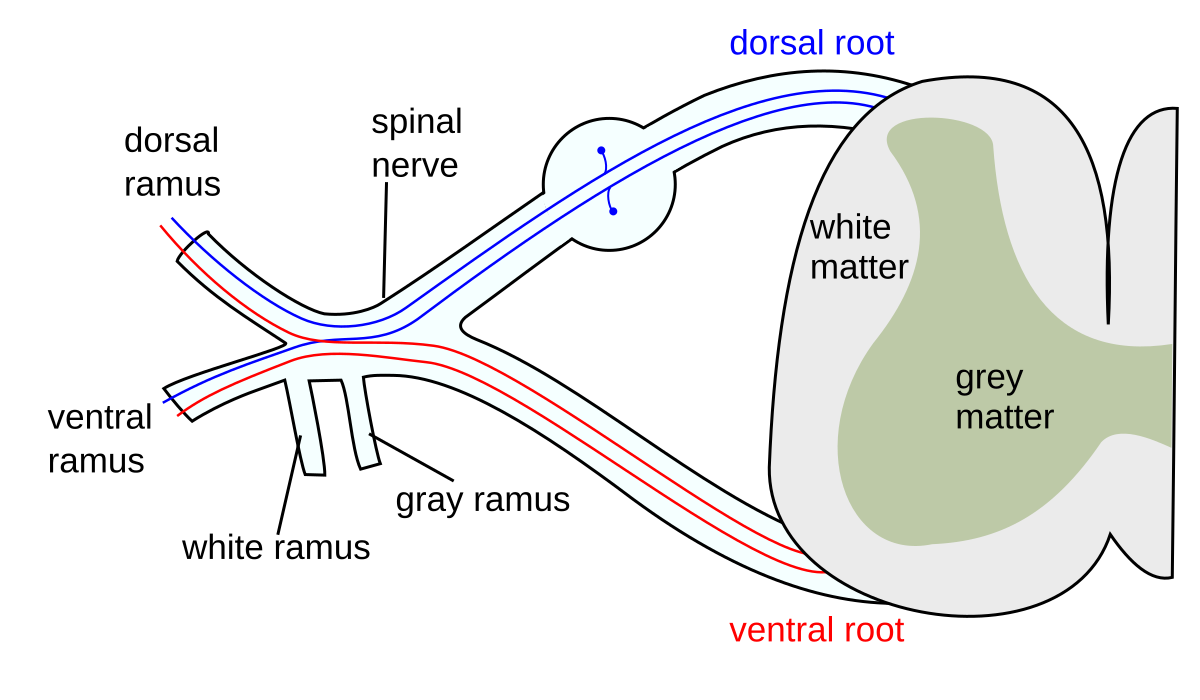
Describe the anatomy of a typical spinal nerve, including its origin and branches. (4)
Originates from dorsal (sensory) and ventral (motor) spinal roots.
Main branches:
Dorsal ramus: Innervates muscles and skin of the back.
Ventral ramus: Forms plexuses and innervates limbs and anterior body wall.
Autonomic components: Sympathetic and parasympathetic fibres for visceral control.
-

Label the Gross Anatomy of the Spinal Cord: (3)
-
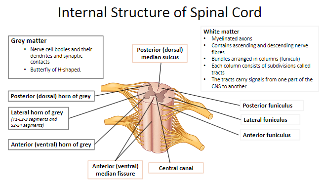
What is the composition of grey matter in the spinal cord? (3)

Contains nerve cell bodies, dendrites, and synaptic contacts (synapses).
Shaped like a butterfly or an "H."
Divided into:
Posterior (dorsal) horn: Receives sensory input.
Anterior (ventral) horn: Contains motor neurons.
Lateral horn: Present in T1-L2/3 and S2-S4 segments; contains autonomic neurons.
-
What are the key landmarks in the grey matter of the spinal cord? (3)
Posterior (dorsal) horn: Sensory processing.
Anterior (ventral) horn: Motor output.
Lateral horn: Autonomic function (only in specific segments).
-
What is the composition of white matter in the spinal cord? (4)

Made of myelinated axons.
Contains ascending and descending nerve fibers.
Organized into columns (funiculi):
Posterior funiculus
Lateral funiculus
Anterior funiculus
Columns are further subdivided into tracts that carry specific signals.
-
What are the main functions of the tracts in white matter? (2)

Ascending tracts: Transmit sensory information to the brain.
Descending tracts: Transmit motor commands from the brain to the body.
-
Identify the external landmarks of the spinal cord. (4)

Posterior (dorsal) median sulcus: Shallow groove on the posterior side.
Anterior (ventral) median fissure: Deep groove on the anterior side.
Central canal: Runs through the center of the spinal cord, contains cerebrospinal fluid.
Funiculi (columns): Bundles of nerve fibers organized for specific functions.
-

Lable the Internal Structure of Spinal Cord: (11)

-
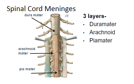
What are the three layers of the spinal cord meninges, from outermost to innermost? (3)

Dura mater: Outermost, tough fibrous layer.
Arachnoid mater: Middle, web-like layer.
Pia mater: Innermost, delicate layer adhering to the spinal cord.
-
What is the relationship of the meninges to the spinal cord and nerve roots? (4)

The dura mater surrounds the spinal cord and nerve roots, forming the dural sac.
The arachnoid mater lines the dura mater and encloses the subarachnoid space containing cerebrospinal fluid (CSF).
The pia mater is directly adhered to the spinal cord and nerve roots, anchoring them to the coccyx via the filum terminale.
The meninges protect the spinal cord and provide a cushion for nerve roots.
-
How does root compression relate to the meninges? (2)

Compression occurs when nerve roots are pressed against the dura mater due to herniated discs, tumors, or inflammation.
Can lead to pain, numbness, or motor deficits depending on the affected nerve root.
-
What is the purpose of an epidural injection, and where is it placed? (3)

Delivers medication (e.g., anesthetics, steroids) into the epidural space outside the dura mater.
Commonly used for pain relief during labor or to treat chronic back pain.
The needle does not penetrate the dura mater.
-
What is the purpose of a lumbar puncture, and where is it performed? (3)

Used to obtain cerebrospinal fluid (CSF) for diagnostic tests or to administer medications.
Performed between L3-L4 or L4-L5 vertebrae to avoid damaging the spinal cord (ends at L1-L2).
Needle passes through:
Skin and subcutaneous tissue.
Supraspinous and interspinous ligaments.
Ligamentum flavum.
Epidural space.
Dura mater and arachnoid mater into the subarachnoid space.
-
What are the landmarks for performing a lumbar puncture? (3)
Iliac crests: Imaginary line connecting the crests crosses L4.
L3-L4 or L4-L5 intervertebral space: Safe region below the spinal cord termination.
Patient positioned in lateral decubitus (side-lying) or sitting position.
-
What is the epidural space, and why is it clinically important? (2)

The space between the dura mater and the walls of the vertebral canal.
Contains fat and venous plexuses, and is the site for epidural anesthesia.
-
What is the subarachnoid space, and what does it contain? (2)

The space between the arachnoid mater and pia mater.
Contains cerebrospinal fluid (CSF), which cushions the spinal cord.
-
What is the extent of the dura mater over the spinal cord? (2)

Extends from the foramen magnum to the S2 vertebral level.
Continues as separate sheaths for each dorsal and ventral root, becoming continuous with the epineurium of spinal nerves.
-
What is the extent of the arachnoid mater over the spinal cord? (1)

Extends to the S2 vertebral level, closely associated with the dura mater.
-
What is the extent of the pia mater over the spinal cord? (2)
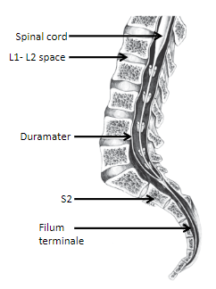
Covers the spinal cord and continues as the filum terminale to the coccyx.
Anchors the spinal cord to the coccyx, providing stability.
-
What is the filum terminale, and what is its composition? (3)

A thin filament extending from the conus medullaris to the coccyx.
Composed of:
Pia mater.
Surrounding continuation of the central canal.
Some neural elements.
-
How does the dura mater relate to the dorsal and ventral roots? (3)

Forms a separate sheath for each dorsal and ventral root.
Extends over the dorsal root ganglion (DRG) posteriorly.
Becomes continuous with the epineurium of the spinal nerve.
-
What is the conus medullaris, and where is it located? (2)

The conical termination of the spinal cord.
Located at the L1-L2 intervertebral space in adults.
-
What is the relationship between the dura mater and spinal nerves? (2)
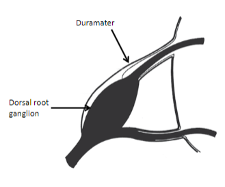
The dura mater extends over the spinal nerve roots.
It transitions into the epineurium, the outer connective tissue covering of peripheral nerves.
-
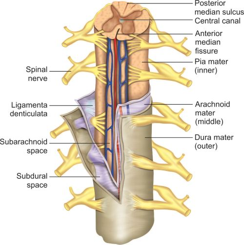
What is the ligamentum denticulatum, and what is it made of? (2)

A thin ridge of pia mater on each side of the spinal cord.
Made up of 21 pointed projections of pia mater extending laterally.
-
What is the extent of the ligamentum denticulatum? (2)

Extends from the foramen magnum to the L1 vertebra.
Anchors the spinal cord within the duramater.
-
What is the function of the ligamentum denticulatum? (1)
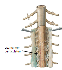
Limits the movement of the spinal cord within the duramater.
-
What does the subarachnoid space contain? (1)

Cerebrospinal fluid (CSF)
-
What is the dura-arachnoid interface also known as, and what is it classified as? (2)

Also known as the subdural (potential) space.
Classified as a potential space.
-
What is the extradural (epidural) space, and what does it contain? (2)
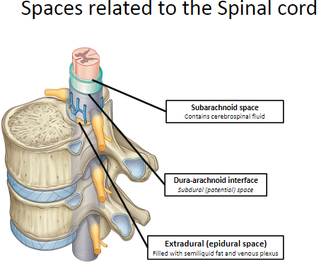
The space outside the dura mater.
Filled with semiliquid fat and a venous plexus.
-
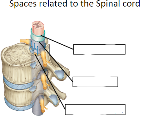
Label the Spaces related to the Spinal cord: (3)

-
What is the purpose of a lumbar puncture? (1)

A needle is used to collect a sample of cerebrospinal fluid from the subarachnoid space.
-
What must be avoided during a lumbar puncture? (1)

Spinal cord.
-
Where is the terminal end of the spinal cord located? (2)

At the lower border of the L1 vertebra or the L1-L2 intervertebral disc.
-
What is the surface landmark used for a lumbar puncture? (2)
The highest point of the iliac crest (L4).
The needle is inserted one or two spaces above this level.
-
What is radiculopathy? (1)

A sensory or motor disturbance caused by pathology of a nerve root.
-
What is the most common cause of radiculopathy? (1)

Intervertebral disc herniation.
-
What are other causes of radiculopathy? (3)
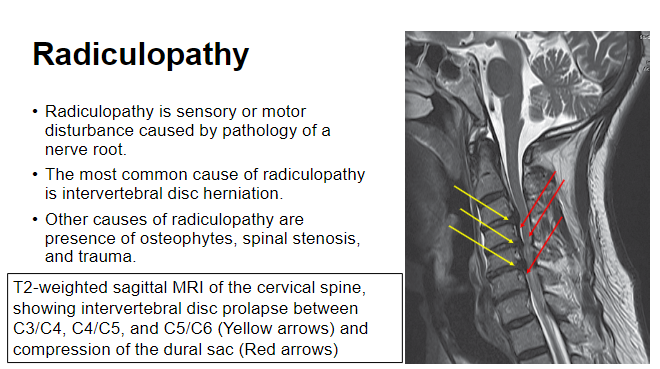
Osteophytes.
Spinal stenosis.
Trauma.
-
What are the main components of a typical spinal nerve? (2)

Contains motor and sensory fibers.
Divides into anterior and posterior ramus.
-
What is the function of the posterior ramus? (2)

Somatic motor to intrinsic back muscles.
Somatic sensory to the skin of the back.
-
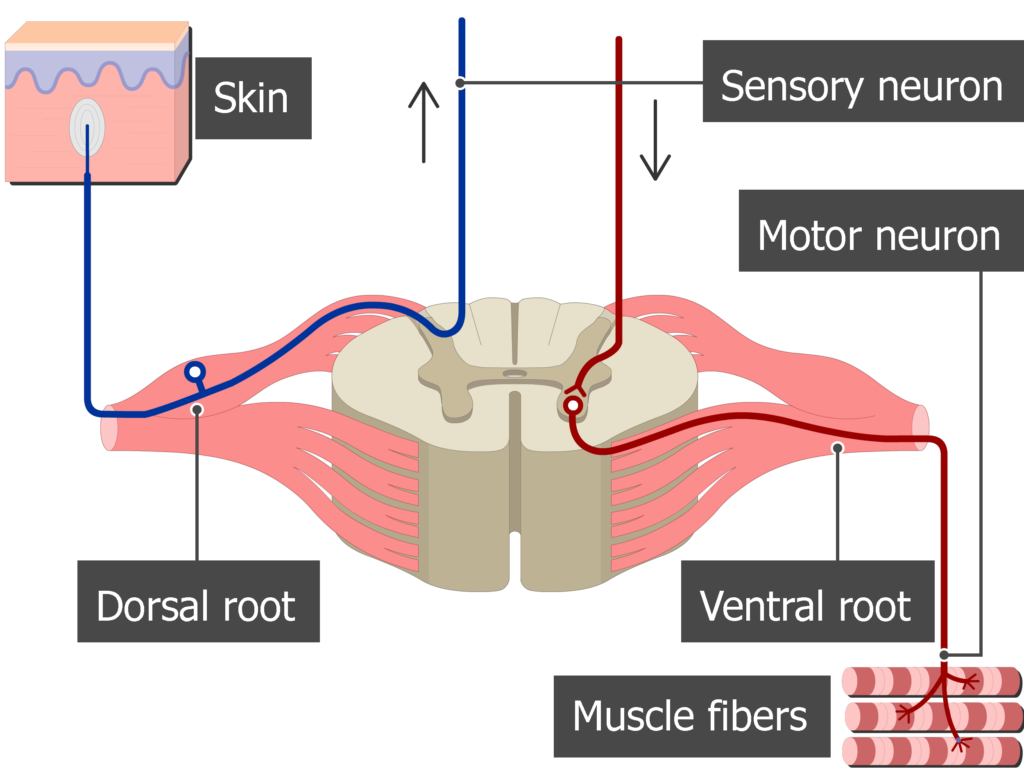
What is the function of the anterior (ventral) root? (2)

Motor (efferent neurons):
Somatic: Muscles.
Visceral: Organs (autonomic nerves).
-
What is the function of the posterior (dorsal) root? (2)

Sensory (afferent neurons):
Somatic: Skin.
Visceral: Organs (e.g., visceral peritoneum).
-
Where are the cell bodies of sensory neurons located in the spinal nerve? (1)

In the dorsal root ganglia.
-
What is the function of the anterior (ventral) ramus? (2)
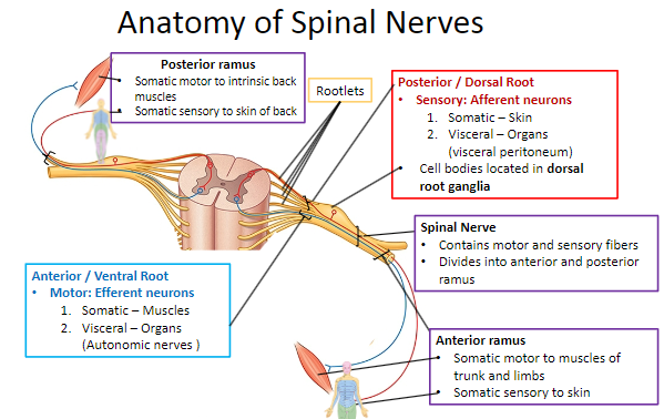
Somatic motor to muscles of the trunk and limbs.
Somatic sensory to the skin.
-
Label the Anatomy of Spinal Nerves, and provide further detail if necessary: (6)

-
How many pairs of spinal nerves are there? (1)
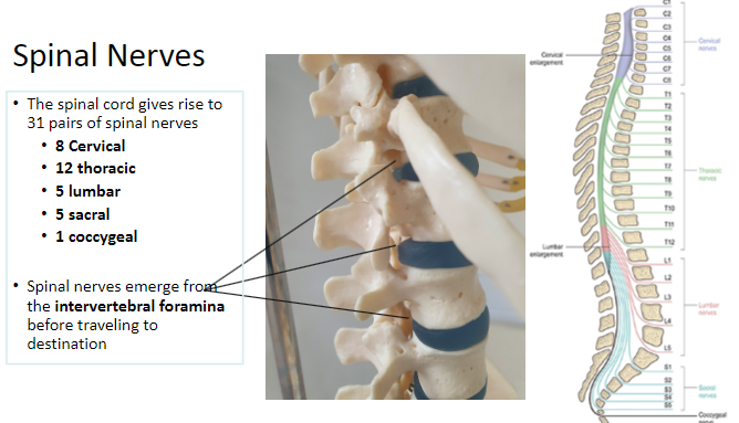
There are 31 pairs of spinal nerves.
-
What are the regions and number of spinal nerves in each region? (5)

8 Cervical
12 Thoracic
5 Lumbar
5 Sacral
1 Coccygeal
-
Where do spinal nerves emerge from? (1)

Spinal nerves emerge from the intervertebral foramina before traveling to their destination.
-
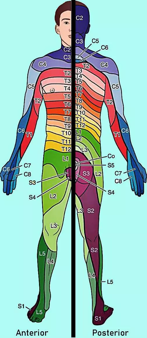
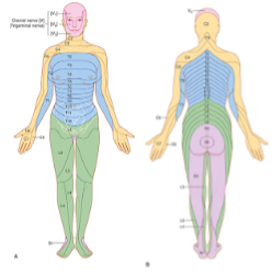
What is a dermatome? (1)

A dermatome is a skin area supplied by the sensory fibers of a single nerve root.
-
What is a myotome? (1)

A myotome is a group of muscles primarily innervated by the motor fibers of a single nerve root.
-
Why are the patterns of distribution of myotomes and dermatomes consistent from person to person? (1)
The patterns are relatively consistent due to the development of the limbs.
-
Where is the neuronal body of the autonomic component located? (1)

The neuronal body of the autonomic component is located in the lateral horn.
-
Where do preganglionic sympathetic fibers travel and what is their role? (2)

Preganglionic sympathetic fibers travel through the ventral root (efferent) and are responsible for carrying signals from the spinal cord to the sympathetic ganglia.
-
What are the two types of ramus communicans and what do they connect? (2)

White ramus communicans (pre-ganglionic): Connects the spinal nerve to the sympathetic ganglia.
Grey ramus communicans (post-ganglionic): Connects the sympathetic ganglia to the spinal nerve.
-
Where are the visceral sensory fibers located? (1)
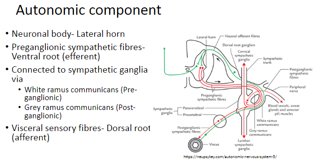
The visceral sensory fibers are located in the dorsal root (afferent).
-
What are the three layers of the meninges surrounding the brain? (3)
Dura Mater: Tough, fibrous outermost covering.
Arachnoid Mater: Consists of the arachnoid membrane and arachnoid trabeculae.
Pia Mater: Blood vessels run along the surface within the subarachnoid space.
-
Describe the two layers of the dura mater around the brain. (2)
Endosteal layer: Periosteum over the skull bones.
Meningeal layer: The proper dura mater that is continuous with the dura mater of the spinal cord.
-
What are the functions of the inward septa sent by the dura mater? (3)
Divide the cranial cavity into freely communicating spaces.
Contain subdivisions of the brain.
Restrict the rotatory displacement of the brain.
-
What is the Falx Cerebri and where is it located? (2)

The Falx Cerebri lies in the midline between the two cerebral hemispheres.
It is a reflection of the dura mater that separates the cerebral hemispheres.
-
What is the Diaphragma Sellae and its function? (2)

The Diaphragma Sellae is a small circular fold of dura.
It allows the passage of the infundibulum (pituitary stalk).
-
Describe the Falx Cerebelli and its location. (2)

The Falx Cerebelli projects forward between the cerebellar hemispheres.
It is another reflection of the dura mater.
-
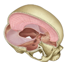
What is the function of the Tentorium Cerebelli? (3)
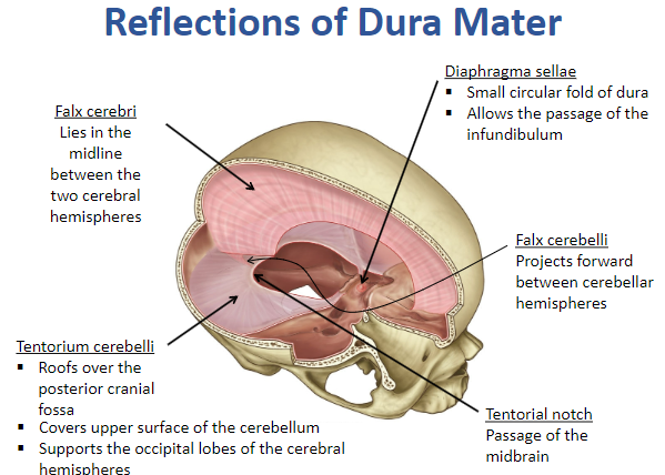
The Tentorium Cerebelli roofs over the posterior cranial fossa.
It covers the upper surface of the cerebellum.
It supports the occipital lobes of the cerebral hemispheres.
-
What is the Tentorial Notch and its significance? (2)

The Tentorial Notch is an opening in the tentorium cerebelli.
It allows the passage of the midbrain.
-
What is the Falx Cerebri and what are its characteristics? (4)

The Falx Cerebri is a vertical fold of dura mater.
It is located in the median longitudinal fissure, separating the two cerebral hemispheres.
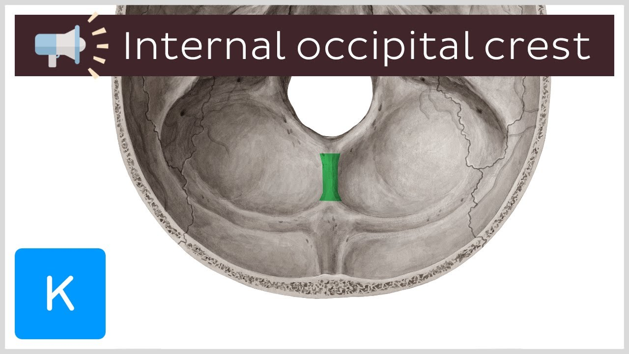
It extends from the crista galli (anteriorly) to the internal occipital protuberance (posteriorly).
Contains the superior sagittal sinus along its superior margin and the inferior sagittal sinus along its inferior margin.
-
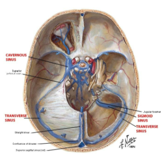
What sinuses are contained within the Falx Cerebri? (3)

Superior Sagittal Sinus
Inferior Sagittal Sinus
Straight Sinus
-
What is the Tentorium Cerebelli and its characteristics? (4)
The Tentorium Cerebelli is a tent-like fold of dura mater.
It lies between the cerebrum and cerebellum.
It is pulled up by its attachment to the Falx Cerebri.
It has attached and free margins.
-
What is the Falx Cerebelli and its characteristics? (2)

The Falx Cerebelli is a small fold of dura mater located along the internal occipital crest.
It contains the occipital sinus.

