 Savannah Tran
-
Cardiovascular System
- A closed system, meaning, the blood is contained within the heart and vessels.
-It functions to connect organs and systems together by pumping blood throughout the body.
-It plays a major role in homeostasis.
-
Cardiovascular System
Two components:
-Heart (cardi-): The pump
-Vessels (-vascular): the "tubes" that contain the blood.
-
Types of Blood Vessels
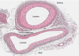
Arteries:
-Vessels that take blood AWAY from heart
-Usually contain blood that is oxygenated
Veins:
-Vessels that return blood back TOWARD the heart.
-Usually they contain the Deoxygenated.
-
Capillaries
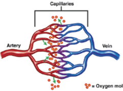
-Located between arteries and veins
-Walls are one-celled thick (simple squamous epithelium)
-Exchange occurs across membrane
-Overall blood pressure in capillary beds decreases b/c overall surface area increases.
-
Blood Pressure
Blood flows through the cardiovascular system due to differences in pressure between one segment of the blood vessel circuit and the next. Blood continues to move through the circuit because it is moving from an area of high pressure to the next area of low pressure.
-
Blood pressure
-The force of the blood against the walls of the blood vessels is blood pressure.
-Stronger when your heart contracts (systole), and weaker when your heart relaxes (diastole).
-Stronger in your arteries and weaker in your veins.
-
Blood pressure
-Typically measured in the brachial artery.
-A sphygmomanometer measures pressure in millimeters (mmHg) of mercury.
-A stethoscope is used to hear the turbulent sounds made as blood begins to flow again through the artery (Korotkoff sounds).
-
Two Circuits
-Pulmonary Circuit
----Circulation of blood through the LUNGS
-Systemic Circuit:
-----Circulation of blood through the body
-
Pulmonary circuit
A function of the RIGHT side of the heart
-Blood is pumped from the heart, to the lungs, and back to the heart.
-
Systemic Circuit
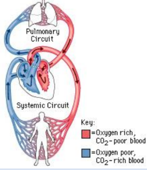
A function of the LEFT side of the heart.
-Blood is pumped from the heart, to the body, and back to the heart.
-
Heart Size
Approximately the size of your fist.
-
Shape
-Base: the TOP, broad, flat surface
-Apex: the BOTTOM, narrow, region
-
Location of the heart
In the center of the thoracic cavity, medial to the lungs and just superior to the diaphragm.
-
Orientation of the heart
The heart is rotated slightly counterclockwise so that the apex is located approximately two inches left of the xiphoid process.
-
Layers of the heart
The heart is enclosed by a two layered serous membrane called the pericardium.
Parietal pericardium (aka PERICARDIUM)
The outermost, loose lining containing the heart and serous fluid.
Visceral Pericardium (aka Epicardium)
The "shrink-wrapped" serous membrane covering the heart wall myocardium.
Myocardium: The thickest portion of the heart wall containing cardiac muscle fibers.
Endocardium: A simple squamous epithelial lining covering the inside of the heart and extending into blood vessels.
-
Chambers of the Heart
The heart has four hollow chambers.
The two SUPERIOR chambers are called: Atria.
The two INFERIOR chambers are called Ventricles.
-
Atria
-Receiving chambers
-Thin-walled
-Separated by the interatrial septum
-
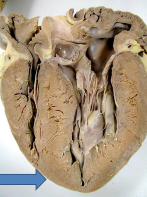
Ventricles
-Pumping chambers
-Thick-walled
-Separated by inter ventricular septum
-
Heart Valves
-There are four valves in the heart.
-Valves are made from endocardium.
-Valves prevent blood from flowing backward.
-An incomplete closure of a valve is a murmur.
-
Atrioventricular (AV) Valves:
-Located between the atria and ventricles.
-The RIGHT AV valve has THREE cusps: Tricuspid.
-The LEFT AV valve has TWO cusps: Bicuspid (aka Mitral).
-Cusps are anchored by chordae tendineae.
-Chordae tendineae attached to papillary muscles.
-
Semilunar Valves
-Semi-lunar valves are located at the EXIT of the heart
-As blood is pumped from the ventricle, it passes through the semi-lunar valve into major arteries.
-The RIGHT semi-lunar valve is located at the exit of the right ventricle and the entrance to the pulmonary trunk.
-The LEFT semilunar valve is located at the exit of the left ventricle and the entrance to the aorta.
-
Heart Sounds pt 1
The Lub-Dub sounds of the heartbeat are caused by the closure of the heart valves.
-The first heartbeat sound is caused by the closure of the atrioventricular (AV) valves.
-Closure of the AV valves is caused by ventricular contraction (systole).
-AV valves are open during ventricular diastole (relaxation) so that blood can fill ventricles.
-
Heart Sounds pt 2
-The second heartbeat sound is caused by the closure of the semi-lunar valves.
-Closure of the semi-lunar valves is caused by ventricular relaxation (diastole).
-Semi-lunar valves are open during ventricular contraction (systole) so that blood is directed out of the heart.
-
Coronary Circulation- Coronary Arteries
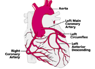
-Right and left coronary arteries branch from the aorta.
-They deliver fresh, oxygenated blood to cardiac muscle tissue (myocardium).
-
Coronary Circulation- Cardiac Veins
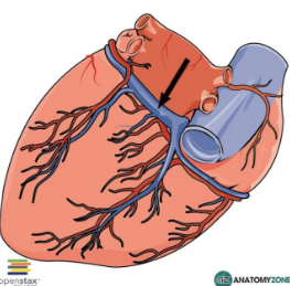
-They return oxygenated blood from cardiac muscle tissue and combine on the posterior aspect of the heart to form the coronary sinus.
-Blood from the coronary sinus then enters the right atrium.
-
Coronary Circulation Problems
Heart Attack, Angina pectoris
-
Heart Attack
(Myocardial Infarction; MI)
-If heart tissue does not get enough blood then death of heart tissue may occur (dead tissue =infarct)
-
Angina Pectoris
-Chest pains accompanied by radiating left arm pain from ischemia (decreased blood supply) to heart muscle.
-
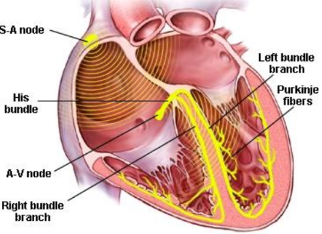
Electrical Conduction Pathway
The heart is auto-excitatory. It produces its spontaneous action potentials at regular intervals from specialized cells called pacemaker cells.
-
Sinoatrial Node (SA node)
Electrical Conduction Pathway
-Located in the top of the right atrium.
-Sets the tempo for the heart called the sinus rhythm.
-It is the heart's natural pacemaker.
-An electrical impulse is generated in the SA node and spread across BOTH atria causing them to contract.
-
Atrioventricular Node (AV Node)
Electrical Conduction Pathway
-Located in the inferior medial wall of the right atrium.
-Electrical impulse from the SA node is passed to the AV node.
-The AV node conducts the electrical impulse through the atrioventricular bundle (Bundle of His).
-Conduction of impulse is slightly delayed to allow contraction of the atria.
-
Atrioventricular Bundle (Bundle of His)
Electrical Conduction Pathway
-Conducts the electrical signal from the AV node downward through the interventricular septum.
-The AV bundle bifurcates (splits in two) into a right and left bundle branch.
-
Purkinje Fibers
Electrical Conduction Pathway
-After the electrical impulse reaches the apex, is sent up the lateral walls of the ventricles through the Purkinje fibers.
-The Purkinje fibers distribute the electrical signals throughout the ventricular myocardium.
-
Electrocardiogram Machine
Monitoring Electrical Activity
-Every time the heart beats, it sends out an electrical signal.
-The heart's electrical signals can be measured with a machine called an electrocardiogram (ECG or EKG)
-
Electrocardiogram Leads
Monitoring Electrical Activity
-Electrodes are specifically placed on different parts of the body (one on each arm and leg and six across the chest).
-
Electrocardiogram Recordings
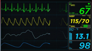
Monitoring Electrical Activity
-Tracings of the heart's electrical activity can be made and permanently recorded on paper or in a computer.
-
Electrocardiogram Readings
Monitoring Electrical Activity
-Doctors assess a patient's heart by studying the shape and size of the waves, the time between waves, and the rate and regularity of beating.
-
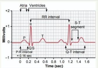
Electrocardiogram Waves
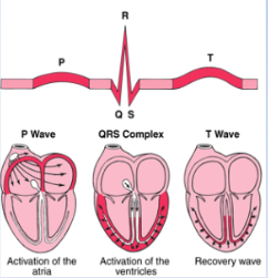
Monitoring Electrical Activity
-Three major waves of electrical signals appear on the ECG. Each one shows a different part of the cardiac cycle.
-
Cardiac Cycle
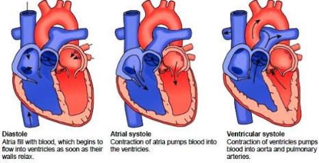
-All of the events that occur during one heartbeat.
-Cycle of systole (contraction) and diastole (relaxation).
-
Cardiac Cycle Phases
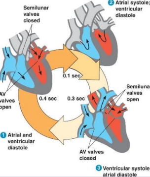
Phase 1: Cardiac Diastole: Atria and ventricles relax
Phase 2: Atrial Systole: Right and left atria contract
Phase 3: Ventricular systole: Right and left ventricles contract
-
Electrocardiogram Waves
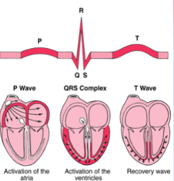
-P-Wave:
atrial contraction
-QRS Complex
ventricular contraction
-T wave:
ventricles reset
-
Arrhythmia
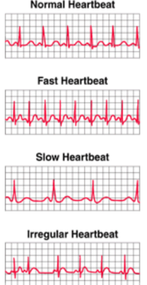
Electrical Conduction Problems
Irregular or abnormal heartbeat
-The SA node doesn't produce the right number of signals
-Another part of the heart takes over as the natural pacemaker
-The electrical pathways are interrupted.
-
The upper number of a blood pressure reading is the _______ pressure.

Systolic
-
The lower number for blood pressure reading is _____ pressure.
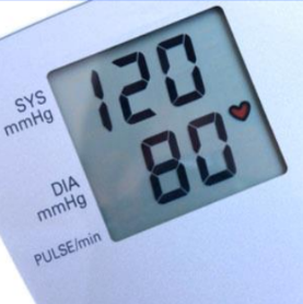
Diastolic
-
What is the medical term for blood pressure cuff?
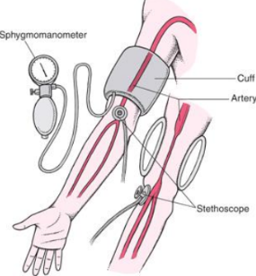
Sphygmomanometer
-
What are that names of the sounds heard when measuring blood pressure?
Korotkoff
-
What artery are you using when taking blood pressure?
Brachial
-
Which of the following is NOT true about hypertension?
-Systolic pressure > 140
-Diastolic pressure is not an indicator
-Hypertension is the same as high BP
-Diastolic pressure > 90
False
Diastolic pressure is not an indicator
Consistent diastolic pressure > 90 indicates hypertension.
-
List the chambers and vessels (in order) of the pulmonary circuit (5).
1.) Right atrium
2.) Right ventricle
3.) Pulmonary trunk
4.) Pulmonary arteries

