 Savannah Tran
-
Parts of Skeletal System
Bones, joints, cartilage, & ligaments
-
Functions of the skeleton for the body
Support and movement
-
How does the skeleton provide support?
Bones and ligaments provide structure for the body.
-
How does the skeleton provide movement?
Bones act as levers and joints as fulcrums to transmit forces exerted by muscles.
-
Skeletal function
Protection- Cover and surround internal organs
Storage-
-Minerals in the matrix (calcium, phosphorus)
-Red marrow in spongy bone.
-Blood cell production (hematopoiesis)
-Yellow marrow in medullary cavity
-Adipose tissue for fat (energy)
-
Mineral Storage-Calcium
Proper levels of calcium in the blood are important for:
-Muscle contraction
-Nerve Conduction
-
Calcium Regulation (negative feedback mechanism)
Hypercalcemia
-Increased levels of calcium in the blood
The thyroid gland releases the hormone calcitonin
-Bones are stimulated to absorb and store calcium
-Calcium levels in the blood decrease
Hypocalcemia
-Decreased levels of calcium in the blood
-Parathyroid gland releases parathyroid hormone (PTH)
-Osteoclasts break down bone and release calcium into the blood
-Calcium levels rise
-
Classification of Bones
Bones may be classified according to their shape
-Long
-Short
-Flat
-Irregular
-Sesamoid
-
Long bones description
-Cylinder-like shape, longer than they are wide
-Used in the body for leverage
Examples: clavicle, metacarpals, metatarsals, phalanges
-
Short bones description
-Cube or block shaped
-Approximately equal in dimension for length, width, height, and thickness
-Provides stability and support while allowing some motion
Examples: carpals, tarsals
-
Flat bones description
-Shallow height, broad width
-Protect internal organs
-Attachment for muscles
Examples: cranial bones, ribs, hip, sternum, scapula
-
Irregular bones description
-Unusually shaped
-Provide structure, muscle attachment, and protection of internal structures
Examples: facial bones, hyoid, vertebrae, sacrum, coccyx.
-
Anatomy of a long bone

Regions:
-Epiphysis (Proximal/Distal)- wider end of long bones containing red bone marrow in spongy bone
-Metaphysis- the area where epiphysis and diaphysis meet which contains the epiphyseal line/plate
Diaphysis-long and narrow middle section of bone containing yellow bone marrow in the medullary cavity
-
Specific structures of long bone
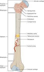
Articular cartilage- hyaline cartilage in the joint protecting the ends of long bones
Spongy bone- open network of bone in the epiphyses (proximal and distal) containing red bone marrow
Red bone marrow-newly produced blood cells in the spongy bone (hematopoiesis)
Epiphyseal line/plate-
--Plate: area of cartilage for longitudinal bone growth
--Line: ossified cartilage indicating completion of longitudinal bone growth.
-
Specific structures of long bone
-Compact bone,
-medullary cavity,
-yellow bone marrow,
-endosteum,
-periosteum,
-nutrient artery
-
Compact Bone
Dense hard layer of osseous tissue around the outside of bones, especially in the diaphyseal region
-
Medullary cavity
Open space in the diaphysis containing bone marrow
-
Yellow bone marrow
Adipose tissue (fat) stored in the medullary cavity
-
Endosteum
Thin membranous lining covering the inner surface of bone
-
Periosteum
Thin membranous lining covering the outer surface of bone
-
Nutrient artery
Supplies and circulates blood to bone
-
Three general classes of bony markings
Articulation- Location where two bone surfaces come together (joint)
Projection- An area of bone that rises above the surface of the bone, attachment point for tendons and ligaments
Foramen- Opening through bone
-
Ossification
The process of forming bony (osseous) tissue
-
Bone Formation-Cells involved
-Osteoblasts: Bone builders
-Osteoclasts: Bone destroyers
-Osteocytes: mature bone cells (anchored in solid bone matrix)
-
Types of osseous tissue
Compact and spongy
-
Compact (cortical) bone
-Dense and hard
-Found on outer surface of bone
-
Spongy (Cancellous or Trabecular) bone
-Inside compact bone
-Many open spaces
-Decreases weight
-Contains red marrow
-
Structure of Compact Bone
-Osteon
-Central Canal
-Lamella
-Osteocytes in lacunae
-Canaliculli
-
Osteon
-Structural unit of compact bone
-Layers of solid bony matrix (lamella) surrounding a central canal which contains blood vessels and nerves
-Osteocytes in lacunae are found between lamellar rings interconnected by canaliculi
-
Central Canal
-Large tunnel through the middle of an osteon
-Contains blood vessels and nerve fibers to serve osteocytes
-
Lamella
-Concentric rings of solid matrix within an osteon surrounding the central canal
-Alternating directions of collagen fibers between each lamellar ring
-Resists torsional (twisting) forces.
-
Osteocytes in Lacunae
-Mature bone cells found in lacuna
-Lacuna: the space within the solid compact bony matrix surrounding an osteocyte
-Canaliculi connect lacunae to each other and to the central canal
-
Structure of Spongy Bone
-Trabecula
-Lamella
-Osteocytes in lacunae
-Canaliculi
-
Trabeculae
-Structural unit of spongy bone
-Contain lamella, osteocytes, and canaliculi but DO NOT contain a central canal
-
Types of Ossification
Endochondral and Intramembranous
-
Intramembranous Ossification
-Process that forms flat bones
-Bone develops between two sheets of fibrous connective tissue
-Spongy bone (diploe) is sandwhiched between two layers of compact bone
Ex.) Scapula, cranium, mandible, clavicle, ribs
-
Endochondral Ossification
-Process that forms long bones
-Bones ossify from a hyaline cartilage model
-Most bones in the body form this way
-
Fun fact!
An embryo is a cartilage skeleton until 8 weeks
-
Bone growth
Bone elongates by ossifying cartilage from the inside of the bone toward the growth plate.
At the end of adolescence chondroblasts top dividing, allowing the plate ossify
-Epiphyseal plate (open)- cartilage present
-Epiphyseal line (closed)- cartilage has ossified
-
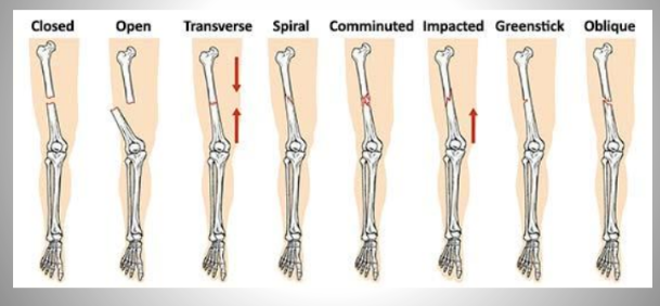
Bone fractures
A break in a bone
-Complete: All the way through
-Incomplete: Partially through
Types of bone fractures
-Closed (simple) fracture- Break that does not penetrate the skin
-Open (compound) fracture- broken bone penetrates through the skin
-
Types of fractures
-
Stages in Fracture Repair PT 1
Fracture Hematoma:
Blood flows into the damaged are from blood vessels broken in the periosteum, osteon, and/or medullary cavity, contributing to inflammation and pain while providing some amount of immobilization.
-
Stages in Fracture Repair PT 2
Fibrocartilaginous (soft) callus formation:
Chondrocytes in the endosteum and periosteum along with osteoblasts form a soft callus of fibrocartilage to stabilize the fracture.
-
Stages in Fracture Repair PT 3
Bony (hard) callus formation
Osteoclasts and osteoblasts replace the soft fibrocartilaginous callus w/ a hard bony callus through endochondral ossification.
-
Stages in Fracture Repair PT 4
Remodeling:
The fully calcified connection in the bone is reshaped according to applied stresses.
-
Bone Remodeling
-Osseous tissue is added or removed in order to balance forces through the bone.
-Osteoblasts deposit bone in greater areas of greater stress
-Osteoclasts break down bone in areas of lesser stress
-
Organization of the Skeleton
Axial Skeleton (80 Bones):
-Skull
-Vertebral Column
-Thoracic Cage
Appendicular Skeleton (126 bones):
-Upper limb and girdle
-Lower limb and girdle
-
Cranium
Eight bones surrounding the brain:
Frontal bone (1): Single bone forming the forehead and upper eye orbits
Parietal bones (2): Paired right and left bones of the upper lateral cranium
Temporal bones (2): Paired right and left bones on the lower lateral sides
-External auditory meatus: ear tunnel
-Zygomatic process: anterior cheek bone
-
Cranium (cont'd)
Occipital bone (1): single bone forming the posterior skull and inferior cranium
-Foramen magnum: large hole
Sphenoid bone (1): single bone of the skull centrally located contacting nearly all other skull bones
Ethmoid bone (1): single bone contributing to the anterior cranium, upper nasal cavity, and medial eye orbits
-
Sutures
-Flat bones of the cranium are locked together w/ immovable joints
-Coronal suture: side to side across the skull joining frontal to the parietal bones
-Sagittal suture: extends posteriorly from the coronal suture joining the two parietal bones
-Lambdoidal suture: extends downard and lateral from the sagittal suture joining the occipital bone w/ the parietal and temporal bones
-Squamous suture: lateral skull joining temporal bones w/ parietal bones
-
Fontanelles
-Fibrous membranes connecting the cranial bones.
-Allow flexibility of the skull for birthing and brain growth.
-Convert to bone within 24 months after birth.
-
Facial Bones
Fourteen bones:
-Maxilla (2): hard palate, medial eye orbit, lateral base of nose, upper jaw.
-Palatine (2): lateral nasal cavity, medial eye orbit, posterior hard palate
-Zygomatic (2): Lateral inferior eye orbit, anterior, zygomatic arch cheekbone'
-Nasal (2): lateral walls of nose (bridge of nose), broken nose bone.
-
Facial bones (cont'd)
-Lacrimal (2): anterior medial eye orbit, tears
-Inferior Nasal Conchae (2): Curved projection into the inferior nasal cavity.
Vomer (1): Posterior inferior nasal septum
-Mandible (1): Only moveable bone in the skull, two pieces fuse together at 1 yr old, lower jaw.
-
Paranasal Sinuses
-Hollow air-filled spaces in certain bones of the skull
-All communicate with the nasal cavity
-Lined w/ mucosal epithelium
-Lighten skull and resonate sound
-
Additional Bones of the Skull
-Hyoid (1):
-"U" shaped bone located above the larynx at the base of the tongue.
-Only bone which does NOT articulate w/ another bone.
-
Additional Bones of the skull
Ear Ossicles (6)
-Malleolus (hammer)
-Incus (anvil)
-Stapes (Stirrup)
-
Vertebral Column
-Extends from skull to pelvis
-Contains 26 irregular bones
-Vertebrae named according to their locations
-
Vertebral Regions
Vertebrae named according to their locations
-Cervical (C1-C7): neck
-Thoracic (T1-T12): Chest
-Lumbar (L1-L5): Low back
-Sacral (5 fused): sacrum (pelvic girdle)
-Coccygeal (4 fused): tailbone
-
General Structure of Vertebrae
-Vertebral body: thick bony anterior for weight bearing
-Vertebrale foramen: opening to house spinal cord
-Spinous process: posterior projection for ligament and muscle attachnent
-Transverse processes: lateral projections for muscle attachment
-
Cervical Vertebrae
-Transvere foramen: opening for vertebral arteries
-Bifid spinous process: split spinous process
-Upper two vertebrae are specialized (C1, C2)
-
Atlas (C1) and Axis (C2)
C1 Atlas:
-No anterior body
-Supports skull
-Skull rocks forward and backward, "yes" motion
C2 Axis:
-Dens (Odontoid process): anterior post
-Skull and C1 rotate around Dens, "no" motion
-
Thoracic Vertebrae
-Spinous processes: long and slender for broad muscle attachment
-Costal facets: attachment locations for ribs
-
Lumbar Vertebrae
-Large, thick, and blunt for increased weight bearing
-
Intevertebral Discs
-Fibrocartilaginous discs between vertebral bodies
-Absorb compression and provide flexibility
-
Herniated Disc
-Bulging or ruptured disc protrudes posteriorly to one side
-May impact spinal nerve and cause radiating pain
-
Spinal Curves
Absorb compressive forces
Lordosis: backward bend (think lean back when kicked)
-Cervical
-Lumbar
Kyphosis: forward bend
-Thoracic
-Sacral
(think already have been kicked so you lean forward)
-
Scoliosis
-Abnormal lateral curve of the spine
-
Thoracic Cage (Rib Cage)
-Forms the thorax (chest) portion of the body
-Consists of 12 pairs of ribs w/ their coastal cartilages and the sternum
-Ribs are anchored posteriorly to each thoracic vertebrae (T1-T12)
-Provides protection of underlying structures
-Allows flexibility for movement and breathing
-
Ribs
Ribs 1-7 are true ribs
-Each has individual attachment to the sternum (vertebrosternal)
Ribs 8-12 are "false" ribs
-Do not connect directly to the sternum, if at all
-
False Ribs
Ribs 8-10
-Attach anteriorly to the costal cartilage of the rib above (vertebrochondral)
Ribs 11-12
-Do NOT attach anteriorly (vertebral or "floating")
-
Sternum
Manubrium: Upper portion of sternum
Body: Middle portion of sternum
Xiphoid Process: lower portion of sternum
-
Appendicular Skeleton
Comprised of 126 bones:
-Pectoral Girdle: bones that attach the arm to the axial skeleton.
-Upper Extremity: bones of the arm
-Pelvic Girdle: bones that attach the leg to the axial skeleton
-Lower Extremity: bones of the leg
-
Bones of the Upper Extremity (Arm)
Clavicle (1)
Scapula (1)
Humerus (1)
Radius (1)
Ulna (1)
Carpals (8)
Metacarpals (5)
Phalanges (14)
-
Pectoral (Shoulder) Girdle
Clavicle (collar bone):
-Articulates medially w/ sternum and laterally w/ scapula.
-Brace for UE
Scapula (shoulder blade):
-Articulates w/ clavicle and humerus
-Glides on thoracic cage.
-
Bony Markings of the Scapula
-Acromion Process: top of shoulder above humeral head where clavicle attaches.
-Spine: long bony ridge
-Glenoid cavity (fossa): socket of the shoulder joint
-Coracoid process: attachment for muscles and ligaments
-
Humerus
-Located in the brachial region
-Articulates proximally w/ the scapula (shoulder joint) and distally w/ the radius and ulna (elbow joint)
Bony markings of the humerus:
-Humeral head: superior rounded surface forming the ball of the shoulder joint.
-Deltoid tuberosity: roughened slightly elevated area where the deltoid muscle attaches laterally
-
Radius and Ulna
-Located in the antebrachial region
-In anatomical postion ulna is medial, radius is lateral
-Ulna functions in elbow flexion/extension, radius functions in hand rotation (distal radius follows the thumb).
Bony markings:
-Olecranon process: bony elbow feature on proximal ulna.
-
Bones of the hand
-Carpals (wrist): eight short bones in two rows of four
-Metacarpals (palm): five long bones in mid-hand region, numbered 1-5 beginning w/ the thumb.
-Phalanges (fingers): bones of the fingers (three) and thumb (two), numbered same as metacarpals 1-5.
-
Bones of the Lower Extremity (Leg)
-Os coxa (1)
-Femur (1)
-Patella (1)
-Tibia (1)
-Fibula (1)
-Tarsals (7)
-Metatarsals (5)
-Phalanges (14)
-
Pelvic (Hip)
-Two coxal bones that connect the LE to axial skeleton
-
Pelvis
-Two coxal bones, the sacrum, the coccyx, and the pubic symphysis
-
Os Coxa
-Hip bone
-Comprised of three bones that have fused together:
---->Ilium = superior
----> Ischium - inferior
----> Pubis --- anterior
-
Bony Markings of the Os Coxa
-Iliac crest: upper ridge of illium, "top of hip" (think top of hill)
-Acetabulum: location where all three portions of the os coxa meet, socket of hip (think ace as all skills come together)
-Obturator foramen: large inferior opening in coxal bone
Ischial tuberosity: enlargement on inferior ischium, "seat" bone
-
Femur
-Larges bone in the body located in the femoral region
-Articulates proximally w/ the acetabulum of the os coxa at the hip joint and distally w/ the tibia and patella at the knee joint
-
Bony markings of the Femur
-Femoral head: rounded ball portion of the hip ball-and-socket joint
-Greater trochanter: enlargement of bone where gluteus medius attaches
-Lesser trochanter: enlarged area of bone where hi adductors attach
-Medial/lateral condyles: rounded ends of the femur that allow it to roll on the menisci of the tibia in the knee joint.
-
Patella
-Bone embedded in the tendon of the quadriceps muscle
-Articulates with the femur
-Prevents rubbing of muscle tendon on the femur
-Increases leverage for the quadriceps muscle
-
Bones of the Lower Leg
Tibia:
-Larger medial bone of the lower leg.
-Weight bearing
Fibula:
-Thinner lateral bone of the lower leg
-Ankle motion
-
Bony Markings of the Tibia and Fibula
Tibia:
-Tibial tuberosity: enlarged roughened surface where patella tendon attaches.
-Medial malleolus: inner ankle bulge
Fibula:
-Lateral malleolus: outer ankle bulge
-
Bones of the Foot
-Tarsals (ankle): seven short bones in the ankle area
-Metatarsals (arch): five long bones in mid-foot region numbered 1-5 beginning medially w/ the big toe.
-Phalanges (toes): bones of the toes (three) and big toe (two), numbered same as metacarpals 1-5
-
Joints
A place where two bones come together (articulate) and make a connection.
Classified according to:
-Function: the amount of movement allowed
-Structure: the way the bones are joined together
-
Structural Classification
-Fibrous joint: connected by fibrous connective tissue
-Cartilaginous joint: joined by hyaline cartilage or fibrocartilage
-Synovial joint (most common): combined through a combination of structures
-
Fibrous Joint Examples
-Suture: between cranial bones
-Syndesmosis: between bones in antebrachium and lower leg
-Gomphosis: between tooth and jaw
-
Cartilaginous Joint Examples
-Synchondrosis: joined by hyaline cartilage
-Symphsis: joined by fibrocartilage
-
Synovial joints
Characteristics:
-Joint cavity: connecting bones do not make direct contact
-Articular capsule: fibrous connective tissue surrounding the entire joint
-Articular cartilage: hyaline cartilage covering the ends of the bones in the joint.
-Synovial membrane: lining the inner surface of the articular capsule, secretes synobial fluid that lubricates joint and helps maintain seperation.
-
Types of Synovial Joints
-Ball and Socket
-Condyloid (Ellipsoidal)
-Plane (Gliding)
-Hinge
-Pivot
-Saddle
-
Ball and Socket
Ball and Socket: rounded head into concave socket
-Hip, shoulder
-
Condyloid
Oval head into oval socket.
-Knuckles (MCP), radiocarpal joint, temporomandibular joint (TMJ)
-
Plane
Two flat surfaces
-Intercarpal and intertarsal joints
-
Hinge
Motion in a single plane.
-Interphalangeal joints (PIP, DIP), elbow, knee
-
Pivot
Rotation around a single axis
-Proximal radioulnar joint, atlantoaxial (C1/C2)

