Youssef Draz
-
Q.Synaptic Transmission Definition:
transmission of impulse (action potential) from one neuron to another along a synapse
-
Q. Synaptic Transmission Synapse
2- junction between 2 neurons: axon terminal of one neuron
(presynaptic neuron) and a second neuron (post synaptic neuron)
-
Q. types of synaptic transmission:
1- Electric: neurons are communicated by low-resistance gap-channel pathway →passage of electric current directly.
N.B.: rare in humans and is common in invertebrates
2- Chemical: release of a chemical substance (neurotransmitter).
-
Q. Functions of Synapses:
a. Facilitating transmission.
b. Blocking transmission.
c. Amplification of impulses.
d. Distribution of impulses.
e. Changing one impulse to repetitive impulses.
f. Integration between different types of neurons.
g. Interpretation, processing and Storage of information.
N.B. synapses are dynamic structures: increasing and decreasing in complexity and number with use and experience.
-
Q. Functional Anatomy of a Synapse
1) Presynaptic Terminal: (or synaptic knob): contains:
a. mitochondria: provides ATP for synthesis and exocytosis of transmitters.
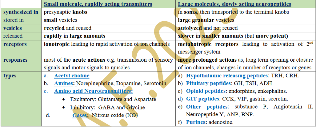
b. A large number of synaptic vesicles contain synaptic transmitters. three types:
1. Small clear for rapidly acting transmitters e.g. acetyl choline, glycine, glutamate and GABA.
2. Small granular containing catecholamines.
3. Large granular contain slowly acting neuropeptides.
(2) synaptic cleft: space containing ECF rich in Na+, Cl- poor in K+.
-
Q. Mechanism of Synaptic Transmission
1- Release of chemical transmitter from presynaptic nerve:
• Arrival of action potential opens voltage-gated Ca++ channels
• Ca++ enters knob according to concentration and electric gradients.
• Influx of Ca++ triggers binding of SNARE proteins:
o V- SNARE (Synaptobrevin) on synaptic vesicle.
o T- SNARE (Syntaxin) on presynaptic membrane
• exocytosis of chemical transmitter into the synaptic cleft.
• N.B.: amount of transmitter is proportional to amount of Ca++ entered.
2- Binding of Transmitter with its Receptors: changes permeability to ions.
-
Q. Functional Anatomy of a Synapse
(3) postsynaptic membrane (PSM): contains Receptors for neurotransmitters. types:
a. Ionotropic receptors: part of Ligand-gated ion channels: affect neuronal
activity directly
b. Metabotropric receptors: (mostly G protein coupled receptors) affect activity
neuron indirectly by second messenger e.g. cAMP: slower and more
prolonged effects, as:
a. Open or close specific ion channels.
b. Activate enzymes e.g. protein kinases: phosphorylation of proteins.
c. Activate gene transcription: formation of new proteins.
-
Q. Mechanism of Synaptic Transmission
3- Generation of Postsynaptic Potential: Change in ion fluxes leads to change in its membrane potential → may become:
a. less negative = excitatory postsynaptic potential. (EPSP).
b. more negative = inhibitory postsynaptic potential. (IPSP).
magnitude and duration of EPSP and IPSP depends on amount of released
Neurotransmitter. “not All or None”.
4- Removal of Neurotransmitters from the Synaptic Cleft:
a. Passive diffusion away from synaptic cleft
b. Inactivation by specific enzymes within synaptic cleft, e.g. acetyl choline
c. Active reuptake into axon terminals, e.g. catecholamines, dopamine, serotonin.
d. Removal by glial cells.
-
Excitatory postsynaptic potential [EPSP]:(Postsynaptic potentials)
Definition: local state of partial depolarization
Ionic basis: excitatory neurotransmitter binding to receptor →
opening of ligand gated Na+(or Ca++) channels
• inside of cell membrane less negative → nearer to firing
level → more excitable.
• membrane facilitated i.e. weaker stimulus to be excited
than at rest.
amplitude: 1- 5 mv
Duration: reaches a maximum after 1-5 msec then gradually declines
N.B.: does not obey all or none rule = graded: Magnitude and duration depends on amount of neurotransmitter
-
Inhibitory Postsynaptic Potential [IPSP](Postsynaptic potentials)
Definition: local state of hyperpolarization
Ionic basis: inhibitory neurotransmitter bind with receptor →
opening of ligand gated K+ or Cl-channels or closure of
Na+ (or Ca++) channels.
• inside of the cell membrane more negative → away
from firing level (threshold) → less excitable.
• membrane is inhibited i.e. needs a stronger stimulus.
amplitude: 1- 5 mv
Duration: reaches a maximum after 1-5 msec then gradually declines
N.B.: does not obey all or none rule = graded: Magnitude and duration depends on amount of neurotransmitter
-
Postsynaptic potentials
Summation: EPSPs have to be summated to reach threshold. Types
1- Temporal (Time): rapid repetitive stimuli originating from ONE presynaptic neuron
2- Spatial (Space): multiple simultaneous stimuli from MANY presynaptic neurons
N.B. IPSP can be summated
3. Grand Post-Synaptic Potential (GPSP): sum of all EPSPs and IPSPs (neuron receives thousands of presynaptic inputs)
possible outcomes:
1. EPSPs = IPSPs → postsynaptic membrane potential will remain at resting level.
2. EPSPs < IPSPs → postsynaptic away from threshold and membrane inhibited
3. EPSPs > IPSPs → postsynaptic neuron doesn’t reach threshold →membrane is facilitated or in an excitatory state.
Example: micturition reflex is influenced by many signals but the response will vary:
a- Sensory signals from receptors in urinary bladder.
b- Facilitatory and Inhibitory signals from brain stem.
c- Voluntary control from the cerebral cortex.
-
Presynaptic Potentials
• release of NT from presynaptic affected by a third neuron.
• N.B.: 3rd neuron synapses axoaxonically with excitatory presynaptic neuron before it reaches the synapse.
-
Presynaptic Inhibition(Presynaptic Potentials)
THIRD NEURON: inhibitory releases an inhibitory transmitter (e.g
GABA), which either opens Cl- or K+ channels.
presynaptic membrane: hyperpolarize → reduced Ca++ influx → reduced
release of NT and transmission from the presynaptic to
the postsynaptic neuron
Example: pain inhibiton
significance: Selective block of synapse without affecting
postsynaptic neuron excitability
-
Presynaptic Facilitation(Presynaptic Potentials)
THIRD NEURON: excitatory neuron releases “serotonin"
→ increases cAMP in the presynaptic terminals → phosphorylates K+ channels and closes them
presynaptic membrane: delays repolarization = prolongation of depolarization: voltage gated Ca++ channels opened for a longer period→ Ca++ influx → increases release of NT
Example: Sensitization of memory
significance: ↑ neurotransmitter release from presynaptic
-
Factors affecting synaptic transmission
1. Changes in internal environment:
a. pH:
• Alkalosis: increases neurons excitability & facilitates transmission: decrease ionized Ca++. (may lead to convulsions: if
epileptic patient hyperventilates, this may precipitate a fit)
• Acidosis: decreases synaptic transmission due to increase ionized Ca++ (may lead to coma, e.g. in diabetic Ketoacidosis).
b. Hypoxia: inhibits synaptic transmission: interruption of cerebral circulation for 3-5 seconds may cause unconsciousness &
prolonged ischemia for minutes cause brain damage.
c. Hypoglycemia: inhibits synaptic transmission (Glucose is the main fuel of brain)
d. Hypocalcemia: facilities transmission as it increases excitability of the post synaptic membrane (leading to tetanic convulsions)
-
Factors affecting synaptic transmission
2- Drugs: Most of the drugs act on the CNS alter synaptic mechanism, e.g.:
a. Theophylline, theobromine, and caffeine ↑ transmission by depolarizing the postsynaptic membrane.
b. Analgesics, hypnotics and anaesthetics: ↓ transmission = stabilizing membrane causing hyperpolarization or by interference
with synthesis, release or reuptake of certain neurotransmitters.
c. Strychnine: ↑↑↑↑ transmission: competes with inhibitory transmitters leaving excitatory pathways unaffected: convulsions.
-
Characters of Synaptic Transmission
I- Forward (uni) direction: from presynaptic to postsynaptic neurons (neurotransmitters released from presynaptic).
II- Synaptic delay: time taken by an impulse to be conducted through the synapse. It equals about 0.5 m. sec: so we can calculate
number of synapses
III- Fatigue: decreased impulses discharged after repetitive stimulation. due to:
1. Exhaustion of synaptic vesicles in the presynaptic terminals.
2. Inactivation of postsynaptic receptors.
• Benefit: stabilize the nervous system e.g. stopping discharge from overexcited areas in CNS during epileptic seizures.
IV- Synaptic plasticity: ability to change function according to demand (synaptic transmission strengthened or weekened)
- represent forms of learning and memory.
Forms of Synaptic Plasticity
-
Q. Chemical Transmitters (Neuro-Transmitters)
• endogenous chemical substances released from presynaptic neurons that transmit signals between neurons across a synapse
• may act as a neurotransmitter in brain & as a hormone elsewhere
Types: More than 100 in two different groups:
-
Q. Organization of Neurons in Neuronal Pools
neuronal pool: collection of neurons carrying the same function in CNS
organizations are
-
Excitation field
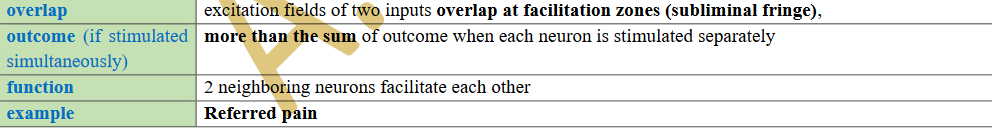
neurons synapsing one afferent neuron.
a) Discharge zone : Neurons in center receive large numbers of knobs: reach threshold and discharge nerve impulses.
b) Facilitated zone( subliminal fringe ): Neurons in periphery receive few numbers of knobs: only facilitated
N.B.: area of discharge zone and subliminal fringe depends on strength of stimulus: the stronger the wider the discharge zone
Facilitation phenomenon
-
Inhibitory Circuits
for stabilizing nervous system by preventing excessive spread of signals.
-
Activating Circuits: (After Discharge):
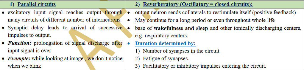
Continued output discharge after stoppage of stimulation of input
-
Sensory Receptors
Q. Definitions:
• Sensory receptor: specialized (modified) structure at peripheral termination of afferent nerve fibers.
• Sensory unit: single sensory afferent with all its receptor endings.
• Receptive field of a neuron: region which contains all the receptor endings of a sensory neuron (stimulus produces a response in that afferent neuron).
Q. Functions:
1- Detectors: detect stimuli (changes in the internal or external environment).
2- Transducers: transform energy in stimulus (chemical, mechanical, etc) into electrical energy (receptor potential).
3- Generators: generation of action potential (nerve impulses) in the sensory nerves
N.B.: Signal Transduction: process by which a stimulus is transformed into an electrical response
-
Q. Properties of receptors:
[1] Specificity:
• Each type of receptor is highly sensitive to one specific type of stimulus = adequate stimulus “e.g. light, sound, pressure, etc.”.
• Receptors respond to other stimuli but:
1) much higher strength
2) sensation perceived is that of adequate stimulus.
e.g. rods and cones in retina can respond to a strong mechanical stimulus, but give sensation of light
[2] Excitability:
• When stimulated, a receptor generates "Receptor (or Generator) Potential": graded electric response:
(as an example: Paccinian corpuscle was chosen . Pressing on capsule by a rod leads to development of receptor potential)
-
Transduction of Sensory Signals = (Mechanism of Receptor Potential = Generator Potential)
1- Stimulation of the receptor:
a. stimulus causes opening of ion channels “transduction channels” → Na+ or Ca++ inflow.
b. number of opened channels is proportional to intensity of the stimulus
2- Creation of Receptor Potential: Na+ or Ca++ influx creates local partial depolarization
3- Electrotonic spread of receptor potential: local circuit of current (current sink) spreads passively to adjacent part of sensory
nerve or 1st node of Ranvier (myelinated neurons) = action potential (A.P.) generating zone (it has low firing level = many voltage
gated Na+ channels)
4- Generation of Action Potential: If amplitude of receptor potential is strong to depolarize [A.P. generating zone] to threshold, it
opens voltage gated Na+ channels and an action potential is produced & propagated along the sensory nerve to CNS.
-
Transduction of Sensory Signals = (Mechanism of Receptor Potential = Generator Potential)
Properties of Receptor Potential (local response):
1. non-propagated
2. does not obey all or none-law, i.e. can be graded.
3. no absolute refractory period, i.e. can be summated.
4. duration (5-10 msec.) is longer than action potential duration (2 msec.) so can cause repeated action potentials.
5. maximum amplitude is 100 mV (at maximum permeability to Na+ ions).
Relationship between strength of stimulus & magnitude of receptor potential: As strength of stimulus increases,
magnitude of the R.P. increases.
• N.B.:
o amplitude increases rapidly at first then less rapidly at high stimulus strengths.
o allows receptor to be sensitive (have an extreme range of response) to very weak sensory stimuli
• Relation of the receptor potential magnitude to number of impulses generated:
o The more the receptor potential rises above threshold level, the greater the frequency of the action potential
-
Adaptation of receptors:
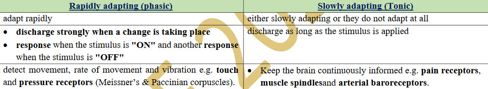
• Definition: gradual decline of receptor potential and frequency of action potentials in spite of the continued presence of
stimulus at constant strength.
• Causes: Each receptor has its own property; adaptation of mechanoreceptors occurs by:
a. Readjustment and remolding in the structure.
b. Accommodation of terminal nerve fiber or 1st node of Ranvier to the stimulus, due to inactivation of Na+ channels.
receptors are classified into:
-
Sensory Code
Definition: ability of CNS to recognize modality (type), locality and intensity of sensation. (All stimuli produce action potentials)
A) Modality of sensation: depends on:
1. Adequate Stimulus: Each receptor is highly sensitive to a particular type of stimulus.
2. Muller’s law of specific nerve energy: Whatever method of stimulation, sensation given is that the receptor is specialized to.
3. Labeled line principle: Each sensation reaches CNS in a specific pathway: Stimulation of this pathway at any point by any form evokes its specific sensation.
-
Sensory Code
B) Locality of the stimulus:
• Law of Projection: There is a separate representation area for each part of the body in the cerebral cortex, (i.e. point to point
representation). When an impulse reaches the specific area in the cortex, it "projects" this stimulus to its original site.
• Phantom limb: a phenomenon in which pressure on the nerves in the stump of an amputated limb makes the patient feel the pain as though it is coming from the absent limb
C) Intensity of sensation: It is encoded by change in:
1- Number of receptors activated: Higher intensity stimuli will recruit more receptors ["recruitment” of receptors]
2- Frequency of impulses: Increasing stimulus intensity increase frequency of impulses with increased intensity of sensation felt
-
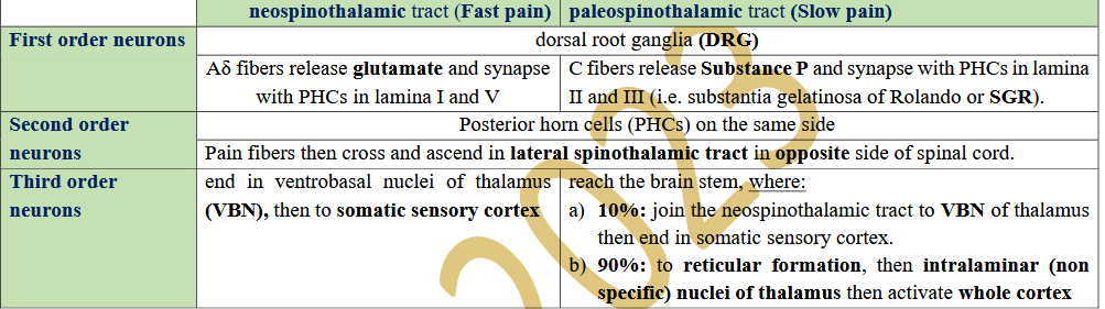
Somatic Sensory Pathways
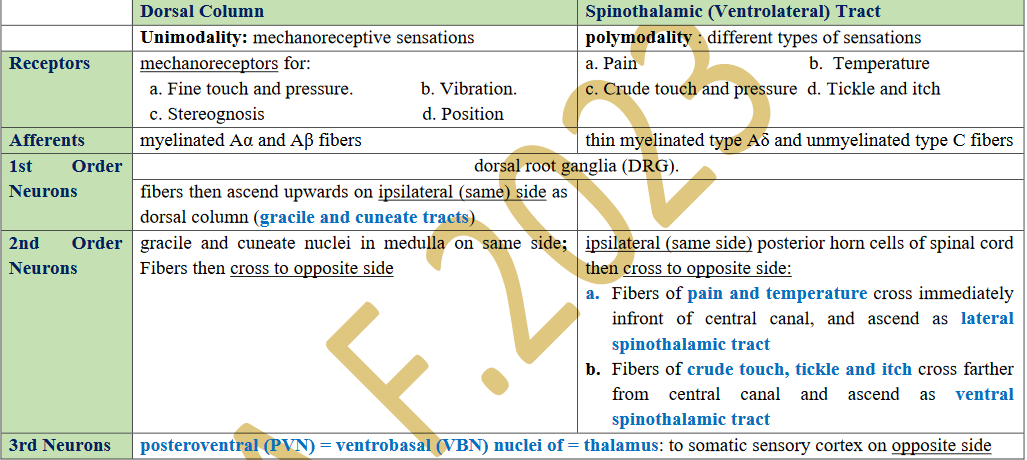
N.B.: passing brain stem, both pathway are joined by additional fibers from nuclei of trigeminal nerve carrying sensations from head.
Role of the thalamus and cortex in sensory perception
a. All sensory tracts, except the olfactory, synapse in the thalamus on their way to the cerebral cortex.
b. subject is crudely aware of sensation but without details: Fine gradations and sensory localization at level of the cortex.
c. Pain perception mainly occurs in thalamus.
-
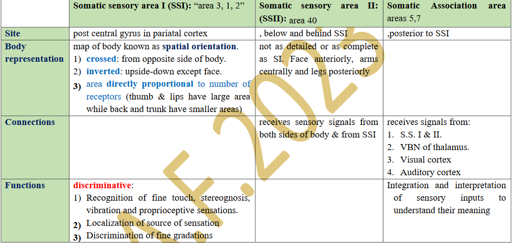
Somatic Sensory Cortex
-
Thermoceptive (temperature )Sensations

Receptors & Afferents & Pathway: Free nerve endings → C fibers (warm) and Ao fibers (cold) → Lateral Spinothalamic tract
At zerooC, no receptors discharge = anaesthesia =.
• between 30oC and 40oC: both cold and warm receptors discharge and are very sensitive to fine gradations
Distribution of thermal receptors:
1. Located in and respond to temperature of the subcutaneous tissue (not to the environmental temperature).
2. numerous in lips & finger tips and least in the trunk
3. Cold receptors 4-10 times more than warm receptors
4. widely separated: wide area of skin has to be exposed to allow spatial summation.
-
Thermoceptive (temperature )Sensations
Adaptation of thermal receptors:
a. between skin temperature 20-40oC: warm receptors adapt faster than cold receptors.
b. 2 components: rapidly at first (phasic component) but continue to generate impulses at low frequency (tonic component).
Mechanism of stimulation:
1- chemically by changes in concentration of metabolites due to change in metabolic rate (each 10oC increase, doubles concentration).
2- membrane ion channels: "Temperature-activated Transient Receptor Potential" (TRP) ion channels
3- respond markedly to changing temperatures rather than steady states:
• Warm receptors increase their firing when temperature is increasing.
• Cold receptors increase their firing when temperature is decreasing.
4- relative Sensation: depends upon original skin temperature. (35oC feel warm if skin is 30oC and feel cold if skin is at 40oC)
-
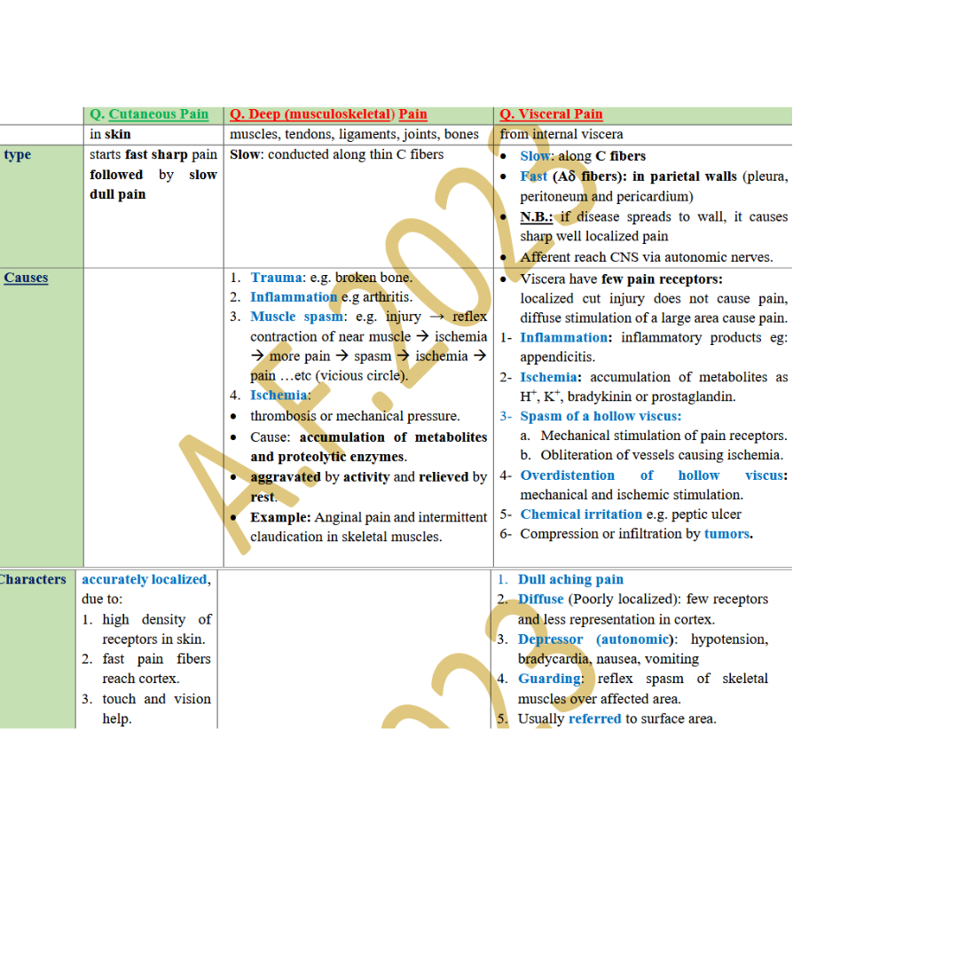
Pain Sensation
Q. Pain:
unpleasant protective sensory and emotional experience if there is physical or potential tissue damage.
➢ Q. Pain Receptors (Nociceptors): Free Nerve Endings types:
1- Mechanical: stimulated by mechanical injurious stimuli, e.g. cuts, bruises.
2- Thermal: respond to extremes of temperature and capsaicin (substance present in hot chilli).
3- Chemical: stimulated by chemical injurious elements.
4- Polymodal: respond to all stimuli.
Distribution of Pain receptors:
• most numerous in superficial layers of the skin, periosteum, peritoneum, pleura, joints, arterial walls, dura and tentorium.
• less distributed in deep tissues and very few in internal viscera.
• absent in liver parenchyma, lung alveoli, and brain tissue (pain insensitive structures).
Adaptation: slowly or non-adaptive receptors.
-
Pain Sensation
Pain Transduction by Nociceptors:
• Noxious stimuli open transduction ion channels in pain receptors: increase membrane permeability to Na+ &/or Ca++ :
a) Mechanical stimuli open "degenerin" ion channels.
b) Thermal stimuli open "temperature sensing TRP" ion channels.
c) Chemical stimuli open "acid sensing" ion channels.
Pain Sensitizers (OR Chemical mediators of pain):
• damaged tissues release a number of pain and inflammation producing chemical substances into ECF. include: histamine,
serotonin, K+, substance P, ATP, bradykinin and prostaglandins.
• Prostaglandins further sensitize nociceptors, lowering their pain threshold, and producing primary hyperalgesia.
N.B: Salicylates and other non steroidal anti-inflammatory analgesics (NSAID) reduce pain by inhibiting prostaglandin synthesis.
-
Types of pain sensation:
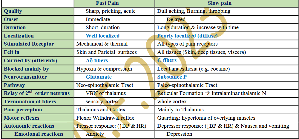
Threshold of Pain: the same for all people but reaction to pain differs: Pain starts if skin temperature reaches 45oC
Q. According to quality (Single painful stimuli may evoke both):
N.B. The two types of pain can be dissociated by:
1. Moderate compression of a nerve trunk block Ao-fibers: fast pain is lost while the slow type remains.
2. Low concentrations of local anaesthetic blocks C-fibers: slow pain is lost while the fast type remains
-
Q. Pain Pathway:
Reaction to Pain: pain fibers give tributaries to:
a. Anterior horn cells in the spinal cord for accompanying motor reflexes.
b. Reticular Activating System for cortical activation and arousal reactions
c. Hypothalamus for accompanying autonomic reactions.
d. Limbic system for emotional reactions.
Arousal reaction to pain signal: intralaminar (non-specific) nuclei of the thalamus and reticular formation of the brain stem
have a strong arousal effect on nervous activities through the reticular activating system (RAS)
Function of cerebral cortex in Perception (conscious awareness) of pain signals:
a. Localization of source of pain: fast pain is very localized.
b. Discrimination of pain modality and intensity.
c. Interpretation of its meaning.
d. prefrontal lobe of the cortex contributes to emotional and behavioral effects of pain
-
Types of Pain Sensation
N.B.: Pain perception can occur subcortically in the Thalamus : cortex not essential
-
Q. Neuropathic Pain:
• chronic type of pain due to damage to or pathological changes in nerve fibers in peripheral or central nervous system.
• Characters:
a. burning, electric, tingling or shooting pain.
b. bouts or paroxysms.
c. accompanied by hyperalgesia and/or parasthesia.
• Examples:
1. Trigeminal neuralgia: (tic douloureux): unilateral facial pain in areas supplied by 2nd and 3rd divisions of the trigeminal nerve.
2. Herpes Zoster:
• viral infection: herpes virus attacks a dorsal root ganglion (DRG) in patients with history of varicella (chicken pox) infection.
• Virus reproduce causing irritation of pain afferents in DRG leading to severe pain felt in dermatomal segment supplied by the
infected ganglion.
• virus also migrates with neuronal cytoplasmic flow to cutaneous terminals → painful skin rash and vesicular formation.
-
Q. Referred Pain:
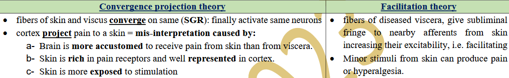
Pain felt in a skin area originating from the same embryonic segment or dermatome as the diseased viscus (or deep structure) = supplied by the same dorsal roots. (Dermatomal rule). Mechanisms:
Examples:
1- Cardiac pain: retrosternal, root of the neck and inner part of the left arm and also in the epigastrium.
2- Gastric pain: between the umbilicus and xiphoid process.
3- Gall bladder pain: in the mid-epigastrium and at tip of right scapula
4- Renal pain: as a back pain radiating to inguinal region and testicles in males
5- Appendicitis pain: around the umbilicus.
-
Q. Headache:
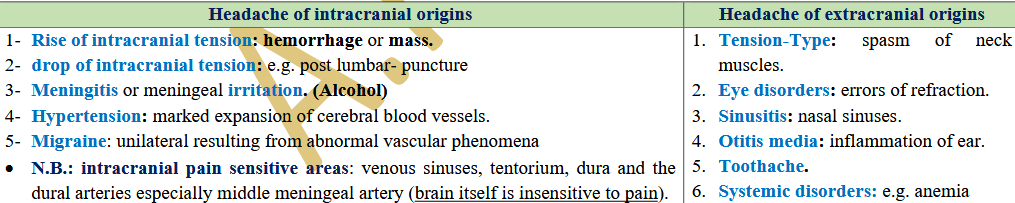
pain referred to surface of head from deep structures. Types:
1) Frontal: Stimulation of pain receptors in areas supplied by trigeminal nerve
2) Occipital: Stimulation of pain receptors in areas supplied by 2nd cervical
-
Q. The Gate Theory(Pain Control)
• dorsal horn cells of the spinal cord, in particular SGR cells, act as a gate for transmission of pain sensation to the brain.
• Impulses coming along C pain fibers cause the release of substance P.
• can be partly or completely closed by Presynaptic Inhibition → prevents release of substance P:
-
Q. Opioid Peptides:(Pain Control)
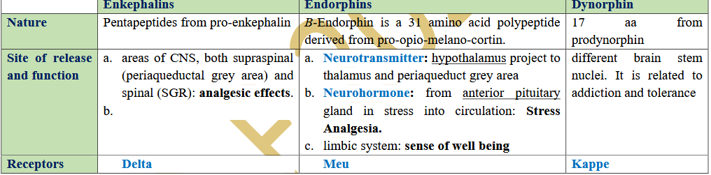
• Morphine is a powerful (analgesic) pain killer found in opium acts by combining with receptors in the CNS.
• Types of endogenous opioid peptides: Among about 12 opioid peptides found in mammals, three are more important:
N.B.: Opiate Receptors: in areas concerned with pain sensation, e.g. Periaqueductal gray area (PGA), SGR, thalamus, hypothalamus
-
Q. Supraspinal Descending Analgesia System: three major components:
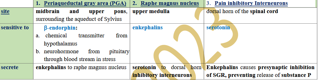
• N.B.; PGA may be stimulated by Collaterals from ascending pain pathway (Lateral spinothalamic tract): noxious stimuli are important for activation of descending pain control system, i.e. pain suppresses pain
-
Q. Peripheral gate inhibition:
• stimulation of large Aβ sensory fibers from peripheral tactile receptors suppress pain transmission:
• tactile fibers send collaterals to activate inhibitory interneurons→ GABA or Enkephalins → presynaptic inhibition of pain
fibers at the SGR → closing the gate.
• Examples:
a. Rubbing or massage of skin inhibits pain.
b. Counter irritation, e.g. mustard plaster
c. Acupuncture.
-
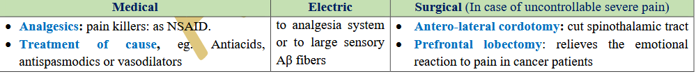
Methods of Treatment of Pain