-
Ganglion
cluster of neurons cell bodies within the PNS
-
Center
Group of CNS neuron cell bodies with a common function
-
Nucleus
Center that displays discrete anatomic boundaries
-
Nerve
Axon bundle extending through the PSN
-
Nerve plexus
Network of nerves
-
Tract
CNS axon bundle in with the axons have a similar function and share a common origin and destination
-
Funiculus
Group of tracts in a specific area of the spinal cord
-
Pathway
Centers and tracts that connect the CNS with body organs and systems
-
Gray vs white matter
Gray matter
-Neuron cell bodies
-Dendrites
-Unmyelinated axons
-Brain (gray outside, white inside)
brain stem
-Spinal cord (white outside, gray inside)
White matter
-Myelinated axons
-
How are nerves organs
made of nervous & connective tissue, and blood vessel
-
Dorsal root vs ventral root of spinal cord
Dorsal root
- sensory neuron axons ONLY
Unipolar neuron cell bodies located in posterior root ganglion
dorsal root ganglian- soma is outside CNS (Afferent)
Ventral root
-motor neuron axons ONLY
- anterior horn of gray matter in cord
-Efferent fibers - soma is inside CNS (nucleus
-
Layers of the Meninges
Dura mater- tough outside layer
Arachnoid Mater- filled with intricate “web” of collagen (barrier for CNS)
subarachnoid- cerebral spinal fluid
Pia Mater- Innermost layer; lines every sulci and gyri of the hemispheres, contours brainstem, and all folds of cerebellum (produces cerebral spinal fluid)
-
Blood brain barrier cells
Endothelial- bodyguard for top people
Pericytes-
Regulate cerebral blood flow and BBB permeability by cell signaling to astrocyte, endothelial and neurons
Astrocyte- communication with endothelial and pericytes
-influence express of pump, receptors, mechanisms
-support energy supply
-
Cerebral spinal fluid
-liquid surrounding brain and spinal cord formed in choroid plexus by ependymal cell and flows in subarachnoid area
-physical support
-controlled chemical environment -> nutrient and waste removal
-
How CSF is produced and travels
Produced in choroid plexus
-flow through ventricles and into subarachnoid space
-absorbed into dural venous sinuses via arachnoid villi
-
Cerebral cortex and function
-thin gray outermost portion of cerebrum, 75% of all neurons in nervous system
-Ultimate control/information processing center
-
Pituitary function
Master endocrine gland
-
Midbrain function
motor movement (eye, auditory, visual)
-
Pons function
Breathing, Hearing Taste, balance
-
Spinal cord function
Pathway to neural fibers traveling to/from brain. Simple reflexes
-
Medulla oblongata function
Regulate breathing, heart and blood vessel
-
Cerebellum function
voluntary movement. Balance and memory
-
Amygdala function
emotions ( reward, fear, social functions)
-
Hippocampus function
memory, cognition (long term memory, map navigation, spatial memory)
-
Fornix function
Carries signal from hippocampus to mammillary bodies and septal nuclei
-
Thalamus and function
relay motor and sensory messages (except smell) between lower brain and cerebral cortex
-large group of nuclei in diencephalon
-left/left lobe connected by interthalamic adhesion/intermediate mass (travel through third ventricle
-
Corpus Callosum function
Axon fibers connecting 2 cerebral hemisphere
-
Cerebrum vs cerebral cortex
Cerebrum
prominent, anterior part of vertebrate brain (2 hemisphere)
-Both gray and white mater
-cell body and nerve fiber
-control voluntary muscle movement
Cerebral cortex
-outer layer of cerebrum
-folded grey matter
-cell bodies and dendrites
-4 lobes
-consciousness
-
The cerebral hemisphere is divided by___
longitudinal fissure
white tract: corpus callosum
-
Gyri and sulci
Gyri- increase surface area
Sulci- some divide into 5 lobes (frontal, central, parietal, occipital)
-
Functional specialization of 2 cerebral hemisphere
cerebral lateralization
left hemisphere: language production
right hemisphere: visuospatial
-
Frontal, Parietal, occipital, temporal lobe
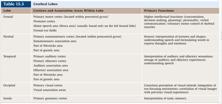
Frontal lobe
Reasoning, planning, speech, movement, emotions, problem solving
Parietal
Movement, orientation, recognition, perception of stimuli
Occipital
Visual
Temporal
Perception, Hearing
-
homunculus and 2 different areas
homunculus- amount of brain tissue devoted to each sonsory/motor functions
Primary somatosensory area -site where sensation originate
Primary motor area - generate action potential for movement, receive info from lobes of brain from d
-
Motor Areas
•Primary Motor Cortex (somatic motor area)—voluntary skeletal muscle activity; located within the precentral gyrus
•Motor Speech Area (Broca’s Area)— movements for vocalization; located within the inferolateral portion of the left frontal lobe
•Frontal Eye Field—regulates eye movements and binocular vision; located immediately anterior to the premotor cortex
-
Sensory Areas
•Primary somatosensory cortex—receives general somatic sensory information from touch, pressure, pain, and temperature receptors; located within the postcentral gyrus
• Primary visual cortex—receives and processes incoming visual information; located in occipital lobe
• Primary auditory cortex—receives and processes auditory information; located in temporal lobe
• Primary gustatory cortex—processes taste information; located in insula
• Primary olfactory cortex—provides conscious awareness of smell; located in medial temporal lobe
-
Limbic system
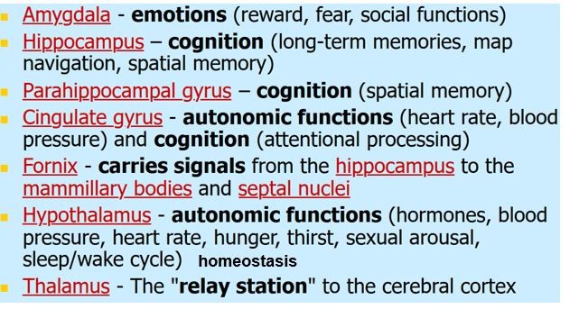
-under cerebrum
-
Functions of the Hypothalamus
Master control of the Autonomic Nervous System
and Endocrine System (emotion and reward)
-body temperature
-certain emotions and sex drive
-thirst and hunger
-hormones, growth
-metabolism
-water and electrolyte balance
blood pressure,
sleep,
homeostasis
-
Basal Nuclei/ basal ganglia
coordinating skeletal muscle contraction
Comprised of nuclei:
-Caudate nucleus
-Lentiform nucleus
-Putamen
-Globus pallidus
parkison disease
-
Cerebellum function
muscle tone, coordination of movement, posture/balance, eye movement, motor learning, cognitive function (language, riding bike)
-influence ipsilateral(same) side of body
-compare motor plan (intent) in cortex with motor performance (from periphery) and smooth them
-synaptic contact with brain stem "motor" and cerebral hemisphere
-
Brain stem
Control info from brain and body, breathing, swallowing heart rate, blood pressure, conscious
Diencephalon of thalamus (relay/process sensory info) and hypothalamus(emotions, autonomic, hormone)
Midbrain-connects pons and medulla oblongata
-process auditory and visual. create reflexive somatic motor response. maintain consciousness
Pons-relay sensory into to cerebellum and thalamus
Medulla oblongata- relay sensory info thalamus and other brainstem. regulation of visceral function (cardiovascular, respiratory, digestive)
-
short term memory vs long term memory
Short term- hold 7-12 info for hours, disappears
Long term- hold large amount of info for days/years.
Converted from short by consolidation
need break and repetition
-
What makes up the PNS
Sensory (afferent) transmit action potential from receptor to CNS
Motor (efferent) transmit action potential from CNS to effectors (muscle, glands)
-
Functions of sensory pathways
Sensory reception (detect stimuli inside and outside body), transduction, transmission, integration
-
Simple, complex, and special sensory receptors
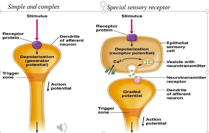
Simple- neurons with free nerve ending (pain, tickle, itch, temp)
Complex- have nerve endings enclosed in connective tissue capsules (touch, pressure, vibration)
Special sensory receptors- cells that release neurotransmitters onto sensory neurons initating action potential (vision, hearing, taste, smell)
-
Transduction
conversion of stimulus energy (light, heat, touch, sound, etc into nerve signal by
EX transducers: sense organ, gasoline engine
-
sensation
subjective awareness of stimulus
most sensory signals delivered to the CNS produce no conscious sensation
-filtered out in the brainstem
-some do not require conscious awareness like pH and body temperature
-
All sensory receptors ______ incoming stimuli into changes in membrane potential (can lead to action potential)
transduce
-
CNS interpretation of stimuli depends on 4 properties
Modality, Location, Intensity, Duration
-
What is modality and the 6 major receptors?
Modality- which type of environmental stimuli do our neurons sense?
Mechanoreceptors-pressure, touch vibration, stretch, itch, proprioception and equilibrium
Thermoreceptors- temperature
Photoreceptors-electromagnetic radiation
Chemoreceptors-chemicals; taste, smell , changes in body fluid chemistry
Nociceptors-tissue damage
Osmoreceptors– detect changes in concentration of solutes
-
3 Location of receptors
Interoceptor- monitor internal systems of organ and blood vessel
(chemoreceptor for O2, baroreceptor for blood pressure, mechanical receptor stretch in organ)
Exteroceptor- external senses (touch, temp, pressure)
-distance sense (seeing, hearing, smelling)
Proprioceptor- monitor position and movement (skeletal muscle and joints)
-
Size of receptive field (sensitivity) is dependent on ___
location
fingers have smaller more # touch receptor than forearm
-
What is lateral inhibition
fine tuning of signal
excited neuron reduce activity of neighbor, depends on location
-
Intensity is dependent on
frequency of action potential
-
Receptor adaptation (duration) Tonic vs phasic
Tonic receptor- slowly adapting receptors that respond for the duration of a stimulus (constant firing)
Ex- stretch receptor, pain receptor, photoreceptor, mechanoreceptor
Phasic receptors- rapidly adapt to a constant stimulus and turn off
Ex-smell, pressure, touch, temp
-
7 senses and their receptors
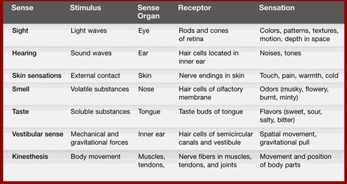
-
Sensation & Perception
Sensation- awareness of changes
-stimulation of receptor
Perception
conscious awareness & interpretation of sensation
generate action potential then to brain
-
Somatosensory pathway
First order neuron- soma in dorsal root ganglion or cranial ganglion (environment to spinal cord/brain stem)
Second order neuron- soma in brain stem, dorsal root and medulla it travels to thalamus
Thirds order neuron- thalamus to sensory area of cerebral cortex
-
Upper motor neuron vs lower motor neuron
Upper motor neuron- Carry motor output from cerebral cortex and brain stem to lower motor neuron in spinal cord/brain stem
Lower motor neuron- in anterior gray horn of spinal cord that innervate muscle
-
What system is involved in movement?
nervous, skeletal, and muscular
-
3 neurons that work together for sensation
sensory neuron, upper motor neuron, lower motor neuron
-
Neurotransmitters involved in ANS
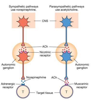
Sympathetic- preganglion release ACH and post release norepinephrine
Parasympathetic- preganglion release Ach- Post release ACH/norepinephrine
-
Parasympathetic vs sympathetic
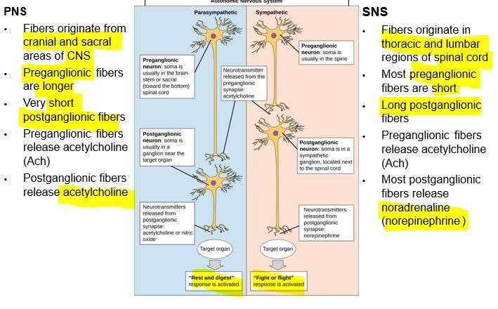
Most visceral organs innervated by both
-
In autonomic nervous system, ____ neuron innervate tissue form and diffuse. ___ are site of neurotransmitter release
postganglionic
Varicosities
-
Single unit (visceral) vs multi unit smooth muscle
Single unit- cells connected by gap junction that act as group. Walls of hallow organs (viscera)
Multi-unit- contract individual cells. iris of eye, walls of blood vessel
-
Organs with sympathetic innervation only
-adrenal medulla
-arrector pilli- goose bump/hair stands up
-blood vessel
-sweat glands
regulation through increase/decrease firing rate by sympathetic neurons
-
Autonomic nervous system maintain homeostasis
through balance of sympathetic "fight or flight"
parasympathetic "rest and digest"
-
What area of brain control autonomic control center?
Hypothalamus- water balance, temp, hunger
Pons- respiration, cardiac, vasoconstriction
Medulla- respiration
-
Location of autonomic nervous system
Preganglionic neurons- in CNS, release ACH(excitatory)
Post ganglionic neuron- ganglia outside CNS
-
Some functions of sympathetic
increase
heart rate, blood pressure, vasodilation in blood vessel, diameter of respiratory airway(max air), glycogen, sweat,
decrease digestion, urine production
-
structure of sympathetic
preganglionic neuron- short, branched, cholinergic neuron
Postganglionic neuron- long, adrenergic neuron
-
Neurotransmitters of sympathetic
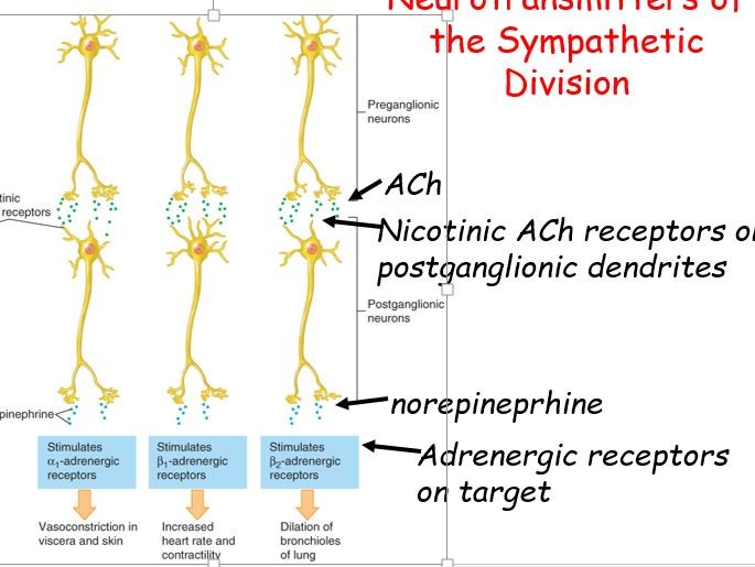
Preganglionic neuron –cholinergic -> release ?
->binds nicotinic Ach receptors on postganglionic neurons
excitatory!
Postganglionic neuron – adrenergic neurons
usually release ?
excitatory or inhibitory depending on type of adrenergic receptor it binds on target
-
sympathetic nervous system and age
↑ age -> ↑ levels of sympathetic activity
may increase hypertension & cardiovascular disease
-
parasympathetic structure
Preganglionic neurons:-origin: brain or spinal cord. long, cholinergic
Postganglionic neurons:-origin: outside CNS near/in effector organ. -short, usually cholinergeric
Vagus nerve –major nerve carrying preganglionic axons of parasympathetic
-
Neurotransmitters of the Parasympathetic Division
Preganglionic neuron -> releases ?
-binds nicotinic receptors on postganglionic cell -excitatory
Postganglionic neuron ->releases ? (some release NO)
-ACh binds muscarinic receptors
-inhibition (IPSPs) in heart
-excitation (EPSPs) in digestive tract
-
Somatic Motor Controls
•Body movement/skeletal muscle
•Appendages
•Locomotion
•Single neuron
-CNS origin
-Myelinated
•Terminus
-Branches
-Neuromuscular junction
-
3 types of muscle
skeletal, smooth, cardiac
chemical energy into mechanical energy
-
Skeletal muscle
repeating sarcomeres (striated)
myosin and actin
attach to bones through tendons
-
smooth muscle
line walls of hallow organs and blood vessel
Nonstriated and under involunatry control
-
cardiac muscle
striated,
intercalated disc
involuntary, autorhythmic
-
skeletal muscle act in antagonistic pairs-
one contract and another return to position
-
Skeletal muscle
contraction = shortens
moves tendons and bones
voluntary control
contract rapidly but tired easily
overall body motility
-
T tubules and SR
T tubules bring action poetntial into muscle fibers
Depolarization cause sarcoplasmic reticulum to release calcium
-
Myofibril
Muscle fiber contains many myofibril (A band, I band, Z line)
myofibril contain many sarcomere
myofilament (action and myosin)
-
Sarcomere
Basic contractile of muscle unit, between 2 Z line
M line, central myosin filament anchor
H zone- only myosin
I band-only actin filament
A band -myosin and actin
-
Thick filament and thin filament
Thick filament (myosin) 2 gobular head and tail
Thin tilament- actin with tropomyosin and troponin
-
Sliding filament model
muscle contract- chemical to mechanical
-Nervous system changes in pulmary potential
-myosin head of thick bind to actin of thin propel toward M line
- H zone, sarcomere, muscle cell, whole muscle shortens
-
How motor neurons influence contraction of muscle
by causing change in membrane potential
Motor cortex- generate motor action potential
cerebellum= smooth action
down to spinal cord then muscle
-
Motor units
muscle made of many motor units
Motor unit: made up of motor neuron and muscle fibers(either fast or slow twitch) within the muscle it innervates
-Groups of motor unit often work together
-All motor units that subserve single muscle are considered a motor unit pool
-
link between the action potential (generated in the sarcolemma) and the start of a muscle contraction
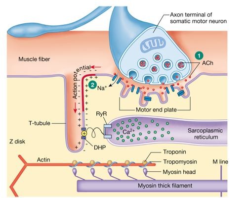
-Action potential gets picked up by voltage gated Ca+ channels.
-Ca+ flows in and it releases Ach. Ach bind to motor end plate and flow of Na+ inside.
-Na+ polarize muscle fibers. Action potential travel to T tubules (bring in action potential.
-DHP activated and linked to RyR that release calcium into cytoplasm
-calcium bind to troponin cause tropomyosin to move and expose myosin. Myosin and actin bind (z disc move closer to m line with calcium and ATP)
-
sliding filament theory steps
1. muscle contractions begin and continues if ATP available and Ca++ in sarcoplasm is high
2. Ca++ released and bind to troponin which cause tropomyosin to release and expose actin
3. Myosin head and actin form cross bridge with help from Ca+
4. ADP and P release, Powerstroke slide thin filament. Sarcomere & Z line shorten (muscle contract)
5. ATP bind to myosin cause detachment
6. Hydrolysis of ATP "cock" myosin head
-
Muscle relaxation
-acetylcholinesterase decomposes acetylcholine
-calcium pump back in SR
-tropomyosin cover myosin binding site
-sarcomere relax and back to normal length
-
Reflexes
involuntary motor response without conscious control.
-short neuronal circuits =quick reaction
-involve spinal cord or brain stem
-fast, higher brain center not involved (few synapses)
-
Reflex purpose
•Protect from tissue damage
•homeostasis
•optimal muscle length for strong contraction
All have similar properties
-stimulus required
-few neurons involved
-preprogrammed response is the same way every time
-involuntary response requires no intent or pre-awareness
-
Reflex Arc
-Stimulus activates receptor
-Nerve impulse travel through sensory neuron to spinal cord
-Nerve impulse processed in by interneurons
-Motor neuron transmits nerve impulse to effector
-Effector responds to impulse from motor neuron
-
Monosynaptic and polysynaptic reflexes
Monosynaptic- direct communication between sensory and motor neuron (knee flex)
Polysynaptic reflex-interneuron faciliates sensory motor communication (hot stove)
-
Autonomic reflexes
Polysynaptic with 1 synapse in CNS and another in Autonomic ganglion
-
3 functions of meninges
- protect from mechanical injury
-provide blood supply to skull
-provide space for cerebrospinal fluid
-
Mechanoreceptors
pressure, touch vibration, stretch, itch, proprioception and equilibrium
-
Thermoreceptors
temperature
-
Photoreceptors
electromagnetic radiation

