-
What types of behavior are supported by the spinal cord and brainstem without requiring the cortex?
reflexes and rythmic movements (walking or breathing)
-
(1) What type of sensory feedback do behaviors supported by the spinal cord and brainstem require?
(2) sensory and motor systems, sensory input
(4) muscle spindles
(1)sensory feedback helps guide involuntary (reflexes) and voluntary (rhythmic) behaviors
(2)sensory and motor systems are closely connected, hard to fully separate them since behavior is continuously motivated and guided by sensory input
(3) muscles have sensory endings called muscle spindles that detect changes in muscle length and send feedback to the nervous system to help regulate movement.
-
(1) What types of sensory receptors and fibers provide this afference (reception of signals from sensory organs by the brain) for behaviors controlled by the spinal cord and brainstem?
(2) gamma motor neurons
(3) Glogi tendon organs
Afferents for reflex circuits is IIa fibers from muscle spindles which send info about muscle stretch to spinal cord as seen in myotatic reflex ( involuntary stretch ex; patellar reflex)
Gamma motor neurons adjust the sensitivity of muscle spindles, allowing dynamic range and control so spindles can accurately signal changes in muscle length
Golgi tendon organs provide feedback on muscle contraction force, helping protect muscles from excessive force
-
what sensory structures are found in the muscles?
muscle splindles that detect change in muscle lenght and send feedback to nervous system, causing movement
-
Anterograde tracing
dye injected into the soma and travels down axon, allowing full reconstruction of axonal pathways
-
Retrograde tracing
dye injected into target area (ex nerve terminal) and travels back to soma which helps trace connection from nerve terminal back to cells control center
-
What are the Pros and cons of Anterograde tracing dye to identify motor unit?
Pro: traces axonal pathway from cell body to muscle, providing complete visualization of axon
Con: harder to visualize the entire motor unit and may not show all terminal connections
-
What are the Pros and cons of Retrograde tracing dye to identify motor unit?
Pro: shows connection from muscle back to cell body, making it easier to identify the neuron controlling the muscle
Con: Won’t show the full axon pathway.
-
Where would you place your injection for anterograde vs retrograde dye?
anterograde: inject into cell body (soma) of neuron
Retrograde: inject into muscle (soles) target zone
-
Assuming you can do more than one experiment, which one would you do first with anterograde vs retrograde dye to identify motor unit?
start with retrograde dye to identify the neurons connection to muscle, then follow up with anterograde dye to trace full axon pathway
-
What is the motor map?
representation of how different areas of the brain (like motor cortex) control specific muscles or body regions
-
What are examples of mapping?
Tontopy: mapping of sound frequencies
Retinotopy: Mapping of visual information from retina
Somatotopy: Mapping of body parts onto the brain or spinal cord
-
What motor structures have maps?
lower motor structures which are organized somatotopically onto the spinal cord
Motor map runs from medial to lateral; motor neurons for distal muscles (like thumb) located laterally, while proximal muscles (triceps) are more medial. Trunk muscles (axial muscles) positioned the most medially
-
How can these maps be made to change?
experience (motor learning) injury, or rehabilitation, allowing for adaptation to new movement or recovery from damage.
-
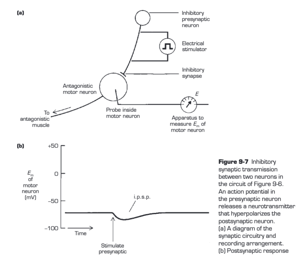
How do antagonists vs. synergist muscles behave during flexor reflex?
Antagonist's muscles are inhibited (relax) to allow movement of agonist's muscles
Synergist muscles contract & work together w/ agonist to create movement
happens on both sides of the body, but the opposite limb, antagonist muscles may be activated for balance
-
Synergist muscle
the muscle that assists the prime mover muscle or agonist during movement
EX: soleus and gastrocnemius muscles both contributed to plantar flexion on the ankle
-
Antagonist muscle
muscle that produces the opposite motor action to another muscle
EX: biceps (flexor) and triceps (extensor) muscles work against each other; when one contracts other relaxes
-
Contralateral muscle
muscles or actions of the opposite limb
EX: if you flex your right arm, the contralateral muscle would be involved in the movement of the left arm
-
recruitment order
order in which motor units are activated based on the force
-
Explain what is meant by the size principle?
states that smaller motor units are recruited first during low-force activities, large motor units are recruited as more force is needed
-
How are recruitment order and size principle related?
recruitment order follows size principle, meaning smaller motor units are recruited before larger ones during muscle activation
-
Oscillator
neural circuit that produces rhythic activity (Ex: walking or breathing)
-
Draw an oscillating circuit

-
Why study leeches or lampreys for central pattern generation (CPG)?
they have simple nervous systems, making it easier to study CPGs, which control rhythic movements like swimming or walking
-
two main types of neurons in the first motor subsystem, and where are they located?
Lower motor neurons: in the spinal cord and brainstem, connected directly to muscles
Local Circuit Neurons: in the spinal cord, provide coordination between muscle and most input of LMN
-
What role do lower motor neurons play in movement?
they send signal directly to muscles, acting as the “final common path“ for movement
-
What functions are controlled by upper motor neurons in the brainstem?
muscle tone, posture, adjusting eyes, head, and body in response to sensory input (ex; visual or auditory)
-
What cortex areas are involved in voluntary movement control?
Primary Motor Cortex (Brodmann's Area 4): initiated voluntary response
Premotor cortex (Broadmann's Area 6): plans and directs movement sequences
-
Main function of cerebellum in motor control?
detects and reduces “motor errors” (differences between intended and actual movements) in real-time and long term
-
How does cerebellum contribute to motor learning?
continuously adjecting to reduce motor errors, helping movements become more precise over time
-
What does basal ganglia do in relation to movement?
prevents unwanted movements and gets the mottor system ready to intiate voluntary movements
-
Name two disorders linked to basal ganglia dysfunction and describe their effects
Parkinsons Disease: difficulty initiating movements
Huntingtons Disease: Problems with movement transitions and control
-
What is motor neuron pool?
group of lower mottor neurons that all innervate a single muscle
-
How are mottor neuron pools aranged in the spinal cord?
organized along the spinal cord to match the muscles they control, forming spatial map
-
Where in the spinal cord are the motor neurons for axial (trunk) muscles located?
located medically in the ventral horn of the spinal cord
-
Describe medial-to-lateral arrangement of mottor neurons in the spinal cord
Medial: Axial (trunk) muscles
Next Lateral: shoulder and pelvis muscles
Further lateral: Arm and leg muscles
Most lateral: distal muscles (ex; hands, fingers)
-
What does retrograde tracing help scientists understand?
helps identify where specific-muscle controlling neurons are located in the spinal cord
-
What do medial descending pathways control?
control posture and balance by targeting medial lower motor neuron pools.
-
Which upper motor neuron pathways control skilled movements?
Lateral descending pathways from the motor cortex control skilled movements, especially for distal muscles like those in the hands.
-
Where are the medial lower motor neuron pools located, and what is their function?
They are located medially in the ventral horn and control axial (trunk) muscles for posture.
-
What is the role of medial circuit neurons?
Medial circuit neurons coordinate bilateral, rhythmic movements and have long axons crossing multiple spinal segments and often the midline.
-
What is the difference between medial and lateral local circuit neurons?
Medial: Long axons, coordinate bilateral movements.
Lateral: Short axons, control fine movements, stay on the same side.
-
What is somatotopic organization in the spinal cord?
It's the arrangement of motor neurons in the spinal cord, with medial neurons controlling trunk muscles and lateral neurons controlling distal muscles.
-
What is the function of alpha motor neurons ?
alpha mottor neurons innervate extrafusal muscle fibers, generating force for posture and movement
-
What is the role of gamma (y) motor neurons?
gamma motor neurons innervate intrafusal muscle fibers in muscle spindles to adjust their sesnsitity, allowing muscle to sense stretch
-
What is the difference between extrafusal and intrafusal muscle fibers?
Extrafusal: Main muscle fibers for force and movement, innervated by α motor neurons.
Intrafusal: Specialized fibers in muscle spindles that detect stretch, innervated by γ motor neurons.
-
where are muscle spindles located, and what is their function?
Muscle spindles are embedded within the muscle and act as sensory receptors to detect changes in muscle length.
-
How do α and γ motor neurons work together?
They coordinate to maintain smooth movement by adjusting muscle length and force as the muscle changes.
-
Where are the lower motor neurons for head, eye, and neck muscles located?
In the brainstem, specifically in eight motor nuclei of cranial nerves.
-
motor unit
A motor unit is a single α motor neuron and all the muscle fibers it innervates, forming the smallest unit of force activation in a muscle.
-
Describe slow (S) motor units.
Slow motor units have small, red fibers, contract slowly, generate small forces, and are fatigue-resistant. They’re important for postural activities.
-
What are fast fatigable (FF) motor units used for?
FF motor units generate large, quick forces but fatigue easily, making them ideal for brief, powerful actions like sprinting or jumping.
-
What is the function of fast fatigue-resistant (FR) motor units?
FR motor units provide moderate force and speed and are fatigue-resistant, suitable for activities requiring moderate, sustained force.
-
What is an innervation ratio, and why does it matter?
The innervation ratio is the number of muscle fibers per α motor neuron, influencing muscle function:
Soleus: 180 fibers for sustained posture.
Gastrocnemius: 1000-2000 fibers for quick movement.
Extraocular: 3 fibers for precise eye movement.
-
What is use-dependent plasticity in motor units?
Motor units adapt based on use; for example, sprinters develop fast, fatigable fibers for power, while marathon runners develop slow, fatigue-resistant fibers for endurance.
-
Define the size principle in motor unit recruitment.
Motor units are recruited in order of size, with small S units activating first, followed by FR units for moderate force, and FF units for maximum force.
-
How does action potential frequency affect muscle tension?
Increased firing rates in motor neurons lead to temporal summation of muscle contractions, increasing overall muscle force.
-
Fused tetanus
state where muscle contractions are smooth and forceful, and no peaks and droughts
-
What firing rate range causes fused tetanus in motor neurons?
max firing rate 20-25 Hz
-
Why does asychronous firing of motor units help movement?
it averages out tension changes, allowing smooth movement
-
What type of response do Group Ia afferents have?
Phasic response to small stretches, sensitive to velocity
-
What fibers are innervated by Group II afferents?
Static nuclear bag and nuclear chain fibers, signaling sustained stretch.
-
What is reciprocal innervation?
Excitatory input to a muscle and inhibitory input to its antagonist.
-
What is the stretch reflex also known as?
Deep tendon or myotatic reflex (e.g., knee-jerk response).
-
Which sensory group primarily mediates muscle tone?
Group II afferents.
-
How does the stretch reflex act as a negative feedback loop?
It adjusts α motor neuron activity to maintain desired muscle length.
-
Role of γ motor neurons in muscle spindles?
Control spindle sensitivity by modulating excitability of intrafusal fibers.
-
Dynamic vs. Static γ Motor Neurons - What's the difference?
Dynamic enhances Ia dynamic response; Static enhances Ia and II static response.
-
What does "gain" in the muscle stretch reflex refer to?
Force generated in response to stretch; high gain = more force for small stretch.
-
Example of reflex gain adjustment?
Standing on a moving bus - modulated by upper motor neuron pathways.
-
Why must the gain of myotatic reflexes be reduced during voluntary stretching?
To allow muscle fiber lengthening, essential for warming up in athletic performance.
-
How do α and γ motor neurons work together during movement?
They are co-activated to prevent muscle spindles from becoming unloaded or overactivated.
-
How is γ motor neuron activity adjusted?
Independently of α motor neurons, for precise movement control and fine adjustments.
-
When is baseline γ motor neuron activity high?
During difficult, precise movements or in unpredictable situations.
-
What factor, besides γ motor neuron activity, affects the gain of the stretch reflex?
The excitability level of α motor neurons.
-
What is the function of Ib inhibitory interneurons in the spinal cord?
They suppress excitatory signals to specific α motor neurons, influencing reflex gain.
-
What role does the Golgi tendon organ play in regulating muscle force?
It acts as a feedback system to reduce muscle force and prevent excessive tension.
-
How does the Golgi tendon organ respond to active muscle contraction?
It increases Ib afferent firing, sensitive to muscle tension.
-
How do muscle spindles and Golgi tendon organs complement each other?
Spindles monitor and maintain muscle length; Golgi tendon organs monitor and maintain muscle force.
-
How do joint receptors contribute to muscle protection?
They signal hyperextension or hyperflexion, enhancing the protective response of Ib interneurons.
-
What does the flexion reflex pathway mediate?
Withdrawal of a limb from a painful stimulus by activating ipsilateral flexor muscles and inhibiting ipsilateral extensor muscles.
-
What is the crossed extension reflex, and why is it important?
the activation of contralateral extensor muscles and inhibition of contralateral flexor muscles, providing postural support during withdrawal.
-
How can descending pathways influence the flexion reflex?
They modulate responsiveness to sensory inputs, sometimes enhancing or suppressing reflex withdrawal based on the stimulus type.
-
What are central pattern generators (CPGs)?
CPGs are specialized local circuits that coordinate rhythmic muscle activation for movements like walking, independent of sensory input or higher brain signals.
-
Describe the two phases of limb movement during locomotion.
The stance phase, where the limb is in contact with the ground, and the swing phase, where the limb is lifted and moved forward.
-
How does locomotion speed affect limb movement patterns in quadrupeds?
At higher speeds, the limb movement pattern changes from a progressive sequence to synchronized movements (e.g., trotting, galloping).
-
How can locomotion continue in animals with a severed spinal cord?
CPGs in the spinal cord allow coordinated movement, and inputs like treadmill speed can regulate the rate of locomotion.
-
What role does dopamine play in spinal locomotion after spinal cord transection?
Dopamine (from l-DOPA injection) activates local circuits, supporting the CPG function even without upper motor neuron input.
-
Why is human rhythmic stepping less effective after spinal cord damage than in quadrupeds?
Human locomotion relies more on upper motor neuron pathways and postural control, which spinal CPGs alone can't fully support.
-
How are CPGs structured for effective movement coordination?
They consist of excitatory and inhibitory neurons and are connected with modular circuits for coordinating left-right and forelimb-hindlimb actions.
-
What is the "Lower Motor Neuron Syndrome"?
It is a set of signs and symptoms resulting from damage to the lower motor neurons in the brainstem or spinal cord, leading to issues such as paralysis, muscle weakness, and loss of reflexes.
-
What are primary symptoms of lower motor neuron syndrome?
Symptoms include paralysis or weakness (paresis), loss of reflexes (areflexia), loss of muscle tone, spontaneous muscle twitches (fibrillations and fasciculations), and eventual muscle atrophy.
-
What causes fibrillations and fasciculations in lower motor neuron syndrome?
Fibrillations result from increased excitability of individual denervated muscle fibers, while fasciculations stem from abnormal activity in injured α motor neuron units.
-
How does lower motor neuron damage lead to muscle atrophy?
Long-term denervation and disuse of the affected muscles result in their gradual wasting or atrophy.
-
Describe the stretch reflex and its significance.
The stretch reflex is a monosynaptic circuit where sensory fibers from muscle spindles connect with α motor neurons, helping control muscle tone and maintaining muscle length.
-
How does lower motor neuron damage affect reflexes and muscle tone?
Damage interrupts reflex pathways, leading to areflexia (loss of reflexes) and decreased muscle tone, as reflex arcs are crucial for maintaining baseline muscle activity.

