 Neil Ivan Visperas
-
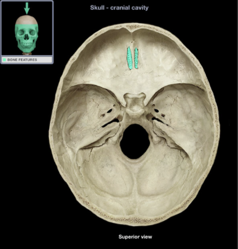
Where is the location of the Cribriform plate
-
What is the Cribriform plate?
Bony plate with foramina (holes) that transmit olfactory nerves (CN I)
-
What does the Cribriform plate contribute to?
The anterior midline portion of anterior cranial fossa
-
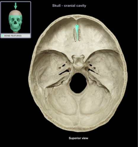
Where is the Crista Galli located
-
What is the Crista Galli?
Triangular process that projects superiorly from the cribriform plate of ethmoid bone
-
What does the Crista Galli contribute to?
the anterior midline portion of anterior cranial fossa
-
What is the Crista Galli the anterior point of attachment for?
the falx cerebri
-
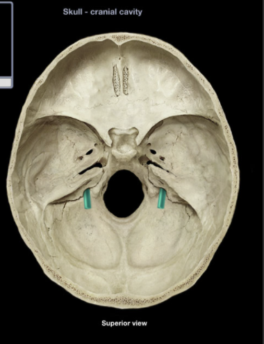
Where is the Foramen Iacerum located?
-
What is the Foramen Iacerum?
-Irregularly shaped opening formed by sphenoid, temporal and occipital bones
- Superior Opening (middle cranial fossa)
- Inferior opening (base of skull)
-
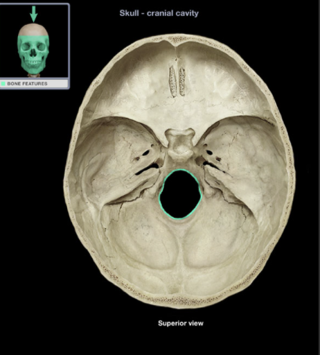
Where is the Foramen Magnum?
-
What is the Foramen Magnum?
- Large opening in inferior part of posterior cranial fossa
- Transversed by spinal root of accessory nerve (CN XI) and vertebral and spinal arteries
- Brainstem and spinal cord continuous through this opening
-
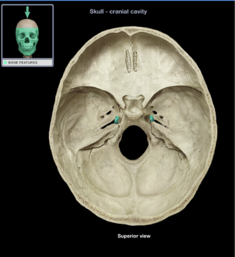
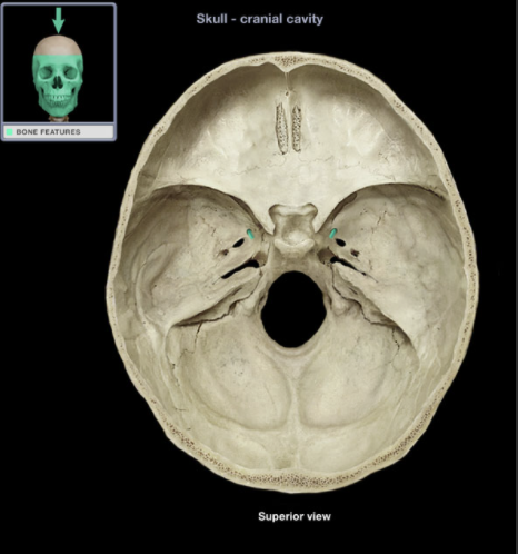
Where is the Foramen Ovale?

-
What is the Foramen Ovale?
Oval-shaped hole
Superior opening in middle cranial fossa
Inferior opening in infratemporal fossa
Mandibular division of trigeminal nerve (CN V3) passes through this opening
-
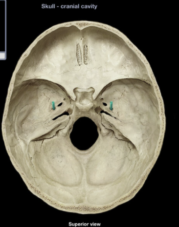
Where is the Foramen Rotundum located?
-
What is the Foramen Rotundum?
Horizontal canal
Connects middle cranial fossa with pterygopalatine fossa
Transmits maxillary (CN V2) nerve
-
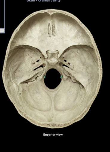
Where is the Foramen Spinosum?
-
What is the Foramen Spinosum?
Round hole
Superior opening in middle cranial fossa
Inferior opening in infratemporal fossa
Middle meningeal artery passes through this opening
-
Where is the Greater Wing of the Sphenoid bone located?
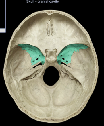
-
What is the Greater Wing of the Sphenoid Bone?
- Paired, Lateral projections
- Forms part of the middle cranial fossa and lateral aspect of skull (temple)
-
Where is the Hypoglassal Canal?
-
What is the Hypoglassal Canal?
Short canal through occipital bone near foramen magnum
External opening on medial aspect of occipital condyle
Traversed by hypoglossal nerve (CN XII)
-
Where is the Jugular Foramen?
-
What is the Jugular Foramen?
Irregularly-shaped opening
Superior opening (posterior cranial fossa)
Inferior opening (base of skull)
Posterior to external opening of carotid canal
Traversed by internal jugular vein, glossopharyngeal nerve (CN IX), vagus nerve (CN X), and accessory nerve (CN XI)
-
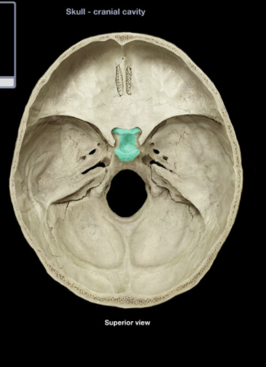
Where is the Sella Tucica?
-
What is the Sella Turcica
Prominent depression on sphenoid body in middle cranial fossa
Formed by tuberculum sellae (anteriorly), hypophysial fossa (central), and dorsum sellae and posterior clinoid processes (posteriorly)
Contains pituitary gland in hypophysial fossa
-
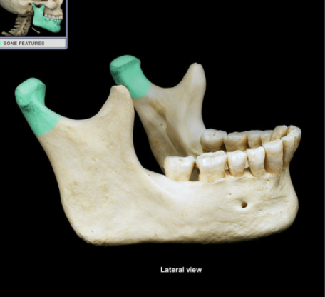
Where is the Condylar Process of Mandible
-
What is the Condylar Process of Mandible
Superior projection from posterior aspect of ramus
Apical end is the expanded mandibular "head" which articulates with mandibular fossa (temporal bone) to form temporomandibular joint (TMJ)
-
What is the function of the CPM
Provides attachment for lateral pterygoid muscle
-
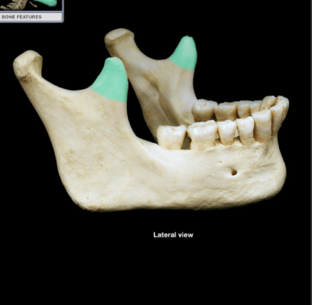
Where is the Coronoid Process of Mandible
-
What is the CoPM
Pointed process on anterior and superior aspect of ramus
-
What is the function of the CoPM
Provides attachment for temporalis muscle
-
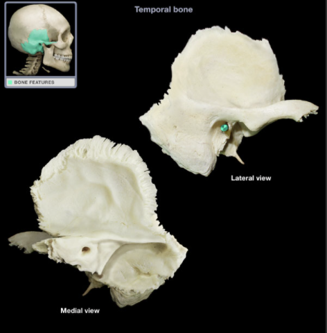
Where is the External Acoustic Meatus?
-
What is the EAM
Short canal (2.5 cm in length) of external ear
Lateral 1/3 is cartilaginous
Medial 2/3 is bony
Ends at tympanic membrane
-
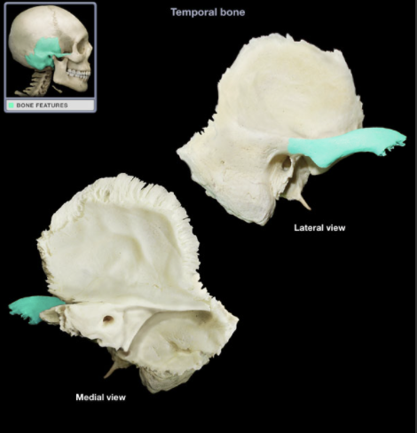
Where is the Zygomatic Process of Temporal Bone
-
What is the ZPTB
Anterior projection of squamous temporal bone
-
What is the function of the ZPTB
Joins temporal process of zygomatic bone to form zygomatic arch
-
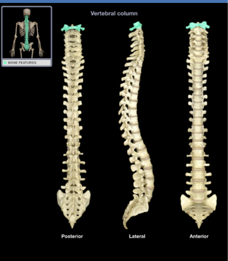
Where is the Atlas
-
What is the Atlas?
Ring-shaped vertebra
Characteristic features include transverse foramen
Lacks vertebral body, spinous process, or lamina
-
What other bones does the Atlas connect with?
Articulates with skull (occipital bone)
No intervertebral disk between C1 vertebra and occipital bone
-
What is the Atlas also known as?
C1 Vertebra
-
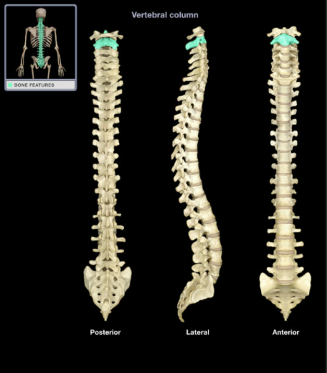
Where is the Axis?
-
What is the Axis?
Characteristic features include transverse foramen, bifid (split) spinous process, and dens (prominent superior projection from body)
-
What is the Axis also known as?
C2 Vertebra
-
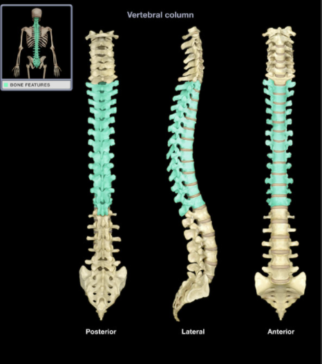
Where is the Cervical Vertebra
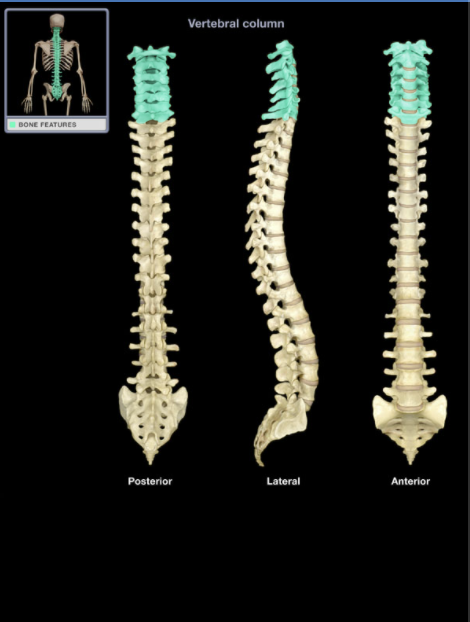
-
What is the Cervical Vertebra?
Seven individual vertebrae
Characteristic features include transverse foramen and bifid (split) spinous process on C3-C6
-
Where is the Coccyx?
-
What is the Coccyx?
Small, triangular bone
Consists of three to five, variably fused, poorly developed vertebrae
Rudiment of the tail in other vertebrates
-
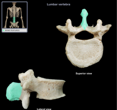
Where is the Lumbar Vertebra
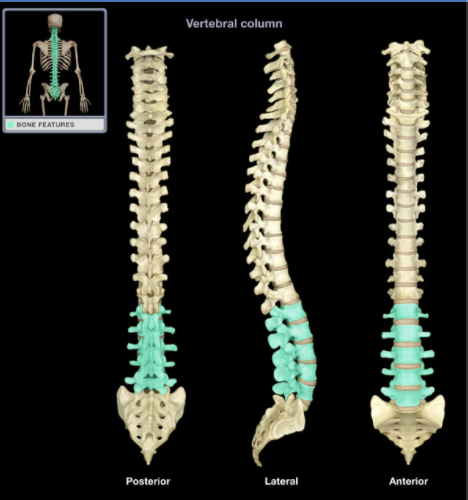
-
What is the Lumbar Vertebra
Five individual vertebrae
Characteristic features include large size, kidney bean-shaped body, and a thick, blunt spinous process
Bodies arranged to form prominent anterior convexity (lumbar curvature; also known as lumbar lordosis, which can be accentuated pathologically)
Intervertebral discs between lumbar vertebrae most commonly herniate ("slipped-disk")
-
Where is the Sacrum?
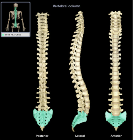
-
What is the Sacrum
Five fused vertebrae
Triangular bone wedged between hip bones
Sacral promontory (the prominent, projecting edge of base of sacrum formed by superior border of S1 vertebral body) is landmark for establishing female pelvic dimensions
-
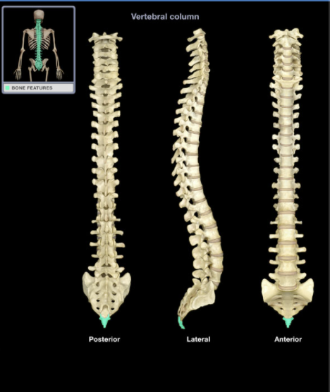
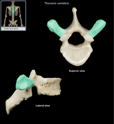
Where is the Thoracic Vertebrae?
-
What is the Thoracic Vertebrae?
12 individual vertebrae
Characteristic features include costal demifacets (or facets) for articulation with head of rib, spinous process slopes inferiorly, and heart-shaped vertebral body
-
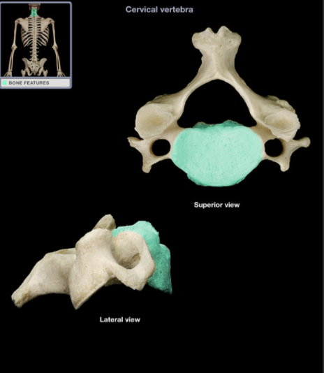
Where is the Cervical Vertebrae?
-
What is the Cervical Vertebrae?
Oval ventral portion
Forms ventral wall of vertebral canal
Supports body weight
Atlas (C1 vertebra) does not have a vertebral body
Progressive increase in size from superior to inferior
-
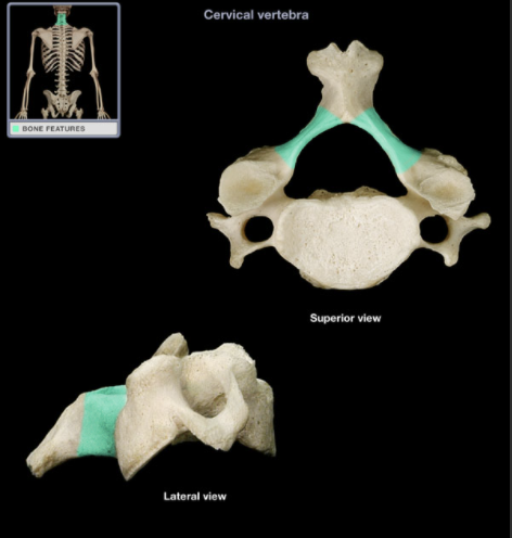
Where is the Lamina of the Cervical Vertebrae?
-
What is the Lamina of the Cervical Vertebrae?
Paired plates that form dorsal wall of vertebral canal
Connects pedicle to spinous process
Atlas (C1 vertebra) does not have laminae
Laminectomy commonly refers to removal of posterior arch of vertebral canal
-
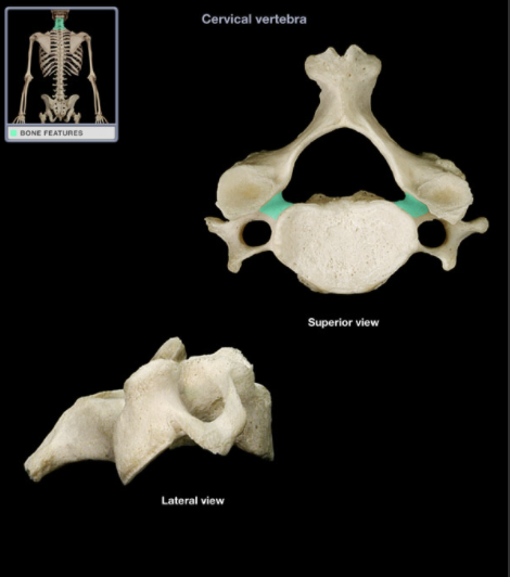
Where is the Pedicle of the Cervical Vertebrae?
-
What is the Pedicle of Cervical Vertebrae?
Short, thick pillar
Connects vertebral body to lamina on each side
Adjacent pedicles contribute to each intervertebral foramen
-
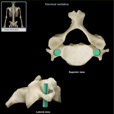
Where is the Transverse Foramen of Cervical Vertebrae?
-
What is the TF of CV
Foramen in each transverse process
Vertebral artery passes through transverse foramina of C1-6 vertebrae (not through C7 transverse foramen)
-
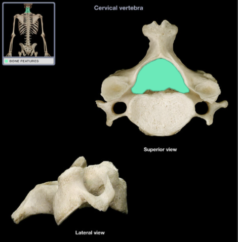
Where is the Vertebral Foramen of Cervical Vertebrae?
-
What is the VF of CV?
Large foramen formed by vertebral arch and posterior aspect of vertebral body
Vertebral arch formed by paired pedicles and laminae
Vertebral canal formed by combined vertebral foramina
Contains spinal cord, meninges, spinal nerve roots, blood vessels, and fat
-
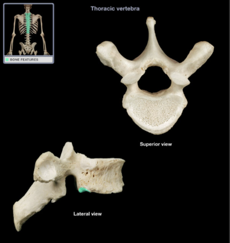
Where is the Inferior Costal Facet of Thoracic Vertebrae?
-
What is the ICF of TV
Each rib articulates with vertebral column at superior and inferior articular costal facets on adjoining thoracic vertebrae
Smooth area
-
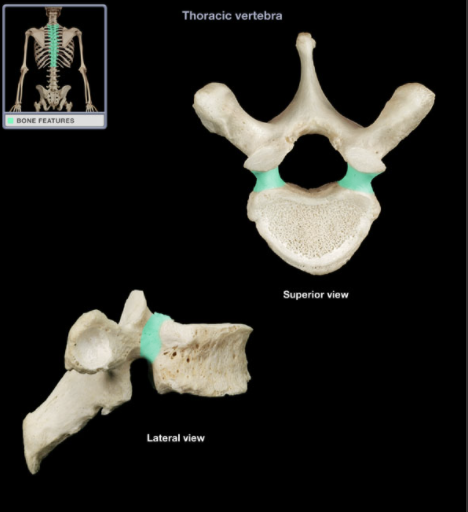
What is the Pedicle of Thoracic Vertebrae?
-
What is the PTV?
Short, thick pillar
Connects vertebral body to lamina on each side
Adjacent pedicles contribute to each intervertebral foramen
-
What is the Transverse Process of Thoracic Vertebrae?
Prominent, paired, laterally-directed process
Provides attachment for intrinsic back muscles and ligaments
-
Where is the Transverse Process of Thoracic Vertebrae?
-
Where is the Spinous Process of Lumbar Vertebrae?
-
What is the SP of LV
Unpaired, posterior projection from midline of vertebral arch
Has characteristic thick, blunt form
Spinous process present on all vertebrae except the atlas (C1 vertebra) and coccygeal vertebrae
Provides attachment for muscles and ligaments
-
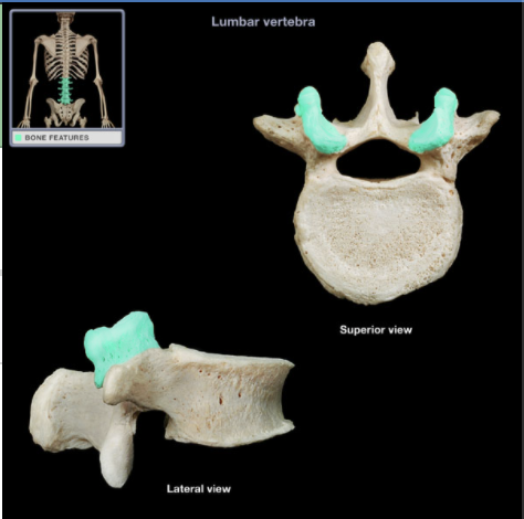
Where is the Superior Articular Process of Lumbar Vertebrae?
-
What is the SAP of LV
Paired process at junction of pedicle and lamina
Has medially-directed articular facet (smooth area)
Articular facet provides articulation between adjacent vertebrae
Lumbar articular facets on articular processes permit lateral flexion and rotation of lumbar vertebral column
-
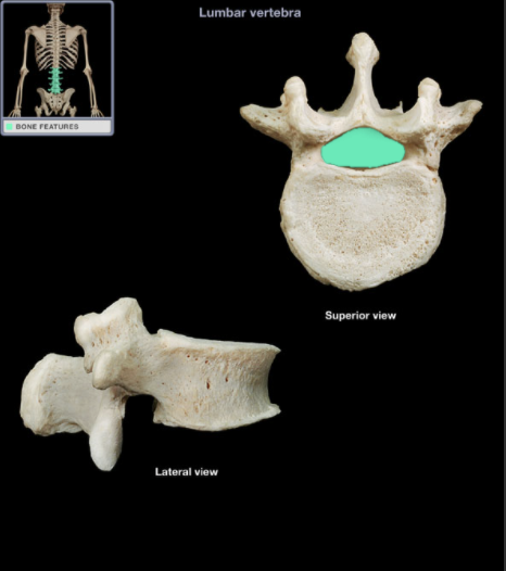
Where is the Vertebral Foramen of Lumbar Vertebrae?
-
What is the VF of LV?
Large foramen formed by vertebral arch and posterior aspect of vertebral body
Vertebral arch formed by paired pedicles and laminae
Vertebral canal formed by combined vertebral foramina
Contains spinal cord (superior to L2 vertebra), meninges, spinal nerve roots (cauda equina inferior to L2 vertebra), blood vessels, and fat

