Fatima Adam
Gastrointestinal System (Anatomy)
The gastrointestinal (or alimentary) tract is a continuous tube that runs from the oral cavity to the anus through the body cavity. It includes the pharynx, oesophagus, stomach, and small and large intestines, as well as accessory digestive organs such as the liver, gall bladder and pancreas. The spleen is not part of the system, but it is often included in any consideration of the anatomy of the area because it also lies in the abdominal cavity. Digestive processes in the G.I. tract include both mechanical and chemical digestion. In this session, you will identify the different parts of the G.I. tract, their blood supply and venous drainage and how the abdominal organs are related to the peritoneum. Why it is important? Without adequate nutrition, the body cannot function. Without food, you die. The G.I. tract provides the means by which food can be broken down into molecules small enough to enter the cells of the body. The different parts of the G.I. tract are specialised for different roles in either mechanical or chemical digestion. Learning Objectives Describe the anatomy of the oral cavity and pharynx Describe the surface anatomy of the abdomen Describe the peritoneum (visceral and parietal peritoneum) Describe and locate the organs of the foregut, midgut and hindgut
-
What is the anatomical span of the gastrointestinal tract?
Mouth to Anus
-
Where is the gastrointestinal tract predominantly located?
Mostly abdominal
-
Name the components of the gastrointestinal tract found in the head and neck region.
Oral cavity + pharynx
-
In which body regions is the oesophagus located?
Neck, thorax, abdomen
-
In which body cavity is the rectum and anal canal situated?
Pelvic cavity
-
Describe the structural nature of the gastrointestinal tract.
Muscle tube with common structure
-
What does the gastrointestinal system comprise?
Tract + accessory organs
-
What is the significance of the terms Upper and Lower GI tract?
They are clinical terms
-
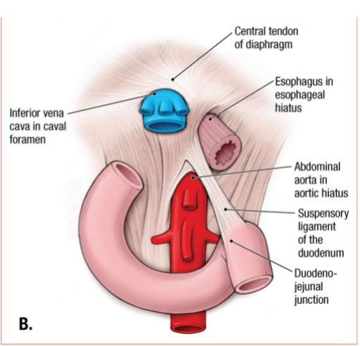
What is the name of the suspensory muscle associated with the duodenum?
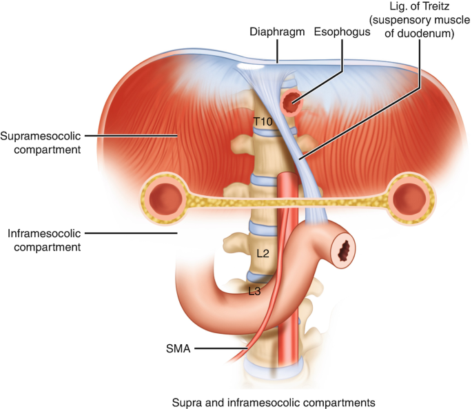
Suspensory muscle of the duodenum
-
Name the components of the Lower GI tract.
Colon (Ascending, Transverse, Descending, Sigmoid), Rectum
-
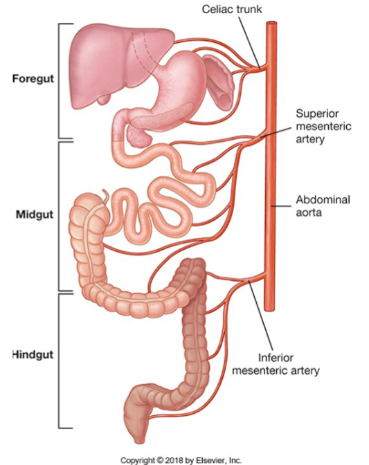
How are the regions of the gastrointestinal tract categorized embryologically?

Foregut, Midgut, Hindgut
-
What are the vasculature and innervation structures related to the Foregut?
Coeliac trunk; Coeliac Plexus
-
What are the arterial and plexus structures associated with the Midgut?

Superior Mesenteric Artery; Superior Mesenteric Plexus
-
What artery and plexus are related to the Hindgut?
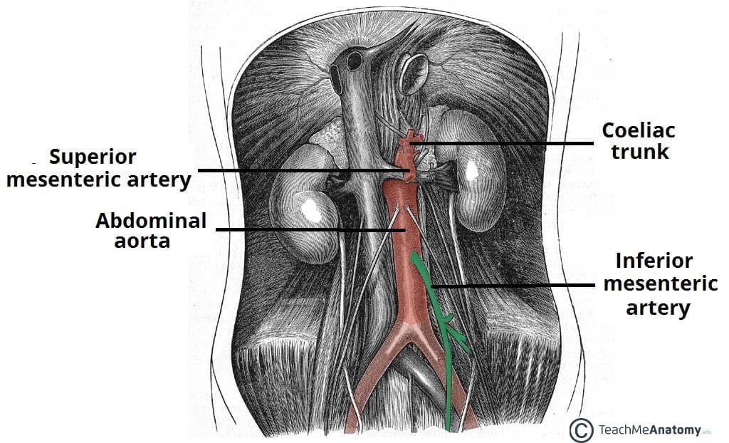
Inferior Mesenteric Artery; Inferior Mesenteric Plexus
-
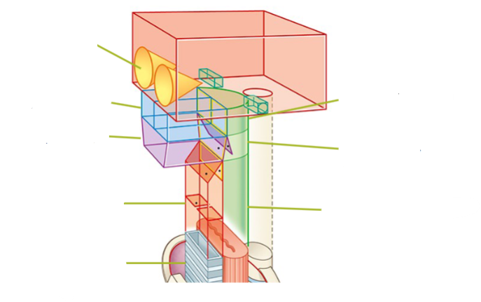
Label these terms of the Upper GI tract:
-
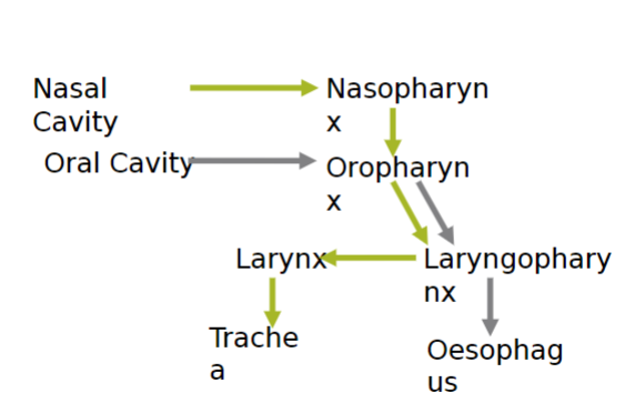
Picture demonstrating the series of events that occurs from the nasal and oral cavities
-
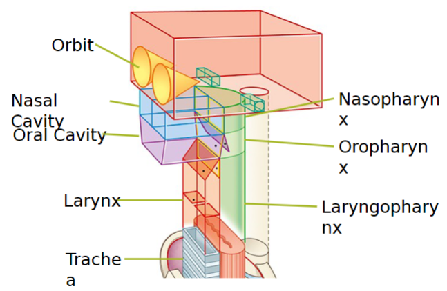
What are the divisions of the mouth and pharynx?
Oral cavity, Oral vestibule, Oral cavity proper, Nasopharynx, Oropharynx, Laryngopharynx
-
What are the functions of the oral cavity?
Mechanical digestion from tongue and teeth
-
Where is the nasopharynx located?

Inferior edge of the soft palate
-
Where is the oropharynx located?

Posterior to the oral cavity; superior border of the epiglottis
-
What function does the laryngopharynx serve?
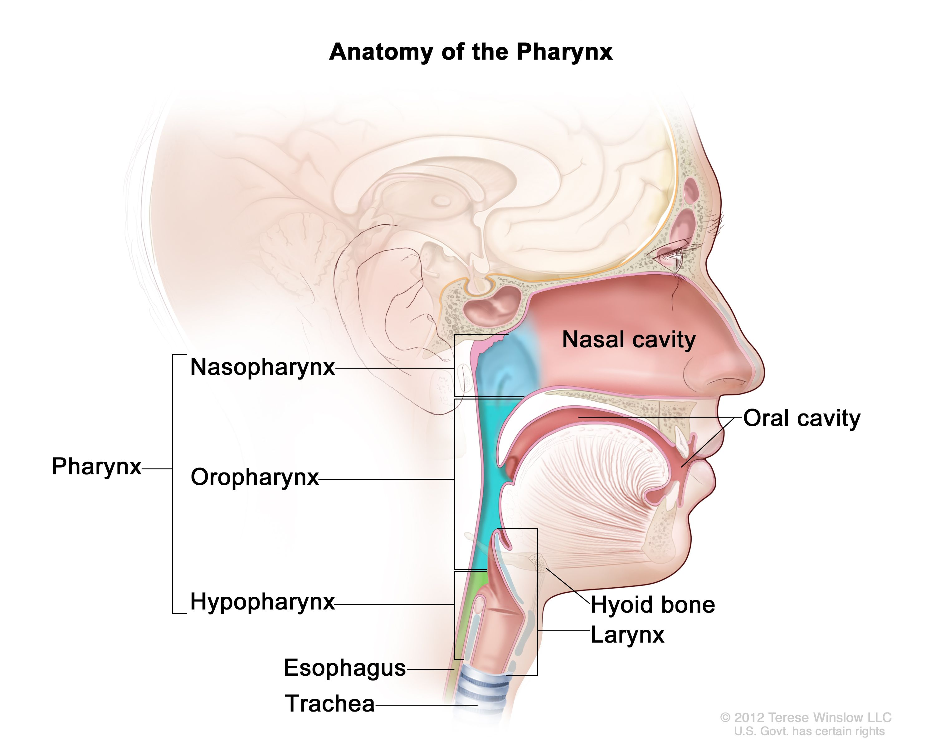
It allows air to pass to the larynx and food to the esophagus.
-
What is the abdomen?
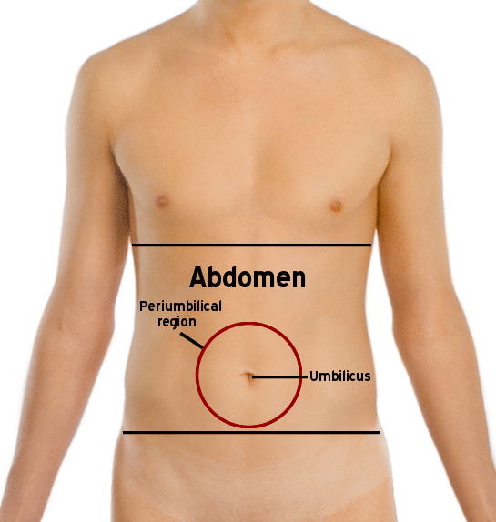
The portion of the trunk between the thorax and pelvis enclosing the abdominal cavity.
-
What structure separates the abdominal cavity from the thorax?
The diaphragm.
-
How is the abdominal cavity related to the pelvic cavity?
It is continuous with the pelvic cavity, forming the abdominopelvic cavity.
-
What protects the abdominal cavity?
The cavity is not protected by bone; it is mostly muscular and contains aponeurosis.
-
How is the abdominal cavity divided sagittally?

Through a medial plane sagittal.
-
What is the orientation of the transumbilical plane?
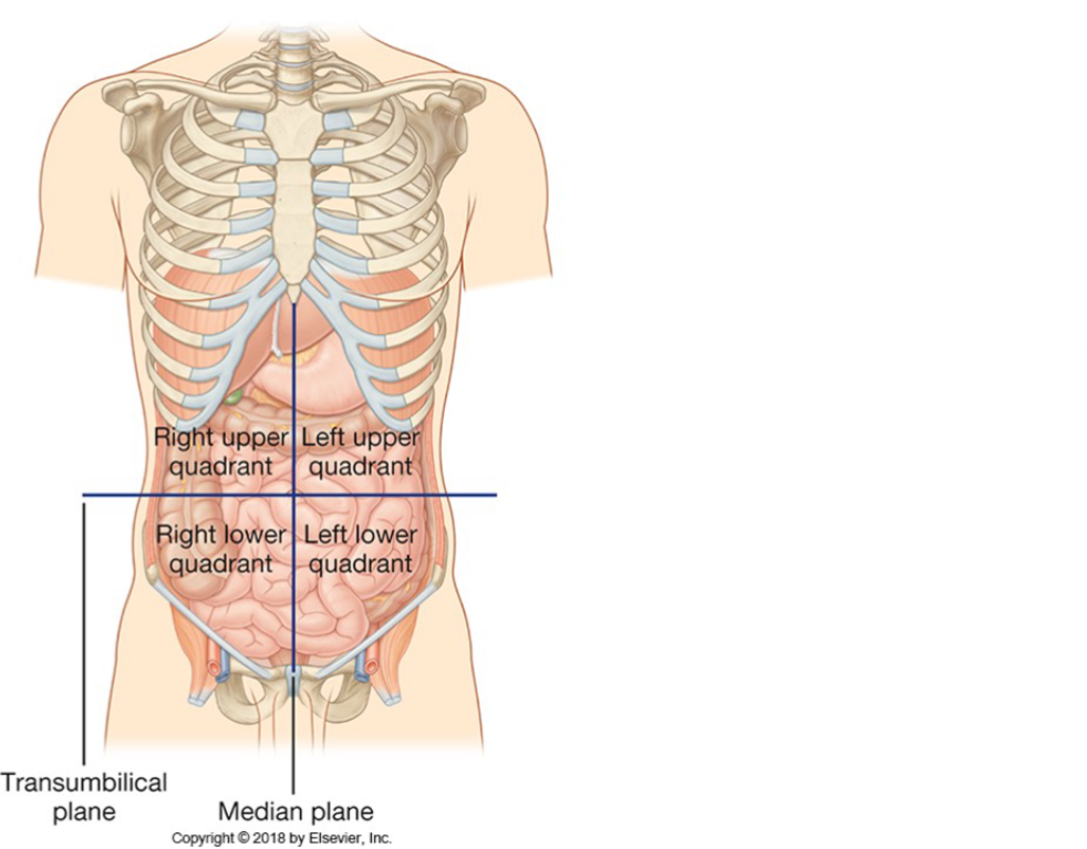
It is transverse and passes through the umbilicus.
-
How many sagittal planes divide the abdomen?

Two sagittal planes.
-
What is another name for the sagittal planes dividing the abdomen?

Midclavicular plane.
-
What landmarks define the subcostal plane?

The inferior borders of the 10th costal cartilage.
-
What landmarks define the transtubercular plane?

Iliac tubercles and the 5th lumbar vertebrae.
-
What are the components of the abdominopelvic cavity?
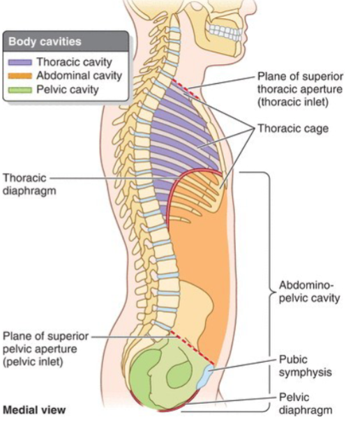
Abdominal cavity, Diaphragm, Anterior and posterior abdominal wall, Pelvic inlet, Pelvic cavity.
-
What structure separates the abdominal cavity from the thorax in the abdominopelvic cavity?

Diaphragm.
-
What structures form the anterior and posterior boundaries of the abdominal cavity?

Anterior and posterior abdominal wall.
-
What is the entryway into the pelvic cavity from the abdominal cavity called?

Pelvic inlet.
-
What is another name for the space overlapping the pelvic cavity?

Greater pelvis; also called false pelvis.
-
What is the function of the diaphragm?

It moves during inspiration and expiration, facilitating the movement of abdominal contents.
-
How is the diaphragm structured?
It is a double muscular domed structure connected by a tendinous section.
-
What does the diaphragm divide from?

The thorax.
-
What are the openings in the diaphragm for?

They are foramina and hiatuses for the passage of the esophagus and vasculature.
-
What mnemonic can be used to remember the order of the diaphragm's major openings?

“I 8 10 EGGs AAT 12“: I 8 = IVC at T8. 10 EGGs = EsophaGus and vaGus at T10. AAT 12 = Aorta, Azygos, and Thoracic duct at T12.
-
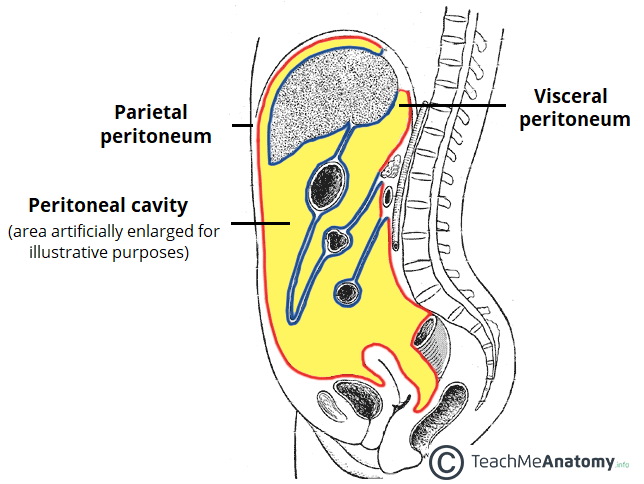
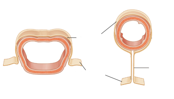
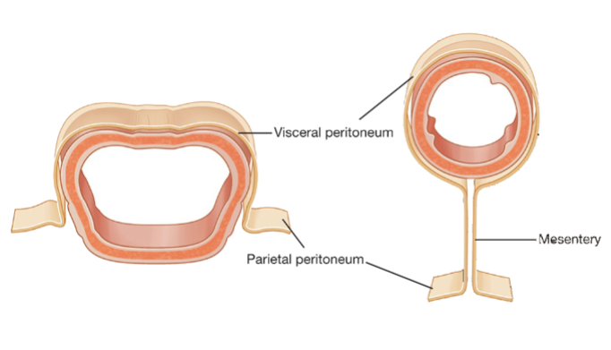
What is the peritoneal cavity?

It is a potential space between the visceral and parietal peritoneum.
-
What are the peritoneal cavity's linings composed of?

Both visceral and parietal peritoneum are from mesoderm and are lined with mesothelium.
-
What type of innervation does the parietal peritoneum have, and what does it line?
It has somatic innervation and lines the walls of the abdominal and pelvic cavities.
-
What type of sensation is associated with the visceral peritoneum, and what does it line?

It can cause referred pain and lines the organs within the abdominal and pelvic cavities.
-
What substance is found in the peritoneal cavity, and in what volume?

Serous fluid, in small volume, is found within the peritoneal cavity.
-
What are mesenteries?

Mesenteries are peritoneal folds that attach viscera to the posterior abdominal wall and serve as conduits for vessels, nerves, and lymphatics.
-
What are omenta?

Omenta are double layers of peritoneum that connect the stomach to other viscera. The greater and lesser omentum correspond to the curvatures of the stomach.
-
What are ligaments in the context of the peritoneal folds?

Ligaments are double folds of peritoneum that connect viscera to the abdominal walls.
-
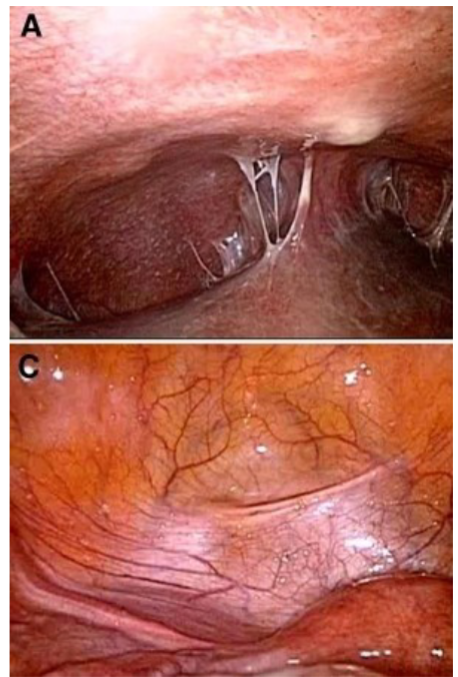
What is a peritoneal recess?

A peritoneal recess is a pouch or concavity of peritoneum formed by surrounding peritoneal structures.
-
What is a peritoneal fold?
A peritoneal fold is a reflection of peritoneum due to underlying structures, typically blood vessels, ducts, or embryonic blood vessels that have since been obliterated.
-
Name these parts of the abdomen:
-
What defines intraperitoneal organs?
They are suspended in the peritoneal cavity and surrounded by visceral peritoneum.
-
What characterizes retroperitoneal organs?
They are located outside the peritoneal cavity, with only one or part of one surface covered by visceral peritoneum.
-
How are secondarily retroperitoneal organs defined?
They initially develop as intraperitoneal organs but later move to a retroperitoneal position.
-
What term describes the process of moving a retroperitoneal organ to an intraperitoneal position for surgery?
Surgical mobilization.
-
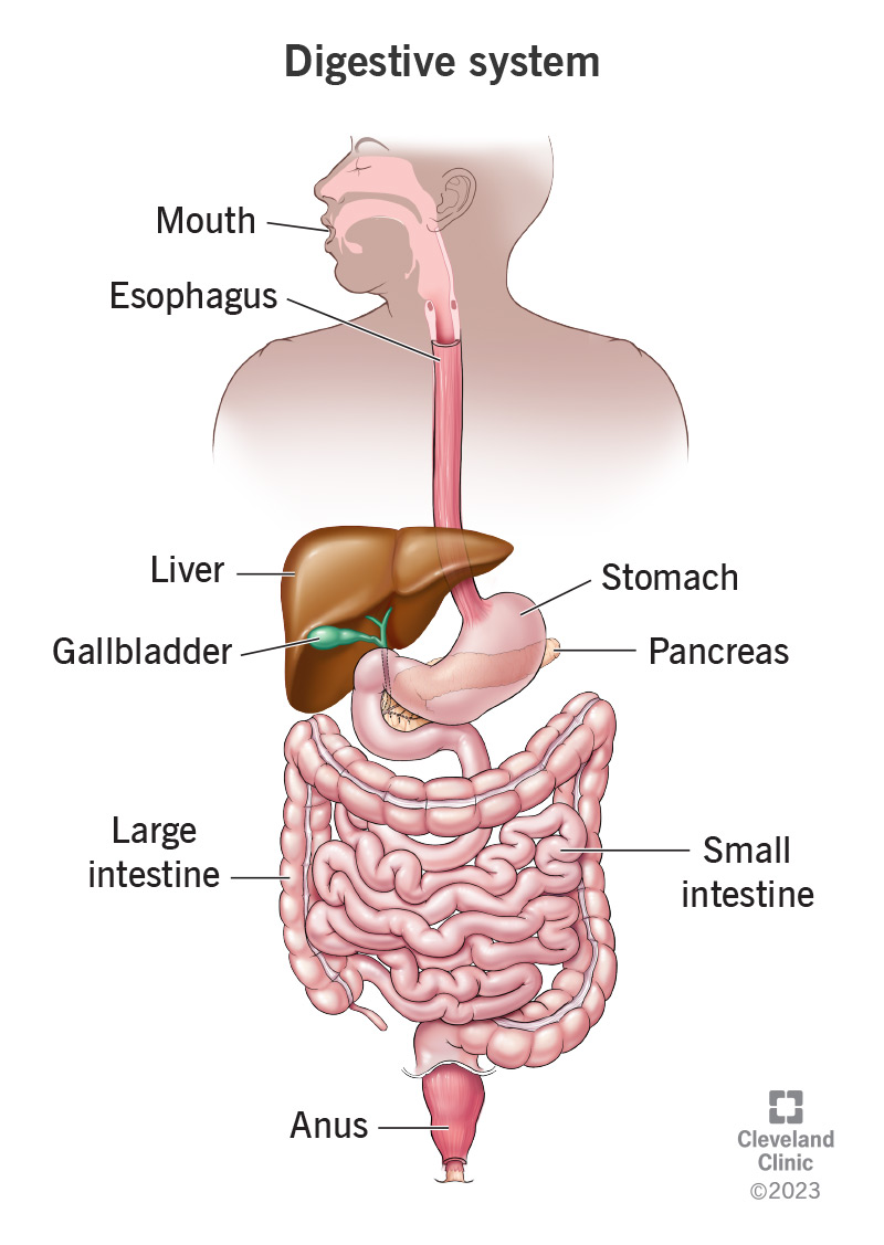
What organs are included in the upper digestive tract?
Oral cavity, Pharynx, Oesophagus, Stomach, Duodenum.
-
What organs constitute the lower digestive tract?

Jejunum, Ileum, Colon.
-
What are the accessory organs of the digestive system?

Accessory organs include the liver, pancreas, and gallbladder.
-
What does the gastrointestinal/alimentary/digestive system comprise?
It encompasses the whole business, including the tract and accessory organs of digestion such as the mouth, liver, and pancreas.
-
What is included in the gastrointestinal/alimentary/digestive tract or canal?
Only the 'tube,' consisting of the esophagus, stomach, small and large intestine, and anal canal.
-
What is the outermost layer of the typical GI tract?

Serosa.
-
Which layer of the GI tract is responsible for peristalsis?

Muscularis externa.
-
What is one component of the muscularis externa?

Longitudinal muscle.
-
Which plexus is found between the longitudinal and circular muscle layers?
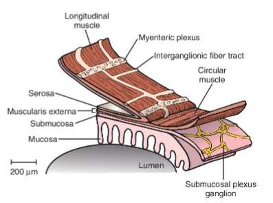
Myenteric plexus.
-
What is another component of the muscularis externa?

Circular muscle.
-
Which plexus is found in the submucosa of the GI tract?

Submucosal plexus.
-
What is the layer of the GI tract that contains blood vessels, lymphatics, and nerve fibers?

Submucosa.
-
What is the innermost layer of the typical GI tract?

Mucosa.
-
What is the approximate length of the esophagus and its composition?

It is approximately 25 cm long and is a muscular tube transitioning from skeletal to smooth muscle.
-
Describe the path of the esophagus through the thorax and abdomen.

It has a thoracic part, passes through the diaphragm, and has a short abdominal part.
-
Where does the esophagus originate, and what does it mark the beginning of?
It originates from the laryngopharynx and marks the beginning of the typical structure of the gastrointestinal tract.
-
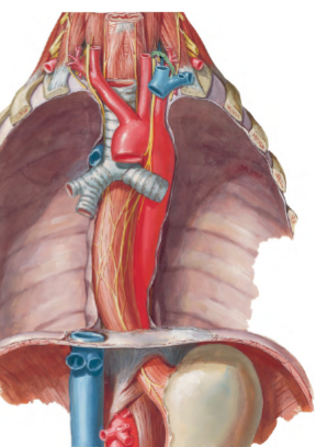
What nerves are associated with the esophagus, and what happens to them as they descend?

The vagal trunks (continuation of Cranial Nerve X) travel down the esophagus, with the right vagus becoming the posterior trunk and the left vagus becoming the anterior trunk.
-
What caution is given regarding the vagal trunks?

Do not cut them.
-
Why is it advised not to cut the vagal trunks during surgical procedures involving the esophagus?

The vagal trunks contain important nerve fibers that contribute to the regulation of various gastrointestinal functions, including motility and secretion. Cutting them could lead to disruption of these functions, causing complications such as dysphagia or gastroesophageal reflux.
-
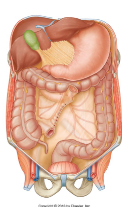
What are the main functions of the stomach, and how is it structured?
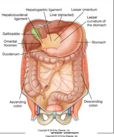
The stomach serves to dilate the GI tract, allowing for storage and chemical digestion. Structurally, it has a curved shape with a lesser curvature and a greater curvature. Its muscular layers include a third oblique layer that facilitates churning of food. The stomach is intraperitoneal and is associated with the greater omentum and lesser omentum. Additionally, it is connected to the liver through the hepatogastric and hepatoduodenal ligaments.
-
What are some distinctive features of the stomach?
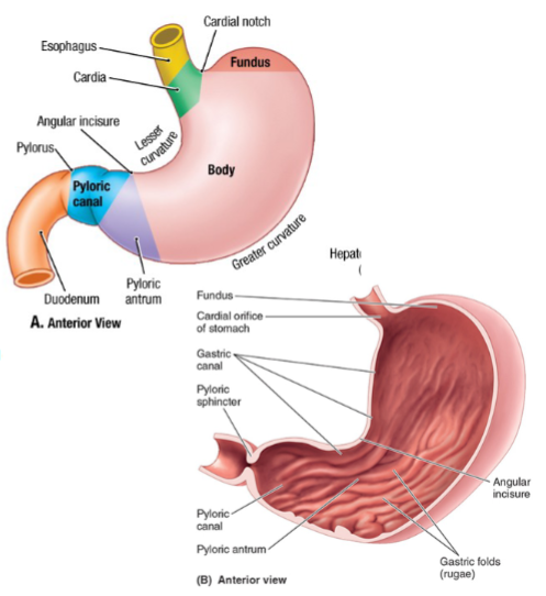
The stomach contains rugae, which are folds in its inner lining that allow for expansion. It has an extra muscle layer, the oblique layer, aiding in mechanical digestion. The gastroesophageal sphincter is a physiological sphincter controlling the passage of food from the esophagus into the stomach. Externally, the stomach is influenced by the diaphragm and exhibits a curved shape. The pyloric sphincter is an anatomical structure at the exit of the stomach, composed of circular fibers.
-
What are the key features of the duodenum?
The duodenum is the first part of the small intestine. It is divided into four parts: superior, descending, inferior, and ascending. It curves around the head of the pancreas and passes behind the greater omentum, making it retroperitoneal. The duodenum receives secretions from the pancreas and liver, aiding in digestion and neutralizing stomach acid.
-
What are the characteristics of the jejunum and ileum?
The jejunum and ileum together make up approximately 5-7 meters of the small intestine, with about two-thirds being the jejunum. The jejunum, located in the left upper quadrant, is responsible for mass absorption. The ileum, situated in the right lower quadrant, absorbs the remaining nutrients, particularly vitamin B12 and bile acids. Both the jejunum and ileum are intraperitoneal and are supported by the mesentery.
-
How can you distinguish between the jejunum and ileum?
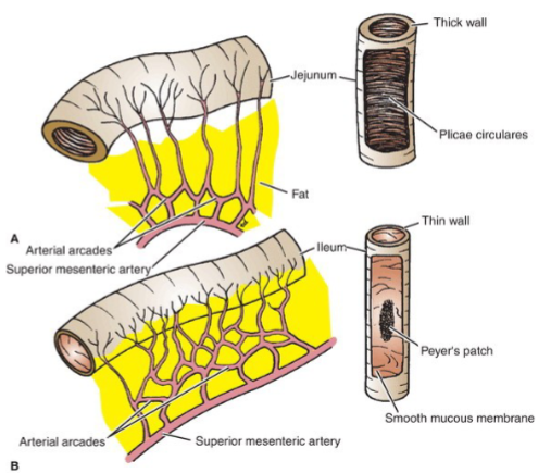
There are no clear boundaries between the jejunum and ileum, but some distinctions can be made based on characteristics as you progress proximally to distally. The jejunum tends to have a redder to pinker color, while the ileum appears lighter. The wall of the jejunum is thicker and becomes thinner and lighter in the ileum. There is more fat in the mesentery of the jejunum compared to the ileum. Circular folds in the jejunum are more abundant and longer, whereas they become more sparse and shorter, eventually absent in the distal ileum. The jejunum tends to have more Peyer's patches than the ileum. Additionally, the jejunum is more vascular, with longer vasa recta and more extended arcades compared to the ileum.
-
What are the characteristics of the cecum and appendix?
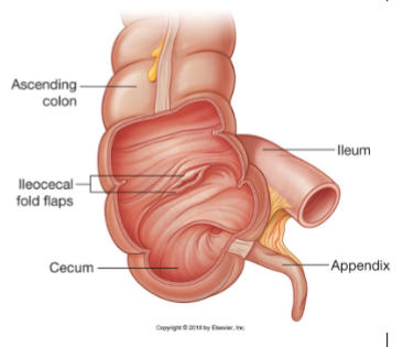
The cecum is a dilated pocket at the beginning of the large intestine, often considered an evolutionary remnant. It is also the location of the appendix, a small blind-ended tube known as the veriform appendix.
-
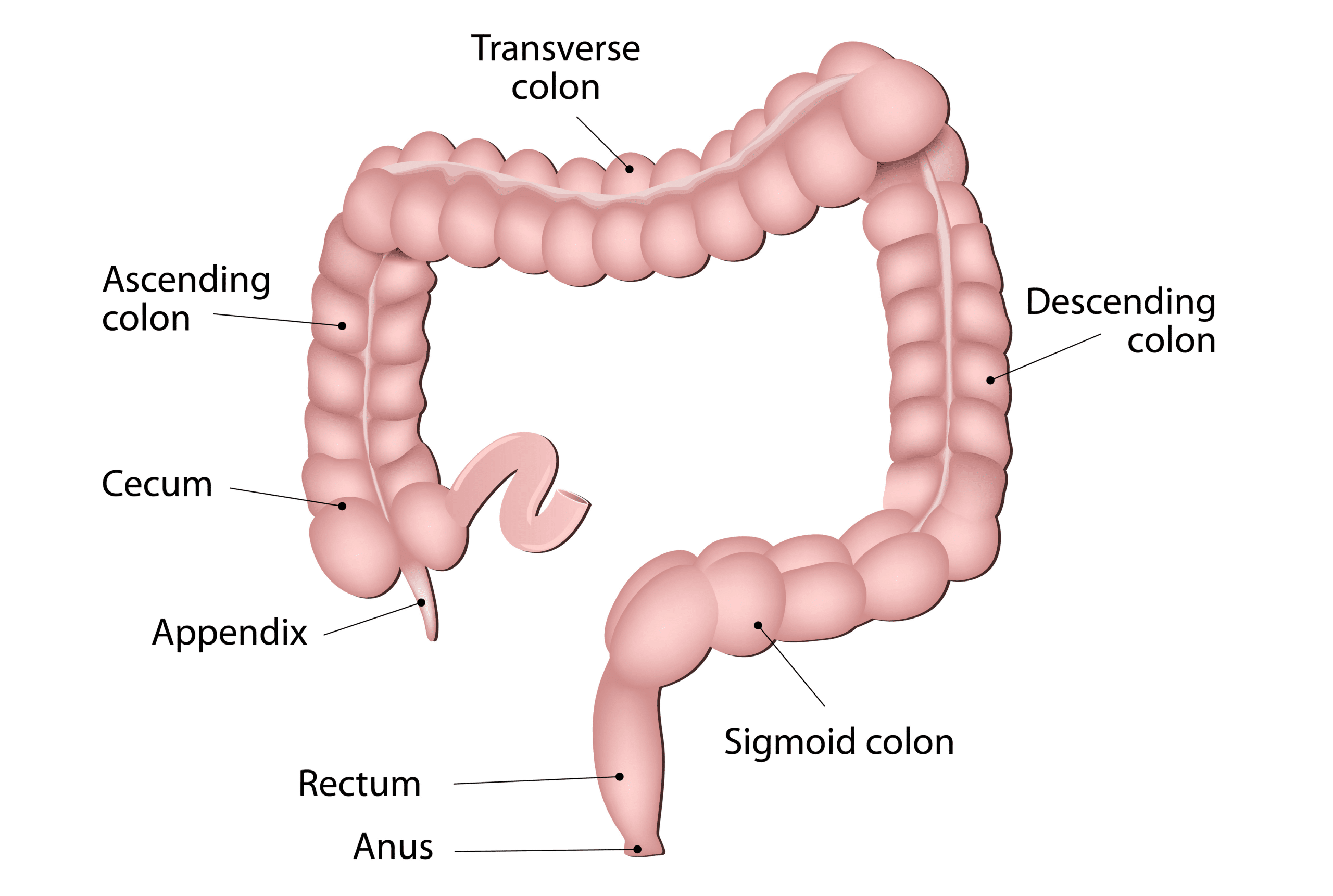
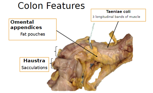
What are some characteristics of the colon?
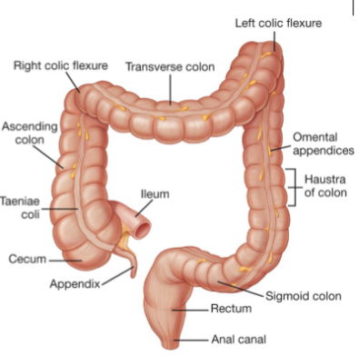
The colon has longitudinal fibers that tighten into three bands called taeniae coli. It transitions back to circular muscle fibers in the rectum. The colon features haustra, which resemble accordion folds. There is a change in structure from the mid to hind gut, occurring approximately two-thirds along the transverse colon. Its relation to the peritoneum varies, and it also contains omental appendices.
-
Picture demonstrating some Colon features:
-
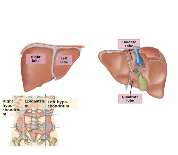
What are some key features of the liver?
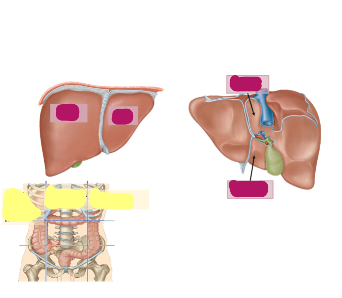
The liver is located in the right upper quadrant of the abdomen and has four lobes. It is partly covered by the thoracic cage. The liver performs a multitude of functions, including bile production. The gallbladder is attached to its inferior surface.
-

State these parts of the Liver:
-
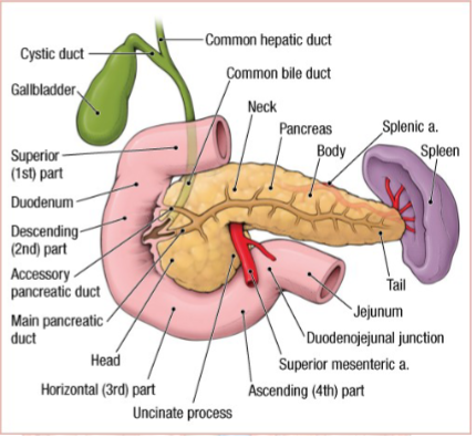
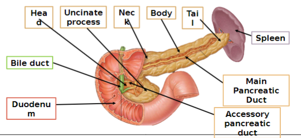
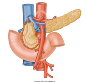
What are the structural characteristics of the pancreas?
The pancreas is a mixed exocrine and endocrine gland. It is located on the posterior abdominal wall, with its head enclosed by the loop of the duodenum and its tail located in the hilum of the spleen. Insulin is produced in the islets of Langerhans within the pancreas.
-
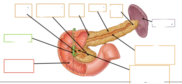
Name these parts of the pancreas:
-
What are some key features of the spleen?
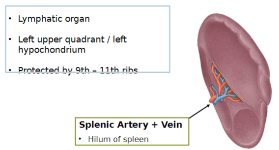
The spleen is located in the left upper quadrant, also known as the left hypochondrium. It is protected by the 9th to 11th ribs. The splenic artery and vein enter and exit the spleen at its hilum. The spleen is a lymphatic organ involved in immune function.
-
What are the main arteries supplying blood to the gastrointestinal regions?
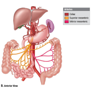
The foregut is supplied by the coeliac trunk, the midgut by the Superior Mesenteric artery, and the hindgut by the Inferior Mesenteric artery.
-
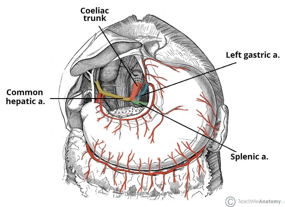
What are the branches of the coeliac trunk?
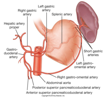
The branches of the coeliac trunk include:
Left Gastric artery
Splenic artery
Left gastro-omental artery
Common Hepatic artery
Proper Hepatic artery
Gastroduodenal artery
-
What are the main vessels of the mesentery and their functions?
The main vessel of the mesentery is the Superior Mesenteric artery. It supplies the midgut, including the jejunum and ileum, and provides branches such as the inferior pancreaticoduodenal artery to the midgut part of the duodenum. The Superior Mesenteric artery also pops over the left renal vein and further branches out to supply the intestines.
-
What are the main arteries supplying blood to the colon?
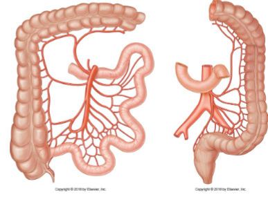
The blood supply of the colon includes:
Marginal artery, which forms anastomoses along the colon.
Ileocolic artery, with branches including the colic branch.
Right colic artery.
Middle colic artery.
Left colic artery, with ascending and descending branches.
Sigmoid branches.
Superior rectal artery.
-
What are the main features of the portal venous system?
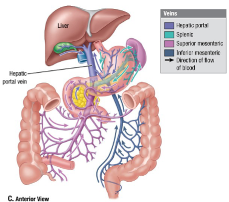
The portal venous system serves as border control for blood from the gastrointestinal tract, as well as blood from accessory organs such as the spleen. All blood from this system passes through the liver, where it is oxygen-poor but nutrient-rich. The hepatic veins drain this blood into the inferior vena cava (IVC).
-
What are the main components of the innervation of the gastrointestinal tract and their effects?
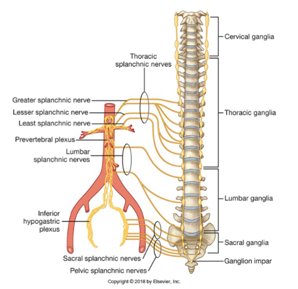
Parasympathetic innervation: Includes vagal branches (from the vagus nerve, CNX) and pelvic splanchnic nerves (S2, S3, S4). It increases secretion and motility of the gastrointestinal tract.
Sympathetic innervation: Slows down motility of the gastrointestinal tract.
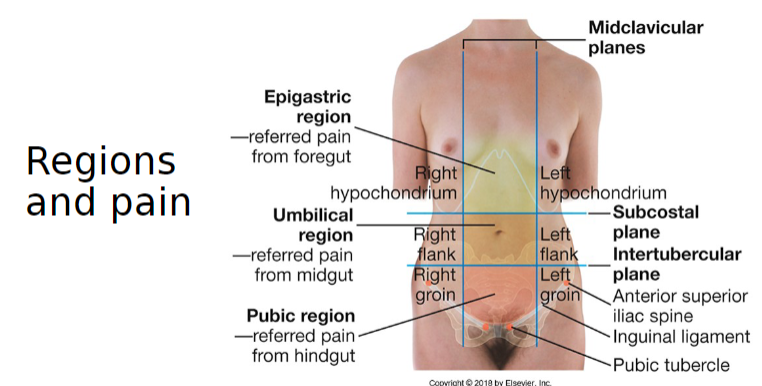
-
Picture demonstrating the regions of pain