Folliculogenesis (Physiology 2) UNFINISHED
Folliculogenesis - Dr Suman Rice Lecture outline The follicle is the structure that supports and protects the egg (oocyte) during its maturation and also produces the majority of the sex steroids necessary for maintenance of the uterus and tubes. In addition the sex steroids feedback onto the hypothalamus and pituitary to control gonadotrophin secretion thereby also regulating the process of follicle growth and development, known as folliculogenesis. Oogenesis, the process by which the oocyte becomes a mature haploid gamete capable of being fertilised, is also inexorably linked to folliculogenesis. Desired Learning Outcomes By the end of this lecture unit, students should be able to: Understand the time span of follicle and oocyte development Describe the structure of the follicle and function of each cellular layer Explain the selection of the dominant follicle that is destined to be ovulated and the mechanisms involved in chromosome number reduction Discuss differing steroid production from both the granulosa and theca cells of the follicle Session resources Download Download Downloadthe lecture slides Session Activities Panopto recording (available after the session) Additional Resources Essential Reproduction (2018) by Johnson M.H. 8th edition: Wiley-Blackwell (Links to an external site.) Glossary Primordial Germ Cell (PGC) – precursor “stem” cells that will become either egg or sperm Oogonia – precursors to eggs, they are diploid and multiply by mitosis Primary oocytes – eggs that have entered meiosis and stopped at meiosis I Secondary oocytes – eggs that have completed meiosis I, entered into meiosis II and stopped Oogenesis – the process of egg development covering the stages from an immature oogonium to a mature ovulated egg ready for fertilization Follicle – oocyte-containing structure containing several cell types i.e. granulosa and theca cells Antrum – fluid-filled space in a follicle Preantral follicle – follicle without an antrum consisting of various stages depending on number of layers of granulosa cells. Antral follicle (AF) – follicle with an antrum filled with follicular fluid Folliculogenesis – the process of follicle development covering the stages of growth from a resting primordial follicle to antral follicles and selection of the dominant follicle destined for ovulation Sex steroids – large group of molecules derived from common sterol precursor: cholesterol. There are 4 main families of steroids – the progestogens; androgens; oestrogens (American spelling estrogen) and corticosteroids. Only the first three are defined as sex steroids. Within each family there are several members.
-
What is folliculogenesis? (4)
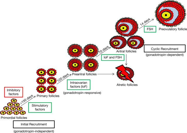
Folliculogenesis is the process of follicle maturation in the ovary.
It involves several stages: Primordial → Primary → Secondary → Mature (Graafian) follicles.
During growth, the oocyte (egg) matures while surrounded by follicular cells.
Follicles secrete estrogen as they grow, which is essential for the reproductive cycle.
-
What are primordial germ cells (PGCs)? (3)
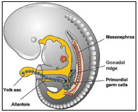
PGCs are the cells that will eventually become eggs (oocytes) in females or sperm in males.
They are first identifiable in the yolk sac of the developing embryo at around 3 weeks after conception.
These cells undergo multiple rounds of mitosis to increase their number.
-
Where do PGCs migrate after formation? (3)
After their formation in the yolk sac, PGCs migrate to the genital ridge, which will eventually form the gonads.
The genital ridge is located near the developing kidneys.
This migration is essential for the further differentiation of PGCs into mature gametes.
-
What is the genital ridge, and how does it contribute to gamete formation? (3)
The genital ridge is a precursor structure that forms the gonads (ovaries in females, testes in males).
It begins as an undifferentiated structure in the embryo.
As PGCs arrive, the genital ridge differentiates into either ovaries or testes depending on the genetic sex of the embryo.
-
How do PGCs become differentiated into male or female gametes? (4)
PGCs migrate to the genital ridge and are exposed to different developmental signals based on genetic sex.
In males, under the influence of SRY gene (Sex-determining Region Y), the genital ridge differentiates into testes.
In females, in the absence of the SRY gene, the genital ridge becomes ovaries.
Males produce sperm in the testes, and females produce oocytes (eggs) in the ovaries.
-
What must be achieved for reproduction to occur? (7)
Differentiation into male or female: This determines the gametes (sperm or eggs).
Sexual maturation: Puberty triggers the ability to produce viable gametes.
Production, storage, and release of gametes: The body must create, store, and release enough eggs or sperm for fertilization.
Correct chromosome number in gametes: Eggs and sperm must contain half the genetic material (haploid) to form a complete embryo when combined.
Gametes must meet: Sperm must reach the egg for fertilization.
Creation of a new embryo: The combination of genes from both parents results in a unique embryo.
Nurturing of the embryo: The embryo must be supported until it can develop into an independent organism.
-
What happens to PGCs when they enter the ovary? (2)
PGCs (primordial germ cells) that enter the ovary differentiate into oogonia.
Oogonia are the precursors to eggs and are diploid cells that multiply by mitosis.
-
What are oogonia, and how do they develop? (3)
Oogonia are the egg precursors formed in the ovary.
These cells are diploid and undergo multiple mitotic divisions to increase their number.
Once mitosis stops, oogonia enter meiosis and become primary oocytes.
-
What happens when oogonia enter meiosis? (3)
Once oogonia enter the first stage of meiosis, they are referred to as primary oocytes.
Primary oocytes are arrested in prophase I of meiosis and remain in this stage until ovulation or death.
No further mitotic divisions occur after they enter meiosis.
-
What is the significance of the mitotic divisions of oogonia? (2)
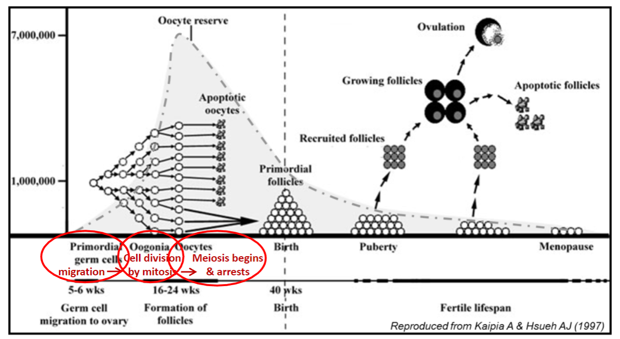
The mitotic divisions of oogonia are crucial because all the eggs a woman will ever have are produced at this stage.
These divisions increase the number of oogonia before they enter meiosis and become primary oocytes.
-
How long do primary oocytes remain arrested in meiosis? (2)
Primary oocytes remain arrested in prophase I of meiosis, sometimes for many years (up to 52 years).
They remain in this arrested state until they are either ovulated or undergo degeneration.
-
Where are primary oocytes located in the ovary? (2)
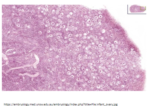
Primary oocytes are located in the outer layer of the ovary, known as the cortex.
This outer region contains the ovarian follicles, each of which houses a primary oocyte.
-
Why is the primary oocyte considered important? (2)
The primary oocyte is arguably the most important cell in the body.
It remains in the first meiotic phase for many years, making it vulnerable.
-
How does the primary oocyte get protection during its arrested phase? (3)
Each primary oocyte becomes surrounded by protective layers and supporting cells.
These cells help protect the oocyte as it remains arrested in meiosis for years.
In the fetal ovary, granulosa cells (GC) differentiate around the oocyte to provide this support.
-
What happens to the surrounding cells in the fetal ovary? (2)
In the fetal ovary, the granulosa cells (GC) condense around the primary oocyte.
These granulosa cells then differentiate into specialized cells that surround and protect the oocyte.
-
What is the basal lamina (BL), and what is its role? (2)
The granulosa cells secrete an acellular layer called the basal lamina (BL).
The basal lamina provides additional structure and protection to the oocyte.
-
What is a primordial follicle? (2)
The structure formed by the primary oocyte surrounded by granulosa cells and the basal lamina is called a primordial follicle.
This is the earliest stage of follicle development in the ovary.
-
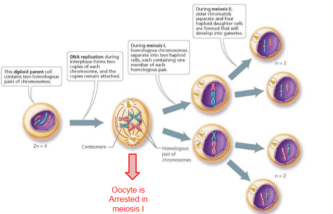
When do chromosomes replicate during the cell cycle? (1)
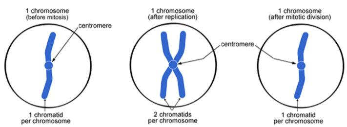
Chromosomes replicate during the S-phase of the cell cycle.
-
How are chromatids formed after chromosome replication? (2)
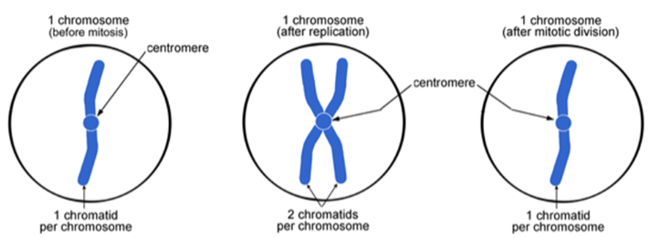
After chromosome replication, the two copies remain attached at the centromere.
Each copy is called a chromatid, and the two copies are identical to each other, known as sister chromatids.
-
What are sister chromatids? (1)
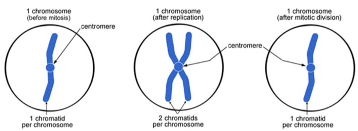
Sister chromatids are the two identical copies of a chromosome, formed after replication.
-
What is the relationship between the original chromosome and the chromatids? (1)
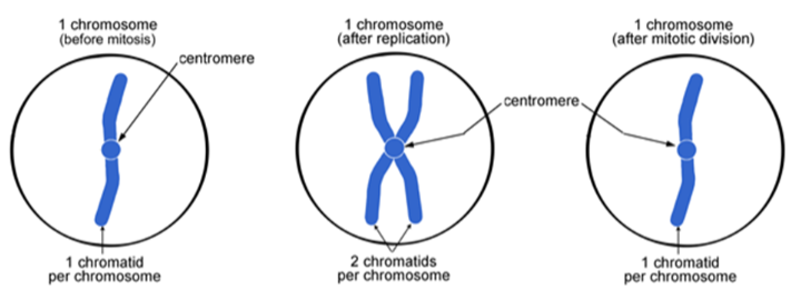
The chromatids are an exact copy of the original chromosomes.
-
What are the 4 stages of Mitosis? (4)
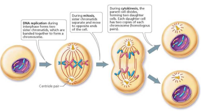
Prophase
Metaphase
Anaphase
Telophase
-
Picture demonstrating an overview of meiosis:
