 Matthew Burn
-
Voluntary vs. Involuntary Muscle
Voluntary = conscious control
Involuntary = not under conscious control
-
3 Types of Muscle Tissue
Skeletal, Smooth, Cardiac
-
Skeletal Muscle Tissue
Voluntary
Used for: movement, posture/support, speech production, defecation/giving birth, breathing
1. Skeletal muscle cells are huge, each cell have 2. multiple nuclei = multinucleated.
3. Shaped liked cylinders.
4. Nuclei are proliferated located. Striated.
-
Smooth Muscle Tissue
Involuntary
1. Located in walls of hallow organs. (ex: small intestine)
2. Spindle shaped cells
3. Prostaglandin: stimulates smooth muscle to contract.
4. Single nucleus
-
Cardiac Muscle Tissue
Branched
1. Located only in the heart. Wall of heart has very thin outer and inner layer, everything in between those two thin layers is Cardiac Muscle Tissue.
2. Extremely specialized
3. Intercalated disc which are special cell junctions that hold the cells together, and facilitate spreading of signal to cell-to-cell.
Heart would destroy itself if not for intercalated discs.
-
Human Heart Pump Rates
Avg. Human @ Rest pumps = 1.39 gpm
Avg. Human @ Exercise pumps = 5.35 gpm
Olympic @ exercise pumps = 10.3 gpm
-
Muscle Origin
point of attachment that moves the least
-
Muscle Insertion
point of attachment that moves the most towards the origin.
-
Tendon vs. Aponeurosis
1. Functionally the same, connect muscle to bone.
2. Made of Dense Irregular CT.
3. Differences at gross/macro level.
Aponeurosis = flat, sheet-like structure. ex: iliotibial tract. Gives stability.
Tendons = cord like structure
-
IT Band Syndrome
inflammation of iliotibial band, pain can range anywhere from iliac crest to knee lateral aspect, often caused by running downhill.
-
Agonist
causes an action
-
Antagonist
prevents an action OR causes opposite action
-
Synergist
The whole is greater than the sum of its parts. Muscles work together to do more than they could individually.
-
Prime mover
Most powerful muscle in an action.
-
4 Characteristics of Muscle Tissue
1. Extensibility - they can be stretched
2. Elasticity - return to normal length after stretching.
3. Irritability - excitability/react
4. Contractility - contracting
-
Innervation
frontal lobe: point of stimulation to send signals by neurons to stimulate muscle contractility
Neurons send signals for muscles to contract.
Muscles stimulated by motor nerves.
Functional units of Muscle = muscle cells/fibers
Functional units of Nerves = nerve cells/neurons
Signals = action potentials, similar to electricity, trillions of signals per movement.
A single neuron innervates a small group of muscle cells (fibers). This single neuron + the muscle cells it innervates constitute a motor unit. Recruit more motor units to meet resistance.
-
Muscle Layers
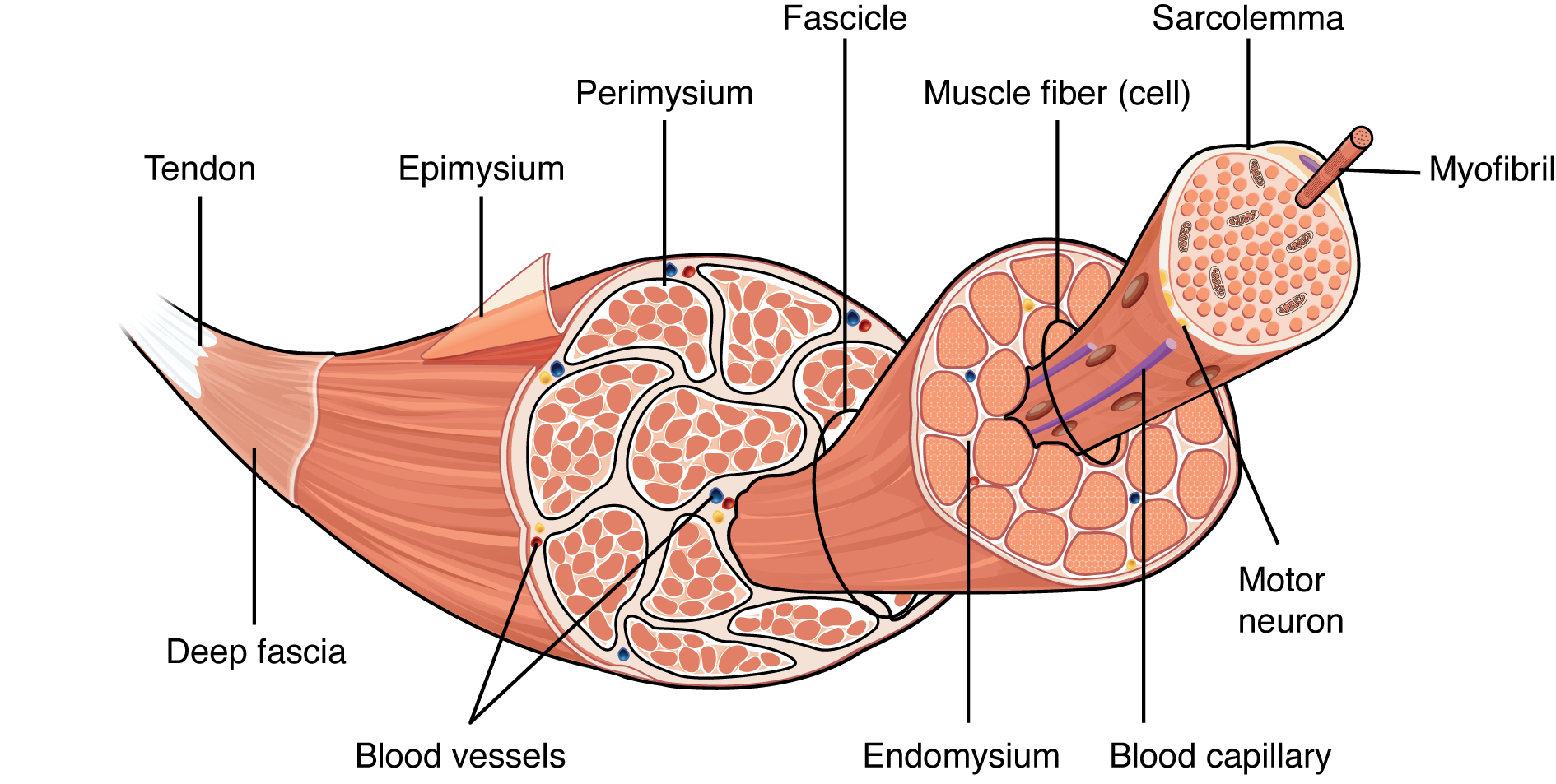
1. Epimysium (muscle) -> Perimysium (fascicle) -> Endomysium (fiber).
Surrounds, binds, holds together subunit
-
Muscle Fascia
superficial layer of muscle tissue (outermost layer).
-
Muscle Subunits
Muscle -> Fascicles -> Fibers/Cells -> Myofibrils -> Myofilaments
-
Muscle Cells Outer Boundaries
Muscle cells have 2 outer boundaries.
1. Sarcolemma = cell membrane
2. Endomysium
-
Sarcomeres
functional units of muscle
(sarcoplasm = endoplasmic reticulum)
sarcomeres shorten to constrict muscles.
-
2 Types of Muscle Filaments (Contractile proteins)
1. Actin = thin
2. Myosin = thick
-
Filament Contraction
-
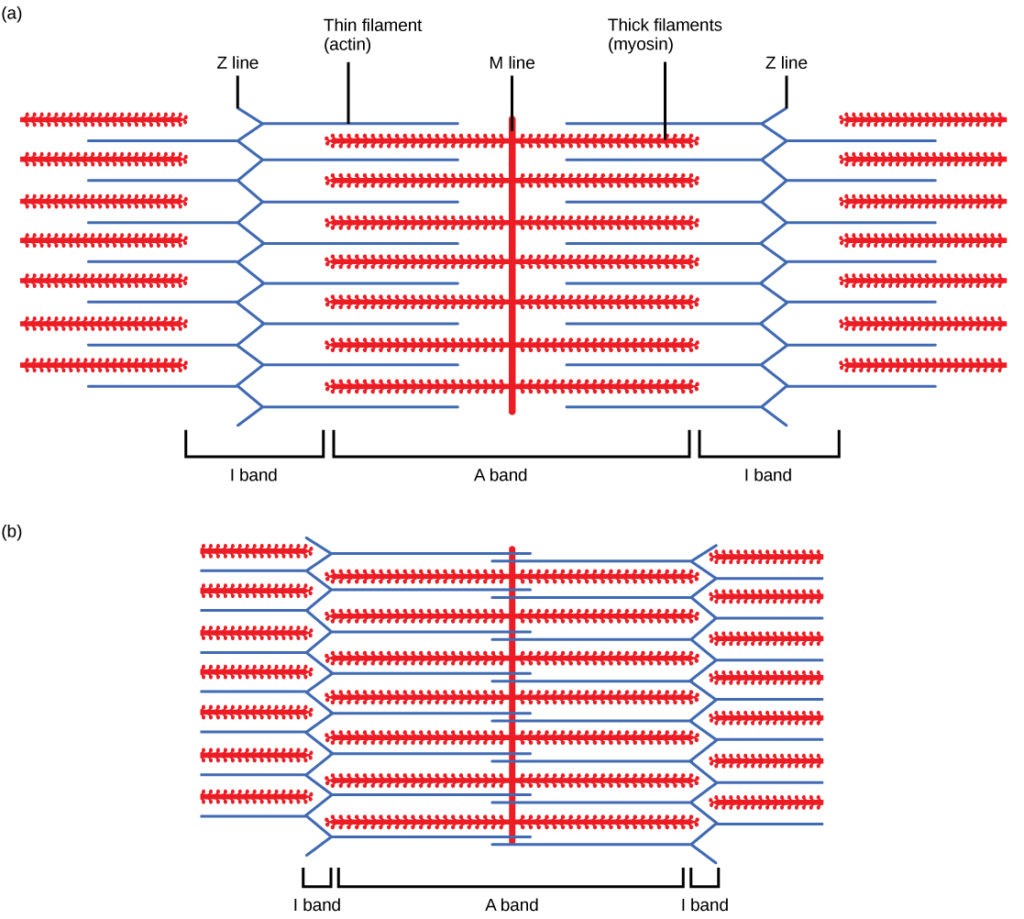
Muscle Striation Illusion
Overlap region between Actin and Myosin filaments stain the darkest, which cause an appearance of striations at low magnifications.
-
I Band
Actin only (thin only)
-
A Band
where actin meets myosin. (where thin meets thick)
-
H-Zone
myosin (THICK) only
-
AOO
Area of overlap actin + myosin (Thin+thick)
-
M-Line
Myomesin, filaments are sliding and as they are sliding the z line is moving closer to myosin, as they are moving closer they need shock absorber which is Titin.
-
Changes in bands upon contraction
A band - stays the same
Z-line - moves closer together
I-band - gets smaller
M-line - doesn't change
H-zone - smaller, negative overlap
Sarcomeres contract, filaments shorten
-
Myosin heads
bind actin, and pull in actin, = why filaments slide Bind, Swivel, Release
-
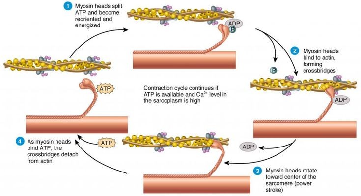
Sliding filament theory
Components - Actin, tropomyosin, and troponin
Binding/active site - where myosin binds
Tropomyosin is blocking binding site.
Calcium bound troponin moves tropomyosin, to allow myosin head to bind to active site, forming a Crossbridge.
Powerstroke - head of myosin swivels
After the powerstroke, crossbridge breaks.
-
Molecular Events of a Contraction
*Sarcomere is resting.
Tropomyosin is blocking binding site.
Myosin heads are holding on ADP + organic phosphate from previously used ATP.
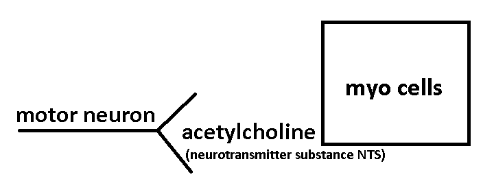
1. Action potential in a motor neuron.
2. Release of acetylcholine at the myoneural junction.
3. The action potential spreads across the sarcolemma into the transverse tubules. (associated with sarcoplasm)
4. Sarcoplasm reticulum releases Ca+.
5. Ca+ binds troponin.
6. The Troponin/Tropomyosin complex moves to the side and exposes the binding site on actin.
7. Myosin head binds actin forming a "cross-bridge".
7.5. - myosin head releases ADP + inorganic Phosphate (The energy for the power-stroke came from the release of ADP + P_i)
8. Power-stroke occurs pulling actin in toward the H-zone.
9. The head must bind a new molecule of ATP to break the crossbridge.
10. Head splits the ATP and is ready to begin another cycle.
-
Uses of ATP
1. Power-stroke
2. Break-crossbridges
3. Action Potential
4. "Pump" Ca+ back into sarcoplasmic reticulum.
-
Sources of ATP
1. Phosphagen energy system - stored energy in cytoplasm. Neither aerobic or anaerobic, already made. 8-10 seconds of energy. Functions immediately.
a.) Stored ATP - ATP -> ADP + P_i + E
b.) Stored Creatine Phosphate (CP) - CP + ADP -> Creatine + ATP -> ADP + P_i + E
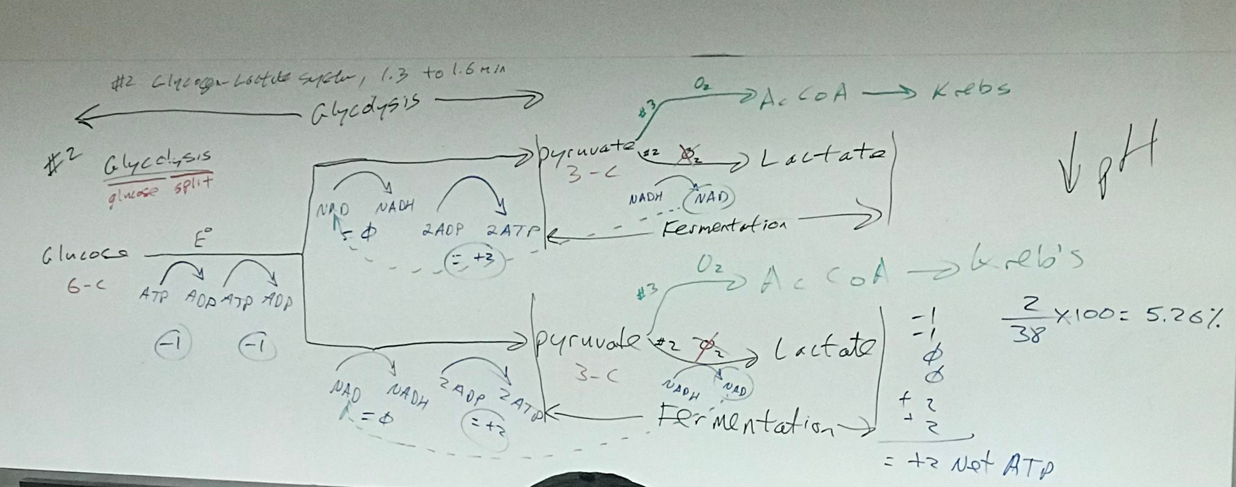
2. Glycogen / Lactate System - (Anaerobic system), 1.3-1.6 minutes of energy
a.) Glycolysis
b.) Fermentation
3. Complete Oxidation of Glucose (Aerobic): 2-4 Hours of energy
i.) Glycolysis - Glucosee - Pyruvate1, Pyruvate2
ii.) Transition Reaction: Pyruvate -> Acetyl CoA
iii.) Kreb's cycle
iv.) Electron transport system
v.) Oxidative Phosphorylation
Range in energy: Depends on amount of glycogen reserves and consumption during activity.
4. Oxidation of Lipids (Aerobic) - Lots of energy, burns fat
-
Metabolism
-
Energizing Glucose molecule
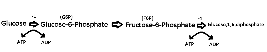
-
Glucose to pyruvate
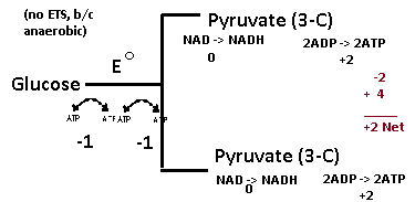
-
Pyruvate Fork
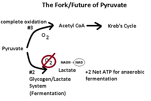
-
Beta-Oxidation Formula
β = ((n/2 - 1) x 5) - 1) + (n/2 x 12), where n = # of C, fatty acid
-
Calories from Macromolecules
Carbs = 4 kcal/g
Protein = 4kcal/g
Fats = 9kcal/g
1 cal = 1 dietary calorie = 1kcal = 1,000 calories
ex: 100 calories = (100*1,000) = 100,000 calories
Carbohydrates are main pathway in animals. Proteins and fats intersect carb pathway.
-
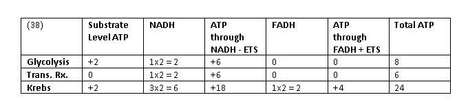
ATP Table
-
Kreb's Cycle
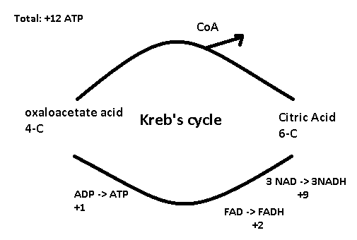
Acetyl CoA is a coupling enzyme, puts together 4-C and 2-C.
-
Red Myo Fibers
Type I
Aerobic
Highly Vascular
Inc. Mitochondria
Inc. Myoglobin
Split ATP Slowly
Resist fatigue
Very good @ burning fat
More intramuscular fat
More Lipoprotein Lipase
Smaller motor units
Endurance
-
White Myo Fibers
Type II
Anaerobic
Less vascular
Less mitochondria
Less myoglobin
Split ATP quickly
Fatigue easily
Depends on carbs
Not much intramuscular fat
Larger motor units
Sprinting
-
Red and White Muscle Fiber Recruitment
1. Recruit all red motor units before recruitment of white motor units.
2. Distribution of red/white fibers is thought to be genetically determined, may be able to convert between the two.
3. Can improve red/white fibers.
-
Exercise related to red/white myo fibers and aerobic vs. anaerobic
1. Send pyruvate to aerobic pathway as long as you have oxygen, remaining pyruvate go to anaerobic pathway.
2. % of aerobic vs. anaerobic usage is a function of your heart rate, that is the % of your Max HR you are functioning at.
-
Exercise Training Zones
1. Classic Aerobic = 70-75%
2. High Aerobic = 75-80%
3. Lactate Threshold = 80-85%
-
Max HR Formula
Max HR = 220-age (males) or Max = HR = 225-age (females),
to find max HR, induce it.
95% Americans sedentary
-
Heart Rate Reserve (HRR) Formula
HRR = Max HR - Resting HR
-
Training HR Formula
Training HR = [(Max - Rest) x %Intensity] + Rest
ex: Sally, age 32 wants to train in Lacatate-threshold zone. Max HR = 192, Rest HR = 61. Calc training zone.
Training HR = [(192-61) x %80)] + 61 = 166
Training HR = [(192-61) x %85)] + 61 = 172
166-172 bpm training zone HR.
-
Benefits of Aerobic Exercise
1. Decrease resting HR
2. Decrease triglycerides
3. Decrease cholesterol
4. Decrease LDL
5. Slight increase in HDL
6. Little increase in myo size
7. Increase myo tone
8. Increase endurance
9. Increase intramuscular mitochondria
10. Increase intramuscular fat
11. Less dependent on carbs
-
Benefits of Anaerobic Exercise
1. Increase HDL
2. Increase intramuscular myofilaments
3. Increase in power and speed
4. Increase in Lactate-Threshold
5. Increase in VO2 Max
6. Increase in size
-
VO₂ Max
VO₂ Max = max volume of O₂ consumed and used per kg bodyweight/minute
-
Isometric muscle movement
the muscle fires (or activates with a force and tension) but there is no movement at a joint. In other words, the joint is static. Muscle length does not change.
-
Isotonic muscle movement
An isotonic muscle contraction occurs when the force or tension in the muscle remains constant while the length of the muscle changes.
-
How do muscles store oxygen?
myoglobin, a protein found in the muscle cells of animals. It functions as an oxygen-storage unit, providing oxygen to the working muscles.
-
Muscle Regulatory Proteins
Troponin and Tropomyosin
-
What are NADH and FADH?
The electron transport chain is located on the inner membrane of the mitochondria, as shown below. The electron transport chain contains a number of electron carriers. These carriers take the electrons from NADH and FADH2, pass them down the chain of complexes and electron carriers, and ultimately produce ATP.
-
How to niacin and riboflavin relate to NAD and FAD?
Two common cofactors that are derived from the B vitamins, niacin and riboflavin, are nicotinamide adenine dinucleotide (NAD) and flavin adenine dinucleotide (FAD), respectively.
-
Steps in a Muscle Contraction Wk Sheet D2L
1. AP in a motor neuron (nerve)2. When signal (AP) reaches the end of the nerve it goes in to the synapse and Ach isreleased @ myoneural (neuromuscular) junction; and the signal is transferred from end of motor neuron to the myo3. AP spreads across sarcolemma into transverse (t) tubules4. SR releases Ca++5. Ca++ binds troponin6. Troponin/tropomyosin complex shifts (changes shape) and exposes binding site on actin for myosin head7. Myosin head, holding an ADP and Pi from a previously split ATP, binds actin8 Myosin head releases ADP and Pi and Epo turns into Eko9 Filaments slide (cross bridge is in place)power stroke10 Myosin head binds a new moleculr of ATP (it is still bound to actin)11 Cross bridge breaks12 Process continues as long as AP's continue13 When AP's stop, Ca++ is pumped back into the SR14 Troponin and tropomyosin shift and block binding site on actin*As speed increases more motor neurons are recruited
-
Give three functions of myo:
Movement, support, ventilation, heat production, protection
-
Every myo is innervated by a
Specific nerve
-
A single nerve cell and the myo fibers it innervates is called a
motor unit
-
Myo features for Skeletal, smooth, cardiac
1. Skeletal myo: voluntary, apperance of striation, multinucleated, very large
2. Smooth myo: involuntary, in walls of hallow organs
3. Cardiac myo: only in heart, appears striated, highly branched, has specialized cell to cell junctions called intercalated discs.
-
How is cardiac myo specialized (Name two ways)?
Conduct the electrical signal between cardiac myo cells and holds cells together.
-
Define Synergist
myo working together to accomplish a particular action (a muscle that assists the prime mover)
-
Are the tibialis anterior and the tibialis posterior agonistic or antagonistic to eachother? Explain
Both. Agonist - both invert foot; Antagonist - tibialis anterior flexes and tibialis posterior extends.
-
What is the microscopic functional unit of a myo?
Sarcomere
-
Give three uses of ATP in a myo contraction
1) Action potential
2) Power stroke & release
3) Pump Ca2+ back into the sarcoplasmic reticulum
-
How many seconds/minutes/hours does the stored energy do the following provide?
Phosphogen energy system, glycogen/lacatate, complete oxidation of glucose
a. phosphogen energy system 8-10 secondsb. glycogen lactate 2-4 minutesc. complete oxidation of glucose 2-4 hours
-
2 Types of Fatigue
1. Inability to continued repeated contractions for running at aerobic pace. (how long can you maintain an aerobic pace or how many times can you lift bar?)
2. Inability to to maintain a sustained contraction. (how long can you main an intense anaerobic pace and how long can you hold the bar?)
-
Causes of fatigue
1. Depleted stored ATP
2. Depleted stored glycogen
3. Lactic acid production -> Lowers intramuscular pH
4. Na+ and K+ are out of balance -> action potentials are inhibited
-
Central Nervous System (CNS)
1. CNS consists of brain and spinal cord
2. Processes information
3. Reflexes are processed in spinal cord
4. Spinal cord conducts info up and down throughout cord.
-
Peripheral Nervous System (PNS)
1. PNS conducts signals
2. Cranial nerves (12 pairs)
3. Spinals Nerves (31) in intervertebral foramen
-
Somatic vs. Autonomic (PNS)
Somatic- voluntary
Autonomic - automatic, ex: pupil constriction, heart rate, urine production, sweating
-
Sympathetic vs. Parasympathetic
Sympathetic-exercise
Parasympathetic-rest
-
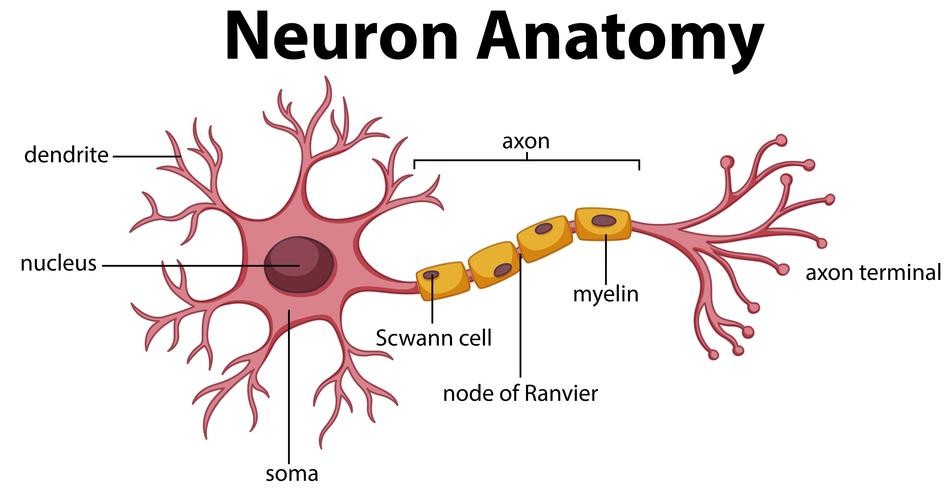
Neurons
1. Conduct signals, action potentials (electricity)
-
Dendrites
1. short processes
2. Dendrites conduct signals TOWARDS cell body
Cell bodies integrate incoming info, majority rules.
-
Axons
1. long processes
2. axons conduct AWAY from cell body
Cell bodies integrate incoming info, majority rules
-
Neuroglia
1. support nervous system
-
Sensory Neurons (Afferent) CNS
Sensory neurons conduct info TOWARDS CNS (Afferent)
-
Motor Neurons (Efferent) CNS
Motor neurons conduct info AWAY CNS (efferent)
-
Ependymal Cells
ciliated cells line the ventricles of the brain and move cerebral spinal fluid
-
Astrocytes
form blood-brain barrier
-
Microglia
phagocytes, devour foreign material in brain, dead cells, injured cells
-
Oligodendrocyte
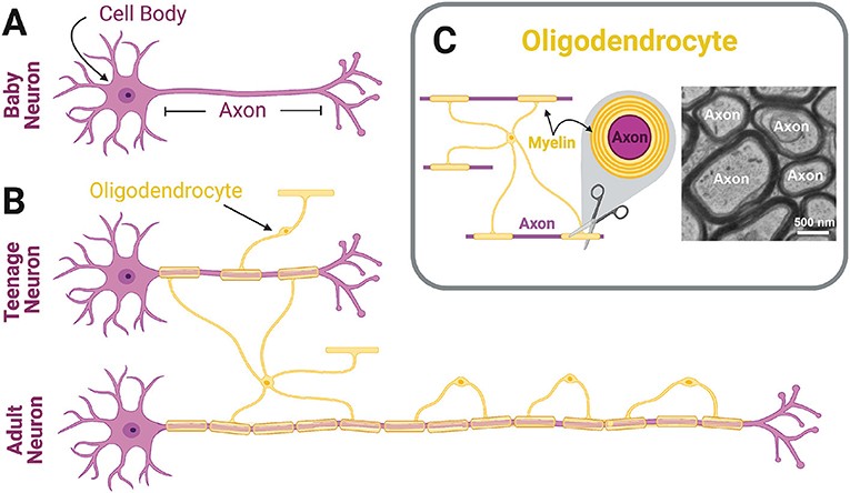
cell web cover
-
Satellite Cells
provide physical support. stabilizing.
-
Schwann Cells
insulate nerve cell processes, primarily axons. This insulation is called myelin sheath. Insulation is discontinuous. Insulation increases action potential by a factor of 10x.
ex: 10 meter/sec without sheath vs. 100 m/s with sheath
MS disorder attacks myosheath
-
Nerve
bundle of neurons or group of nerve cell process in PNS
ex: Sensory Nerves, Motor Nerves, or Mixed Nerves
-
Tract
1. Spinal Cord Tracts
i.) Ascending Tracts (sensory)
ii.) Descending Tracts (motor)
2. Brain Tracts - CPA
i.) Commissural Tracts - connect left/right hemispheres
ii.) Projection Tracts - connect cerebrum w/inferior parts of brain and primitive parts.
iii.) Association Tracts - connect different lobes of the brain
-
Activity vs. Lobe
Motor Activity = Frontal Lobe
Sensory Activity = Parietal Lobe
Vision = Occipital Lobe
Hearing = Temporal Lobe

