 rika
rika-
His studies are significant for us because they represent a shift from superstition and conjecture toward observation
Aristotle
-
Learned much about the structure of relatively advanced fetuses when the development of the microscope allowed the study of early stages of embryos
Galen
-
In Holland, the human sperm was first seen in 1677 by
Hamm and Leeuwenhoek
-
In 1672, ovarian follicles were described by
de Graaf
-
Contended that the sperm contained the new individual in miniature and was merely nourished in the ovum
Spermists
-
Argued that the ovum contained a minute body, which was stimulated to grow by the seminal fluid
Ovists
-
Discovered in 1745 that the eggs of some insects can develop parthenogenetically (without the participation of sperm), which strengthened the ovists' cause
Bonnet
-
A German biologist that wrote a thesis setting forth the conception of epigenesis
Kaspar Friedrich Wolff
-
His work during 1828 in Estonia first emphasized the fact that the more general basic features of any animal group appear earlier in development than do the special features that are peculiar to different members of the group (von Baer's law)
Karl Ernst von Baer
-
The foundation of modem embryology was laid down and embryology as a science began because of the formulation of the cell theory by German biologists
Matthias Schleiden and Theodor Schwann
-
The zygote has a dual origin from two gametes
spermatozoon and ovum
-
Refers to the time of fertilization that represents the starting point in an individual's life history; Individual's entire life span
Ontogeny
-
German biologist that made the important distinction between the soma (body) and the germ-cell line (gametes)
August Weismann
-
Weismann: All-important for perpetuation of the species
Germ-cell line
-
Weismann: Vehicle for protecting and perpetuation the germ plasm
Soma
-
The period starting with fertilization and ending with metamorphosis in Amphibia, hatching in birds, and birth in mammals.
Also deals with the development and maturation of gametes
Embryology
-
The development and maturation of gametes
Gametogenesis
-
Between 1880 and 1890, the new techniques of serial sections and of making three-dimensional wax plate reconstructions from them provided the basis for
Descriptive Embryology
-
Arose late in the 19th century. A great interest in evolution was a driving force behind the development of this field. Provided the insight for the concept that "ontogeny recapitulates phylogeny"
Comparative Embryology
-
The acquisition of detailed structural information on embryos paved the way for the growth of this field. They seek to understand causative factors in development by posing hypotheses and testing them by manipulating the embryos.
Experimental Embryology
-
One of the pioneers of experimental embryology who performed an experiment that ushered in the era of this field. He destroyed one cell (blastomere) of a two-cell frog embryo with a hot needle.
This showed that if the cells of a two-cell frog embryo are entirely separated, each cell is capable of giving rise to a complete individual. This experiment provided proof of the untenability of the preformationist doctrine and laid the foundations for a new field
Wilhelm Roux
-
Roux coined the German word Entwicklungsmechanik which means
Developmental Mechanics
-
Development is brought about by a series of causal interactions between the various parts; and also reminds one that genetic factors are among the most important determinants of development
Epigenetics
-
During the 1930s and 1940s newly emerging chemical and biochemical techniques led to the establishment of this field, which provided descriptive information about chemical and physiological events in the embryo
Chemical Embryology
-
The branch of embryology concerned with the study of malformations
Teratology
-
The rapid growth of research related to problems of conception and contraception has led to the establishment of a discipline that is commonly called
Reproductive Biology
-
A popular way of looking at embryonic development is through the approach known as ____. This field includes not only embryonic development, but also postnatal processes such as normal and neoplastic growth, metamorphosis, regeneration, and tissue repair at levels of complexity ranging from the molecular to the organismal
Developmental Biology
-
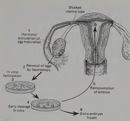
A technique that allowed childless couples to have children from their own genetic heritage. It is used in cases where both the mother and father are capable of producing viable eggs and sperm cells, but because of a blockage in the women’s uterine tubes the ovulated eggs are unable to be fertilized in her body and then become transported to her uterus
In Vitro Fertilization and Embryo Transfer
-
Just before the eggs would normally be shed from the ovary, a doctor, using a technique called
Laparoscopy
-
Some women who are able to produce fertile eggs but are unable to carry an embryo to term in their own uteri have made arrangements with other women to act as
surrogate mothers
-
This technique makes it possible to determine the sex of a baby before it is born and to detect the presence of genetic conditions that could lead to a defective child
Examination of a small amount of the amniotic fluid
-
This technique allows the diagnosis of many anatomical defects in fetuses. Some of these can be dealt with by means of intrauterine surgery
Application of Ultrasound and New X-Ray Imaging
-
[Intracellular Synthesis and Its Regulation] In an interphase cell (one between mitotic divisions) certain portions of the nuclear DNA molecule are free of restricting proteins that bind to the DNA and can direct the synthesis of messenger RNA (mRNA).
The newly formed RNA molecules commonly contain regions (exons) that code for specific segments of a protein molecule and other regions (introns or intervening sequences) that appear to contain no information directly involved in the amino acid sequence of the protein to be formed
Transcription
-
[Intracellular Synthesis and Its Regulation] The introns are enzymatically cut out and the remaining exons are spliced together to form the definitive mRNA molecule. After processing, the newly formed mRNA molecules migrate from the nucleus into the cytoplasm of the cell via pores in the nuclear membrane
mRNA Processing
-
[Intracellular Synthesis and Its Regulation] Once in the cytoplasm, the mRNA molecules may follow either of two chief pathways, depending on the type of
molecule and cell
-
[Intracellular Synthesis and Its Regulation] For the formation of protein molecules that are destined to function within the cell (structural proteins and most enzymes), the mRNA molecules link up with ribosomes to form _______, the length of which varies according to the size of the protein that is being made
polyribosomes
-
[Intracellular Synthesis and Its Regulation] If, however, the mRNAs are coding for proteins that will be secreted from the cell (e.g., collagen, immunoglobulins), the mRNA forms complexes with the rough endoplasmic reticulum. These are called
polypeptide chains
-
The polypeptide chains that are formed in the rough endoplasmic reticulum are then transported to the Golgi apparatus, where they are commonly linked with ________.
From the Golgi complex, the finished proteins are then brought to the cell membrane within vesicles and emptied into the medium surrounding the cell
polysaccharide molecules
-
Many extracellular influences of regulatory mechanisms are mediated by ______ located at the cell surface
receptor molecules
-
In __________, messenger RNA is first transcribed on the DNA template
(1). After processing within the nucleus
(2), the mRNA leaves the nucleus through nuclear pores
(3). The synthesis of intracellular proteins
(4) is accomplished by polysomes, which consist of molecules of mRNA associated with ribosomes. Synthesis of proteins for export from the cell is accomplished on the rough endoplasmic reticulum
(5). From there they are transported
(6) to the Golgi apparatus
(7), where they may be complexed with newly synthesized polysaccharides. Small membrane-bound vesicles containing the proteins leave the Golgi apparatus
(8) and, when they reach the cell membrane
(9), fuse with it and release the protein molecules by a process called exocytosis
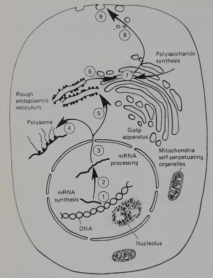
protein synthesis
-
Extracellular signaling substance
ligand
-
The overall sequence of events from the binding of an extracellular signaling substance (called a ligand) by cell-surface receptors to the activation of specific genes or the stimulation of other intracellular processes is commonly called
Signal Transduction
-
Receptors that develop active intracellular enzyme activity (e.g., tyrosine-specific protein kinase) when bound to their ligand
Catalytic Receptors
-
Receptors that activate cellular processes through an intermediate protein
G-protein Receptors
-
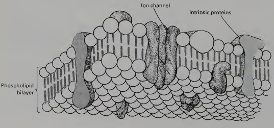
A cell's plasma membrane is composed of a
phospholipid bilayer
-
A component of the cell surface that has at least 130 varieties, constitute about 5% of the lipid molecules in the outer surface of the plasma membrane
glycosphingolipid molecules
-
A type of junctional complexes that bind epithelial cells together in small spots. Also serve as focal points for the attachment of fibrillar intracellular proteins
Desmosomes
-
Another spot-like junction which mediates communication and the exchange of small molecules between two cells
Gap Junction
-
Found along the surface of many epithelia which bind adjacent cells together, forming an impermeable barrier to the outside. It also prevents the mingling of membrane proteins on either side of the junction
Tight Junction
-
An important property of most embryonic structures. A number of crucial experiments have shown that like cells tend to stick together and sort out from cells of a different sort.
Cell Adhesion
-
This person demonstrated cell adhesion by squeezing a sponge through a silk mesh and dissociating it into individual cells. The dissociated cells later reaggregated and ultimately formed a new sponge
In later work, when two species of sponges were thus treated, the disaggregated cells sorted out according to species, and the two original types of sponge re-formed
H.V. Wilson
-
1. Ca++-mediated adhesion
2. N-CAM (neural cell adhesion molecule)
3. Lock and key fashion (heterophilic binding) between complementary saccharides
Main molecular mechanisms of cell adhesion
-
The surface morphology of cells can be examined with
scanning electron microscopy
-
At a finer level, the surface morphology of cells is examined using the technique
freeze fracture
-
Cells are embedded in or rest upon this part which is a macromolecular meshwork that varies in composition from one tissue to the next and from one developmental period to the next
Extracellular Matrix
-
Epithelial-cell layers rest upon a thin sheet-like form of extracellular matrix
Basal Lamina
-
Embedded in a massive extracellular matrix designed to support great weight
Cartilage cells and bone cells
-
The spaces between different tissues are filled with this extracellular matrix that serves as both a biological packing material and a means of transmitting mechanical tension
Fascia
-
Represents an extreme example of an extracellular matrix designed to transmit powerful mechanical forces from a muscle to a bone
Tendon
-
The generic term for a family of glycoproteins that are characterized by having glycine as every third amino acid and also by possessing two amino acids, hydroxyproline, and hydroxylysine, which are rarely found in other proteins
Collagen
-
Basic unit of collagen
Tropocollagen
-
Involved in attaching cells to other components of the extracellular matrix. In developmental processes characterized by the migration or extension of cells, these are an important feature of the substrates through which the cells move
attachment glycoproteins
-
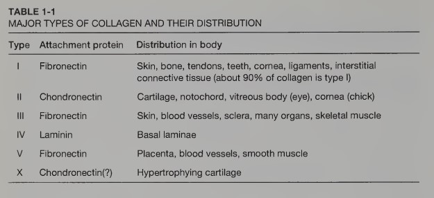
I. Fibronectin (skin, bone, tendons, teeth, cornea, ligaments, interstitial connective tissue (about 90% of collagen is type I))
II. Chondronectin (Cartilage, notochord, vitreous body (eye), cornea (chick))
III. Fibronectin (Skin, blood vessels, sclera, many organs, skeletal muscle)
IV. Laminin (Basal laminae)
V. Fibronectin (Placenta, blood vessels, smooth muscle)
X. Chondronectin (Hypertrophying cartilage)
major types of collagen
-
Best understood of the attachment glycoproteins; a dimer with similar polypeptide subunits of 220-250,000 daltons.
Fibronectin
-
Accounts for the fibronectin's cell-binding properties
RGD (arginine-glycine-aspartic acid) sequence of fibronectin
-
The RGD sequence of fibronectin attaches to a specific cell-surface binding protein that is a member of a large family of binding proteins
Integrins
-
Sites of fibronectin attachment to cells are also areas upon which bundles of ___, an important intracellular contractile protein, converge
Actin
-
A glycoprotein with an analogous function; mediates the attachment of chondrocytes (cartilage cells) to type II collagen in cartilage matrix
Chondronectin
-
A major attachment glycoprotein that is cross-shaped molecule composed of three A chains of 200,000 daltons each and one B chain of 400,000 daltons. Laminin is a major component of basal laminae, where it binds cells to type IV collagen and other matrix molecules
Laminin
-
Shaped like an irregular 6-pointed star which is found in much more restricted circumstances in development than either fibronectin or laminin, displays different degrees of adhesiveness to several types of cells
Tenascin
-
It is formerly called mucopolysaccharides, constitute another of the fundamental groups of extracellular matrix molecules. Although they are large molecules, most consist of repeated disaccharide units.
It bind large amounts of water, which is important in maintaining the physical and mechanical properties of different types of extracellular matrix. The water-binding properties of hyaluronic acid make it particularly important in early developmental processes
Glycosaminoglycans (GAGs)
-
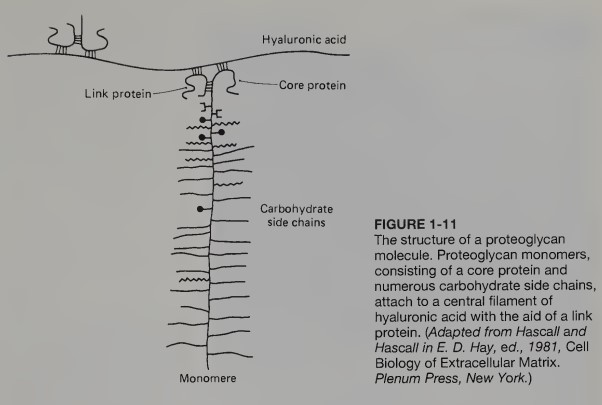
These are immense molecules of the extracellular matrix with molecular weights in the millions. Consists of a brush-like monomer, with a protein core and numerous glycosaminoglycan branches
Proteoglycan

