 Youssef Draz
-
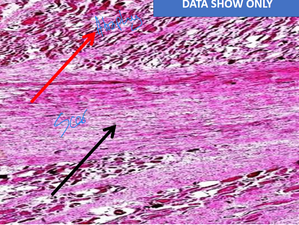
Section in the MYOCARDIUM (cardiac muscle) showing:
• Areas of SCAR TISSUE
(homogenous pink with few capillaries and some fibroblasts).
• The ADJACENT CARDIAC MUSCLE fibers are red and slightly atrophic with pyknotic nuclei.
Arrows?
Black: Scar
Red: Atrophic cardiac muscle
-
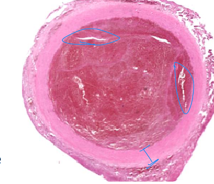
ARTERIAL THROMBUS
Transverse section in a BLOODVESSEL:
• The lumen is occluded by a thrombus appearing as a red mass traversed by paler structureless lines called the lines of Zahn formed of fused platelets.
• The thrombus in between the lines of Zhan is formed by a network of fibrin entangling the red and white blood cells.
-
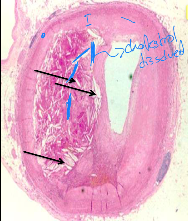
Atheroma, small artery
Transverse section in a SMALL ARTERY
showing:
• Thickened and hyalinosed
SUBENDOTHELIAL connective tissue with needle and spindle shaped clefts (arrow).
• These empty CLEFTS were occupied by cholesterol and lipids which dissolve during preparation of the section. Blue granules of calcification are also present.
• The INTERNAL ELASTIC LAMINA at the site of the atheroma is thinned out and
fragmented and the MEDIA is thin (atrophic).
Empty spaces? Cholesterol that dissolved
during the preparation
-
Describe the picture.
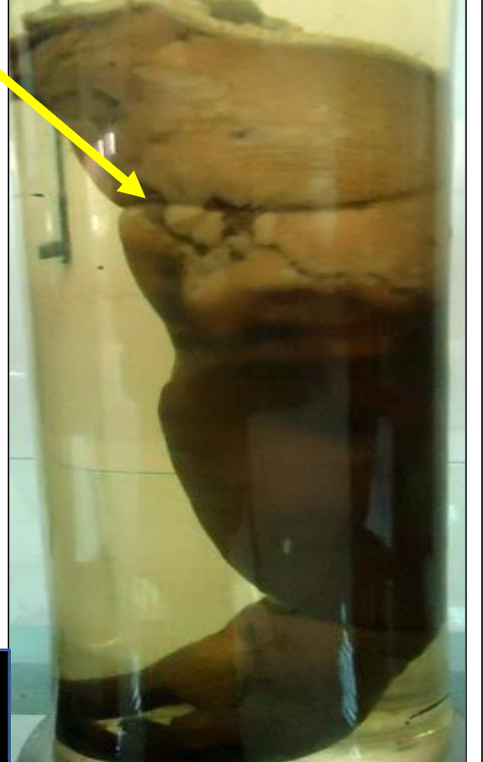
Specimen: Inferior vena cava and the common iliac veins.
Gross pathology:
The lumen of the vessel is occluded by dark red thrombi.
What is your diagnosis ?
Thrombosis of the inferior vena cava and the common iliac veins.
Name the vessels in the picture?
Inferior vena cava and common iliac veins
-
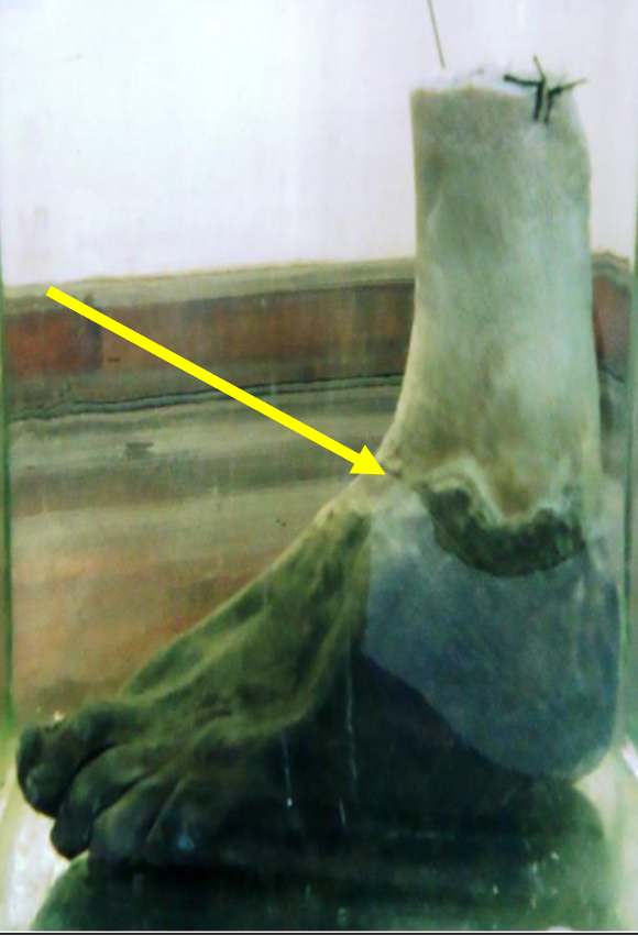
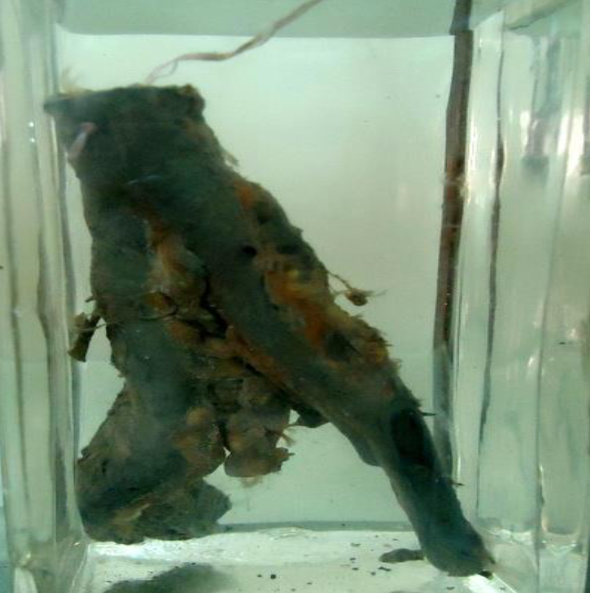
Specimen: Left foot.
Gross Pathology:
1. The FOOT is black, shrunken and mummified with wrinkled skin (dry gangrene).
2. An irregular groove (line of separation) is seen at the level of malleoli.
3. The cross section of the ANTERIOR TIBIAL ARTERY in the cut section of the leg shows yellow thickened crescent due to atherosclerosis and an occluding
thrombus.
Diagnosis:
1. Dry gangrene of left foot (senile gangrene).
2. Atherosclerosis and thrombosis of anterior tibial
artery.
Arrow? Line of separation / Obstruction of vein or
artery? Artery / Can natural amputation occur? Yes
-
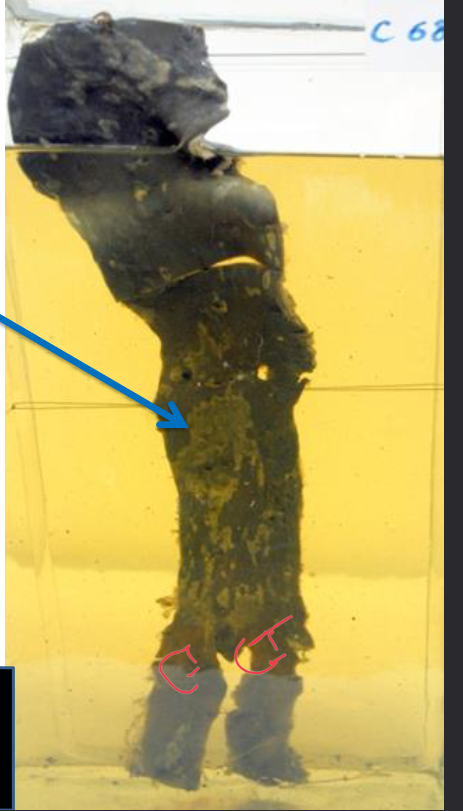
Specimen: Right upper limb.
Gross Pathology:
1. The distal part of the arm, forearm and hand are dark brown with skin maceration.
2. The circular groove, proximal to the gangrenous part represents the site of a tightly applied tourniquet.
3. No line of separation was identified.
Diagnosis:
Moist gangrene of the right upper limb (due to tightly applied tourniquet).
Arrow? site of a tightly applied tourniquet / Obstruction of
vein or artery? Both / Can natural amputation occur? No
-
Specimen: An aortic segment with the two common iliac arteries.
Gross Pathology:
1. The intima of the aorta and the common iliac arteries shows multiple raised yellow irregular atherosclerotic patches.
2. Some of these lesions are ulcerated.
3. Small brownish thrombi are seen covering some of the atherosclerotic lesions.
Diagnosis:
Atherosclerosis of abdominal aorta and the common
iliac arteries associated with ulceration and thrombosis.
Arrow? Atherosclerotic patches,
Name the vessels in the picture? Aorta and two common iliac arteries / Two complications of the
lesion seen in the picture? Ulceration and thrombosis.
-
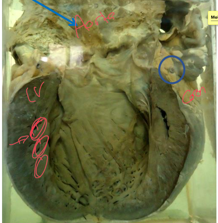
Specimen: Heart with open left side.
Gross Pathology:
1. The AORTA shows yellow atherosclerotic plaques and ulcers.
2. The CORONARY ARTERY shows yellow thickening of the wall and
narrowed lumen.
3. The LEFT VENTRICLE shows marked hypertrophy, dilatation and greyish
white subendocardial fibrosis.
What is your diagnosis ?
1-Atherosclerosis of the aorta and
coronary arteries
2. Arteriosclerotic heart disease.
Arrow? Atherosclerotic plaques and ulcers.
Name the opened ventricle? Left
-
Specimen: Heart with aorta (till aortic arch) and spleen.
Gross Pathology:
1. The intima of the aorta shows multiple raised yellow irregular ATHEROSCLEROTIC patches. Some patches are ulcerated and covered with thrombi.
2. The spleen shows a large pyramidal INFARCT,with its base towards the surface. It is brownish incolor with dark congested margins and bulging
bases covered with opaque fibrin deposits (peri-splenitis).
Diagnosis:
1. Atherosclerosis of aorta (with ulceration and
thrombosis).
2. Recent splenic infarct.
Arrow? Atherosclerotic patches, / Two
complications of the lesion seen in the picture?
Ulceration and thrombosis.
-
Describe:
Specimen: Open left ventricle, thoracic aorta and thoracic part
of vertebral column.
Gross Pathology:
1. The thoracic AORTA and aortic arch show yellow, raised
irregular atherosclerotic patches
2. The DESCENDING THORACIC AORTA is affected by a fusiform
aneurysms containing a laminated thrombus, filling its cavity.
4. The adjacent THORACIC VERTEBRAE show pressure atrophy,
but the intervening discs are normal.
5. The left ventricle is hypertrophied.
Diagnosis:
1. Aortic atherosclerosis associated with fusiform aneurysms
and thrombosis causing pressure atrophy of thoracic vertebrae.
2. Left ventricular hypertrophy
Lesion pointed by the Arrow: Etiology?
Atherosclerosis / Shape ? Fusiform / Site? Descending
aorta / Two complications in the picture? Thrombosis
and pressure atrophy of vertebral bodies
-
Describe:
Specimen: Heart with ascending aorta and arch.
Gross Pathology:
1. The AORTIC intima shows multiple raised yellow
atherosclerotic patches.
2. The ASCENDING AORTA AND THE ARCH show saccular aneurysm with a thin fibrotic wall and laminated brown thrombus filling its cavity.
Diagnosis:
1. Syphilitic aortitis
2. Aortic atherosclerosis
3. Saccular aortic aneurysm with thrombosis.
Lesion pointed by the Arrow: Etiology? Syphilitic /
Shape ? Saccular / Site? Ascending aorta and arch /
One complication in the picture? Thrombosis
-
Describe:
Specimen: A transverse section of both ventricles and inter-ventricular septum.
Gross Pathology:
1. The wall of the left ventricle shows marked hypertrophy.
2. The inter-ventricular septum is hypertrophied and bulging towards the right
ventricle.
3. The wall of right ventricle is hypertrophied.
Diagnosis:
Concentric hypertrophy of the heart.
-
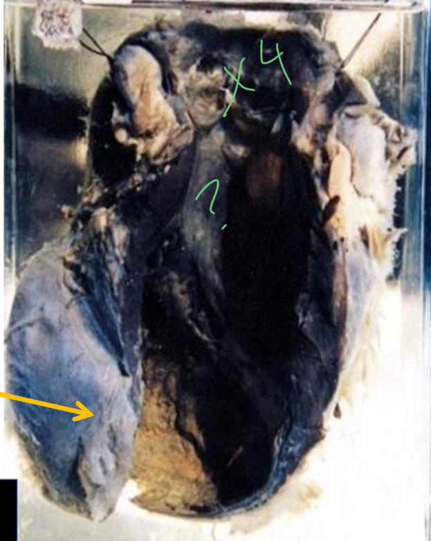
Describe.
Specimen: Heart of a child with an open left ventricle and aorta.
Gross Pathology:
1. The mitral cusps are opaque.
2. Multiple small greyish white vegetations are seen on the atrial surface of the mitral cusps, at their lines of contact.
Diagnose.
Rheumatic Endocarditis of the mitral valve.
Valve pointed by arrow? Mitral / Lesion
pointed by Arrow? Vegetations / Age? Child
-
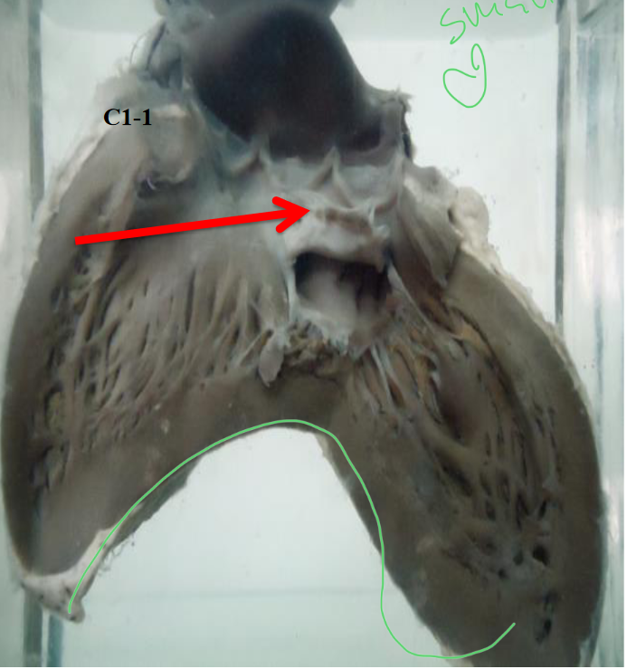
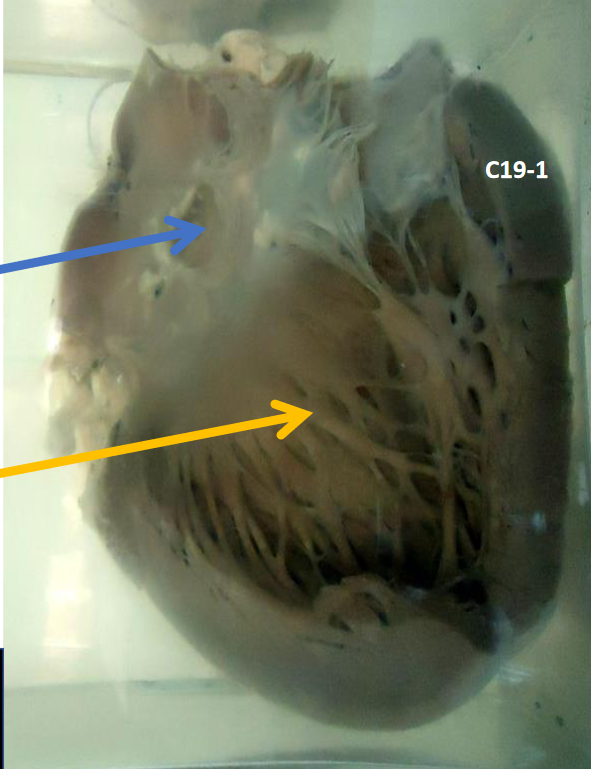
Describe:
Specimen: A transversely cut slice of the heart showing mitral and tricuspid valves as well as parts of the atria and ventricles
Gross Pathology:
1.The MITRAL VALVE shows:
• Stenosed (button hole) mitral orifice and thickened opaque fused cusps.
• Hypertrophied papillary muscle (seen from the back of the specimen).
2.The TRICUSPID VALVE shows:
• Thickened grayish white, opaque retracted cusps.
• Widely patent tricuspid orifice.
Diagnosis:
Chronic rheumatic Endocarditis of the mitral valve with
stenosis and tricuspid valve with incompetence.
Valve pointed by arrow?
Red: Tricuspid, Blue: Mitral
-
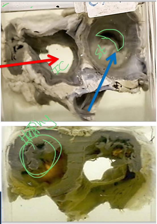
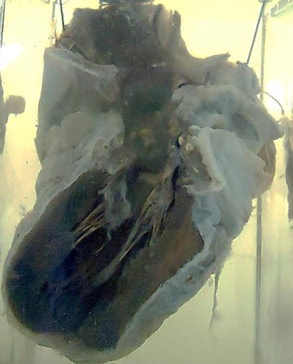
Specimen: Part of the left ventricle, aortic valve and root of the aorta.
Gross Pathology:
1. The aortic cusps are yellowish.
2. One of the aortic cusps shows
irregular perforation.
What is your diagnosis ?
Acute infective endocarditis of the
aortic valve with cusp perforation.
Circle? Cusp perforation
-
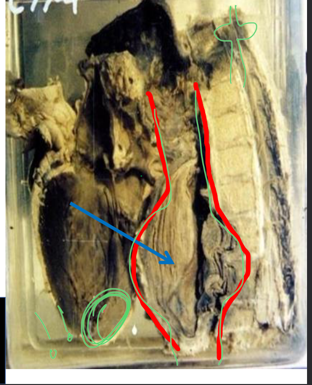
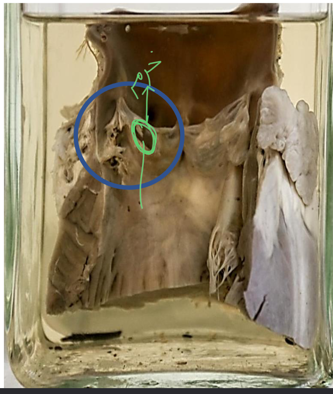
Describe the picture.
Specimen: Heart with open left side.
Gross:
* The anterior wall of left ventricle at the apex of the heart and inter-ventricular septum appears thinned and replaced by grayish white fibrous tissue (healed infarct).
* The endocardial surface of the infarct is covered by a large grayish brown mural
thrombus.
*The pericardium over the infarct shows whitish fibrosis and adhesions.
What is your diagnosis ?
1.Healed anterior infarct of left ventricle.
2. Mural thrombus.
3. Pericardial fibrosis and adhesions
Arrow? Mural thrombus
-
Specimen: Heart with open left side.
Gross Pathology:
1. The anterior wall of left ventricle, apex and adjacent part of the inter-ventricular septum are replaced by thin greyish white fibrous tissue (healed infarct).
2. This healed infarction shows ruptured aneurysm (cardiac aneurysm).
3. Left ventricular shows greyish white subendocardial fibrosis.
4. The pericardium over the infarct shows whitish fibrosis and adhesions
5. Yellow atherosclerotic patches are seen on the aorta and the anterior descending branch of left coronary.
Diagnosis:
1. Healed left ventricular infarct.
2. Rupture of cardiac aneurysm.
3. Pericardial fibrosis and adhesions.
4. Atherosclerosis of aorta and left coronary artery.
Pathology pointed by the arrow? Ruptured cardiac aneurysm
-
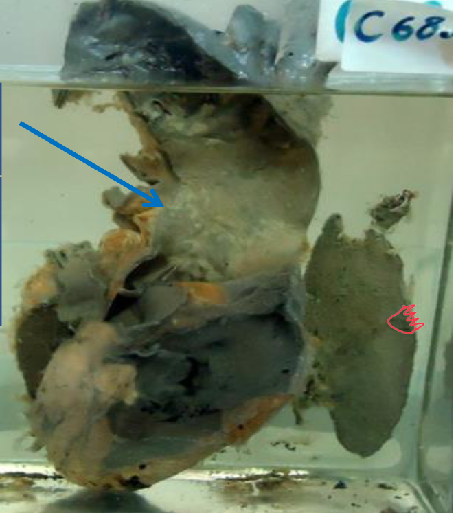
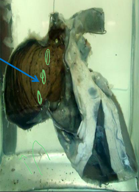
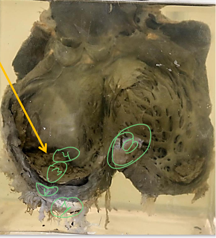
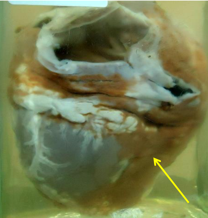
Describe.
Specimen: Heart with open chambers.
Gross pathology:
1. The MYOCARDIUM is yellow in color. The Columnae carneae show brown dots alternating with yellow ones (Tabby cat appearance).
2. The LEFT VENTRICLE shows hypertrophy and dilatation.
3. The AORTA and mitral valve shows yellow atherosclerotic patches.
What is your diagnosis ?
1- Fatty change of the heart (Tabby cat).
2. Hypertrophy and dilatation of left ventricle.
3. Atherosclerosis of aorta and mitral valve
The appearance in the yellow Arrow? Tabby cat / Blue
Arrow? Atherosclerotic patches
-
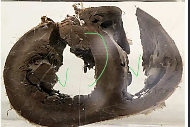
Describe.
Specimen: Heart with open left side.
Gross Pathology:
1. HEART is reduced in size (compare to the size of the aorta) with tortuous Coronaries (seen at the outer surface).
2. The myocardium is dark brown.
3. PERICARDIAL FAT is replaced by edematous tissue.
4. AORTA shows yellow atherosclerotic patches.
Diagnosis.
- Brown atrophy of the heart
- Serous atrophy of pericardial fat
- Aortic atherosclerosis.
What is the color of the myocardium and mention
why? Dark brown due to lipofuscin pigment
deposition
-
Describe.
Specimen: Unopened Heart
Gross Pathology:
• The visceral pericardium is rough, reticulated and appears opaque
yellowish due to fibrin deposition.
• 2. Part of this fibrin is removed to show the underlying smooth surface
of the heart.
Diagnose:
Fibrinous pericarditis.
Arrow? Fibrin deposit / Type of
inflammation? Acute non-suppurative
fibrinous inflammation
-
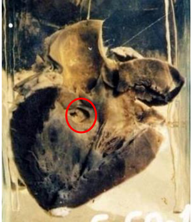
Describe:
Specimen: Heart of a child with open left and right sides.
Gross Pathology:
1. The membranous part of the interventricular septum shows a rounded
defect; less than 1 in diameter.
2. The right and left ventricles are dilated and hypertrophied.
Diagnosis:
1. Inter-ventricular septal defect
2. Hypertrophy and dilatation of the
right & left ventricles
Circle? VSD / Age ? Child

