-
brain is composed of four major regions:
•Cerebrum
•Diencephalon
•Brainstem
•Cerebellum
-
folds and depressions between folds called
•Gyri: folds
•Sulci: depressions between folds
-
Directional terms for brain
•Anterior (rostral): toward the nose
•Posterior (caudal): toward the tail
-
late fourth week of development, three primary brain vesicles have formed:
•Prosencephalon (forebrain)
•Mesencephalon (midbrain)
•Rhombencephalon (hindbrain)
-
fifth week of development, there are five secondary brain vesicles:
•Telencephalon: from prosencephalon; forms cerebrum
•Diencephalon: from prosencephalon; forms thalamus, hypothalamus, and epithalamus
•Mesencephalon: only primary vesicle that does not form a new secondary vesicle
•Metencephalon: from rhombencephalon; forms pons and cerebrum
•Myelencephalon: from rhombencephalon; forms medulla oblongata
-
Gray matter in brain:
motor neuron and interneuron cell bodies, dendrites, telodendria, unmyelinated axons
•Forms cerebral cortex, surface of most of adult brain
•Within white matter, gray deep clusters of neuron cell bodies called cerebral nuclei
-
White matter in brain:
•Made up of myelinated axons
•Deep to the gray matter
-
Brain is protected by 4 structures:
•Bony cranium
•Protective connective tissue (meninges)
•CSF
•Blood–brain barrier
-
What are Cranial meninges and their function?
Cranial meninges are connective tissues
•Separating brain from bones of cranium
•Protecting blood vessels of brain; form some large veins that drain blood from brain
•Containing and circulating cerebrospinal fluid
-
Three layers of meninges and location? does what do they form when separate?
•Pia mater: thin, innermost layer adhering to brain
•Subarachnoid space is area between arachnoid and pia
•Arachnoid mater: has web of fibers called arachnoid trabeculae
•Dura mater: tough outer layer with two sublayers
•Deeper meningeal layer (superficial to subdural space)
•More superficial periosteal layer
•Layers usually fused except where they separate to form dural venous sinuses
-
Cranial Dural Septa
The meningeal layer extends into cranial cavity at four locations to form double-layered dura called cranial dural septa:
-partition the brain and provide support
-
4 Cranial Dural Septa & location
•Falx cerebri: projects into longitudinal fissure; separates left and right cerebral hemispheres
•Tentorium cerebelli: horizontal fold that separates cerebrum from cerebellum
•Falx cerebelli: separates left and right cerebellar hemispheres
•Diaphragma sellae: small septum between pituitary and hypothalamus
-
what is Dural venous sinuses?
form in locations where the two layers of the dura mater have separated:
-
4 Dural venous sinuses and location
1.Superior sagittal sinus: located within the superior margin of the falx cerebri
2.Inferior sagittal sinus: located within the inferior margin of the falx cerebri
3.Transverse sinuses: located within the posterior border of the tentorium cerebelli
4.Occipital sinus: located in the posterior vertical border of the falx cerebelli
-
Brain Ventricles filled with
cavities within brain that contain cerebrospinal fluid (CSF)
-
Four ventricles in the brain & location:
•Two lateral ventricles, one in each cerebral hemisphere, separated by a thin septum pellucidum
•A third ventricle in diencephalon
-Communicates with lateral ventricles through interventricular foramen
-Communiates with fourth ventricle through cerebral aqueduct
•A fourth ventricle between pons and cerebellum
-Merges with central canal of spinal cord
-
what is Cerebrospinal fluid (CSF) and function?
clear liquid that circulates in ventricles and subarachnoid space
•Buoyancy: brain “floats” in the CSF
•Protection: provides liquid cushion
•Environmental stability: transports nutrients and removes waste from brain
-
Cerebrospinal fluid is formed by?
Formed by the choroid plexus in each ventricle
•Composed of a layer of ependymal cells and the capillaries that lie within the pia mater
•Blood from capillaries is filtered to form CSF
-
Production and Circulation of Cerebrospinal Fluid
1)CSF is produced by the choroid plexus in the ventricles.
2)CSF flows from the lateral ventricles, through the interventricular foramen into the third ventricle, and then through the cerebral aqueduct into the fourth ventricle.
3)CSF in the fourth ventricle passes through the paired lateral apertures or the single median aperture, and into the subarachnoid space as well as the central canal of the spinal cord.
4)As the CSF flows through the subarachnoid space, it provides buoyancy to support the brain.
5)Excess CSF flows into the arachnoid villi, then drains into the dural venous sinuses. The greater pressure on the CSF in the subarachnoid space ensures that CSF moves into the dural venous sinuses without permitting venous blood to enter the subarachnoid space.
-
Blood–brain barrier (BBB), feets
regulates what substances can enter interstitial fluid of brain
Capillary endothelial cells, astrocyte perivascular feet, and continuous basement membrane contribute to the BBB
-
BBB is missing or reduced in three locations of CNS:
•Choroid plexus (because the capillaries must be permeable to make CSF)
•Hypothalamus (hormones are made that need ready access to blood)
•Pineal gland (hormones are made that need ready access to blood)
-
Cerebrum is in charge of
conscious thought and intellectual functions
•Contains many neurons for complex, analytical functions
-Contains an outer cortex, inner white matter, and deep regions of gray matter called cerebral nuclei
- gyri (ridges), sulci (grooves), and deep fissures
-
two halves of cerebrum called left and right:
It is divided by:
cerebral hemispheres
longitudinal fissure that extends along the midsagittal plane
-
Largest tract that connects the two hemispheres of cerebral
corpus callosum
-
hemispheric lateralization
The two hemispheres appear to be mirror images but display some functional differences
-
Cerebral Hemispheres receive and project info and?
Both cerebral hemispheres generally receive their sensory information from and project motor commands to the opposite sides of the body
-
Where is memory or consciousness
Some functions, such as memory or consciousness, cannot easily be assigned to a single region
-
Cerebrum five lobes:
1.Frontal lobe
2.Parietal lobe
3.Temporal lobe
4.Occipital lobe
5.Insula
-
Where is frontal lobe? what part is inside?
Frontal lobe is located deep to the frontal bone and forms anterior part of cerebral hemisphere
•Ends posteriorly at central sulcus; inferior border marked by lateral sulcus
•includes Precentral gyrus anterior to central sulcus
-
function of frontal lobe?
•Frontal lobe functions: voluntary movement, concentration, verbal communication, decision making, planning, and personality
-
Parietal lobe location and function. what part is inside?
superoposterior part of each hemisphere and is under parietal bone
•Terminates anteriorly at the central sulcus, laterally at the lateral sulcus, and posteriorly at the parieto-occipital sulcus
•includes Postcentral gyrus(posterior to central sulcus)
-general sensory functions
-
Temporal lobe location and function
inferior to lateral sulcus, under the temporal bone
•Involved with hearing and smell
-
Occipital lobe location and function
posterior region of each hemisphere underneath occipital bone
•Processes incoming visual information and stores visual memories
-
Insula location and function
deep to the lateral sulcus
•Involved in interoceptive awareness, emotion, empathy, taste
-
Three categories of functional areas of the Cerebrum
•Motor areas: control voluntary motor functions
•Sensory areas: provide conscious awareness of sensation
•Association areas: integrate and store information
-
Primary motor cortex aka, location & control? diagram called?
(somatic motor area) controls voluntary skeletal muscle activity
•Located within precentral gyrus; axons project contralaterally to brainstem and spinal cord
diagrammed as a motor homunculus on the precentral gyrus
-
Motor speech area aka, location and control?
(Broca area) controls muscular movements for vocalization
•Located in most individuals within the inferolateral portion of the left frontal lobe
Frontal eye field helps control and regulate eye movements
•Located on superior surface of middle frontal gyrus, immediately anterior to premotor cortex
-
Primary somatosensory cortex location, function? diagram?
located within the postcentral gyrus
•This area receives general somatic sensory information from touch, pressure, pain, and temperature receptors
•Sensory homunculus
-
Primary visual, auditory, gustatory, olfactory cortex function and location
Primary visual cortex processes visual information; located in occipital lobe
Primary auditory cortex processes auditory information; located in temporal lobe
Primary gustatory cortex processes taste information; located in insula
Primary olfactory cortex provides awareness of smell; located in temporal lobe
-
what is Premotor cortex and location?
coordinates skilled motor activities
•Located in frontal lobe just anterior to precentral gyrus
-
what is Somatosensory association area and location?
integrates and interprets sensory information
•Located in parietal lobe just posterior to post central gyrus
-
Auditory & Visual association?location?
Auditory association area interprets characteristics of sound and stores memories of sound
•Located in temporal lobe, posteroinferior to primary auditory cortex
Visual association area processes visual information
•Located in occipital lobe, surrounding primary visual cortex
-
Wernicke area is? location?
recognizes and comprehends spoken and written language
•Typically located in left hemisphere overlapping the parietal and temporal lobes
-
Higher-Order Processing Centers
Direct complicated analytical functions or complex motor activity
- speech, cognition, understanding spatial relationships, and general interpretation
Housed in both cerebral hemispheres
-
Where is brain White Matter? what is it made of?
deep to cortical gray matter
Composed primarily of myelinated axons
-
what does Association tracts connect? and 2 kinds
connect areas within one hemisphere
•Arcuate fibers within a lobe
•Longitudinal fasciculi connect different lobes of same hemisphere
-
what does Commissural tracts connect?
•connect two hemispheres
-
what does Projection tracts connect?
connect cerebrum to lower areas (e.g., spinal cord)
-
Cerebral nuclei aka, is? some include and their function
(basal nuclei) are masses of gray matter located deep within brain’s white matter
Specific cerebral nuclei include:
•Caudate nucleus—helps coordinate walking
•Amygdaloid body—participates in emotional expression
•Lentiform nucleus—involved in movement and muscle tone
-Composed of putamen and globus pallidus
•Claustrum—consciousness and processing of multiple sensory stimuli
-
Components of the diencephalon include:
•Epithalamus
•Thalamus
•Hypothalamus
-
epithalamus location, includes 2 things and function?
partially forms posterior roof of diencephalon and covers third ventricle
Components include:
•Pineal gland: secretes melatonin, a hormone that regulates circadian rhythm
•Habenular nuclei: relays signals from limbic system to midbrain; involved in visceral and emotional responses to odor
-
Thalamus location, connected by , impulse except?
Paired masses of gray matter on each side of third ventricle
•These masses are connected by the midline interthalamic adhesion
Each mass is composed of about a dozen thalamic nuclei with axons projecting to regions of cerebral cortex
-Sensory impulses except olfaction
•Main relay point for sensory information that will be projected to somatosensory cortex
-
Hypothalamus location and control?
is the anteroinferior region of diencephalon
Thin, stalklike infundibulum extends inferiorly from hypothalamus to attach to pituitary gland
Specific nuclei control various functions in body:
•Master control of the autonomic nervous system
•Master control of the endocrine system
•Regulation of body temperature
•Control of emotional behavior, food intake, water intake
•Regulation of sleep–wake (circadian) rhythms
-
Brainstem
connects forebrain and cerebellum to spinal cord
•Passageway for all tracts between cerebrum and spinal cord
Contains many autonomic and reflex centers required for survival
Houses nuclei of many of the cranial nerves
-
Midbrain location
superior portion of brainstem
Nuclei of oculomotor nerve (CN III) and trochlear nerve (CN IV)
Somatic motor axons descend from primary motor cortex through cerebral peduncles to spinal cord
-
Cerebral aqueduct
extends through midbrain and connects third and fourth ventricles
•Surrounded by periaqueductal gray matter
-
Superior cerebellar peduncles connects?
connect cerebellum to midbrain
-
Tegmentum
between substantia nigra and periaqueductal gray; relays information between cerebrum and cerebellum
-
Substantia nigra
produce dopamine; involved in motor control, emotion, pleasure, and pain
•Degeneration of substantia nigra=Parkinson’s disease
-
Superior and inferior colliculi
visual and auditory reflex centers
-
Pons location, includes, and nerve
Pons is bulging region on anterior brainstem
Middle cerebellar peduncles are transverse fibers that connect pons to cerebellum
Contains autonomic nuclei in pontine respiratory center that help regulate breathing
Houses sensory and motor cranial nerve nuclei for trigeminal (CN V), abducens (CN VI), and facial (CN VII) nerves
Superior olivary complex nuclei receive auditory input and help localize sound source
-
Medulla oblongata (medulla) location and contains
most inferior part of brainstem
Pyramids are composed of motor projection tracts called the corticospinal tracts
•Most axons in pyramids cross midline at decussation of the pyramids
Contains inferior olivary nucleus that relays proprioceptive information to cerebellum
•Inferior cerebellar peduncles connect medulla to cerebellum
Contains nucleus cuneatus and nucleus gracilis which relay somatic sensory information to thalamus
-
Medulla oblongata autonomic nuclei
•Cardiac center: regulates heart rate and its strength of contraction
•Vasomotor center: controls blood pressure by regulating contraction and relaxation of smooth muscle in walls of arterioles
•Medullary respiratory center: regulates respiratory rate
•Other nuclei involved in coughing, sneezing, salivation, swallowing, gagging, and vomiting
-
Cerebellum size, and 3 regions
second largest part of brain
Partitioned into three regions:
•Outer gray matter layer of cerebellar cortex
•Internal region of white matter, called the arbor vitae
•Cerebellar nuclei in deepest layer
-
Cerebellum contains
Cerebellum has left and right cerebellar hemispheres
•Each hemisphere has an anterior and posterior lobe
•A narrow vermis sits on midline between hemispheres
Folds called folia
-
Cerebellum in charge of
coordinates and fine-tunes movements
Stores memories of previously learned movement patterns
Adjusts muscle activity to maintain equilibrium and posture
Uses proprioceptive information from muscles and joints and to regulate the body’s position
•Monitors position of each body joint and its muscle tone
-
•Superior cerebellar peduncles: connect
•Middle cerebellar peduncles:
Inferior cerebellar peduncles:
•Superior cerebellar peduncles: connect midbrain to cerebellum
•Middle cerebellar peduncles: connect pons to cerebellum
•Inferior cerebellar peduncles: connect medulla oblongata to cerebellum
-
Limbic system location, function
cerebral and diencephalic structures that form a ring around the diencephalon
-process and experience emotions
-Affects memory formation through integration of past memories of physical sensations with emotional states
-
Components of the limbic system:
•Cingulate gyrus: ridge superior to corpus callosum; brings emotions into consciousness
•Parahippocampal gyrus: tissue associated with hippocampus (functions in memory)
•Hippocampus: nucleus shaped like a seahorse; essential in consolidating long-term memories
•Amygdaloid body: involved in emotion, especially fear; helps sort and code memories based on how they are emotionally perceived
•Olfactory bulbs, olfactory tract, olfactory cortex: odors can provoke emotions/memories
•Fornix: thin tract of white matter connecting hippocampus with other limbic structures
•Various nuclei in diencephalon also contribute to emotional function
-
cranial nerves
Twelve pairs Roman numerals
Some only motor or sensory; mixed nerves)
The motor components of a cranial nerve may be somatic motor and/or parasympathetic motor in function
-
I

olfactory
Sensory
smell
Damage: anosmia (loss smell)
-
I I

optic
Sensory
Vision
damage: anopsia (visual defect)
-
I I I
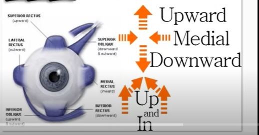
oculomotor
Motor
Four extrinsic eye muscles (medial rectus, superior rectus, inferior rectus, inferior oblique); levator palpebrae superioris muscle (elevates eyelid)
-pupil constrict;
damage: ptosis eyelid droop, strabismus(eye not parallel)
-
IV
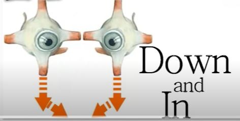
trochlear
Motor
Superior oblique eye muscle
damage:strabismus(eye not parallel), diplopia double vision
-
V
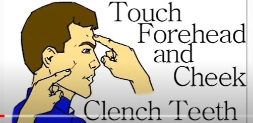
trigeminal
Both
-General sensory from anterior scalp, nasal cavity, nasopharynx, entire face, most of oral cavity, teeth, anterior two-thirds of tongue; part of auricle of ear; meninges
-Muscles of mastication, mylohyoid, digastric (anterior belly), tensor tympani, tensor veli palatini
damage:trigeminal neuralgia pain
-
VI
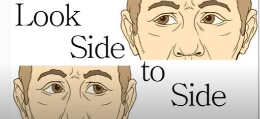
abducens
Motor
Lateral rectus eye muscle
damage: paralysis of lateral rectus limits, diplopia double vision
-
VII

facial
Both
-Taste from anterior two-thirds of tongue
-Muscles of facial expression, digastric (posterior belly), stylohyoid, stapedius
-secretion from silva and tears
damage: decrease tears, dry mouth, bell palsy
-
VIII
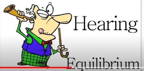
vestibulocochlear
Sensory
Hearing (cochlear branch); equilibrium (vestibular branch)
damage:loss of balance, nausea
-
IX

glossopharyngeal
Both
-General sensory and taste to posterior one-third of tongue, general sensory to part of pharynx, visceral sensory from carotid bodies
-One pharyngeal muscle (stylopharyngeus)
-Increases secretion from parotid salivary gland
damage:dry mouth, lost of taste posterior 1 third of tongue
-
X
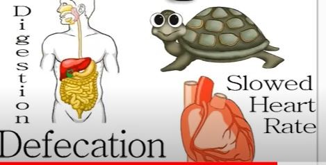
vagus
Both
-Visceral sensory information from heart, lungs, most abdominal organs
General sensory information from external acoustic meatus, tympanic membrane, part of the pharynx, laryngopharynx, and larynx
-Most pharyngeal muscles; laryngeal muscles
-Innervates smooth muscle and glands of heart, lungs, larynx, trachea, most abdominal organs
damage: larynx issue, hoarseness, monotone, lost voice, swallowing
-
XI

accessory
Motor
Trapezius muscle, sternocleidomastoid muscle
damage: difficult to elevate shoulder
-
XII

hypoglossal
Motor
Intrinsic tongue muscles and extrinsic tongue muscles
damage: swallowing and speech difficulties
-
•Cingulate gyrus
•Cingulate gyrus: ridge superior to corpus callosum; brings emotions into consciousness
-
•Parahippocampal gyrus:
•Parahippocampal gyrus: tissue associated with hippocampus (functions in memory)
-
•Hippocampus:
nucleus shaped like a seahorse; essential in consolidating long-term memories
-
•Amygdaloid body
involved in emotion, especially fear; helps sort and code memories based on how they are emotionally perceived
-
•Olfactory bulbs, olfactory tract, olfactory cortex
odors can provoke emotions/memories
-
•Fornix
thin tract of white matter connecting hippocampus with other limbic structures
-
•Pia mater:
•Pia mater: thin, innermost layer adhering to brain
-
•Subarachnoid space
area between arachnoid and pia
-
•Arachnoid mater
has web of fibers called arachnoid trabeculae
-
•Dura mater
tough outer layer with two sublayers
•Deeper meningeal layer (superficial to subdural space)
•More superficial periosteal layer
•Layers usually fused except where they separate to form dural venous sinuses

