 Fatima Adam

Cardiac Cycle (Physiology)
Session Summary This lecture will examine how the heart works as an effective pump. We will examine action potentials and conduction pathways. The central theme will be how the conduction pathway comes together to form the cardiac cycle which is the coordination of all the different contractions of the chambers and how they produce the cardiac output. A few general heart sounds will be considered. Learning outcomes At the end of this session you will be able to: Explain cardiac action potentials – generation of electrical activity. Describe the conduction pathway of the heart. Outline mechanical events during phases of cardiac cycle. Focusing on the left ventricle describe changes in volume, pressure and stroke work during cardiac cycle. Appreciate associated sounds of cardiac cycle.
-
What do ECGs/EKGs stand for?
Electrocardiogram
-
When was the first artificial cardiac pacemaker developed and by who?
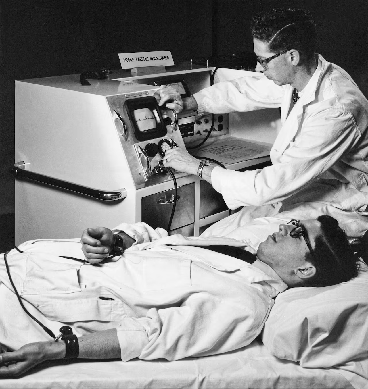
1950s by John Hopps
-
What is membrane potential measured in?
Millivolts mV
-
State some information about the SA node: (4)
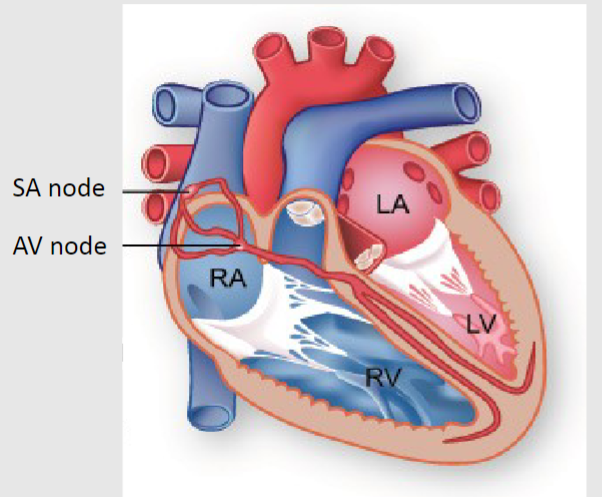
-SA node is a group of cells located in the wall of the right atrium
-Ability to spontaneously produce action potential that travels through the heart via the electrical conduction system
-Sets the rhythm of the heart and so is known as the heart's natural pacemaker
-The rate of action potential production (and therefore the heart rate) is influenced by nerves that supply it
-
Why do the ventricles delay impulses?
So that atria have time to eject their blood into ventricles before ventricular contraction
-
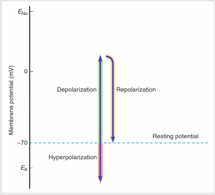
Picture demonstrating ions. Depolarisation and repolarisation
-
What does efflux mean?
The flowing out of a substance or particle
-
What is the membrane potential of cells at rest usually?
-70mV
-
What is Depolarisation?
When the electrical charge inside a neuron becomes less negative, making it more likely to send a signal
-
What is Hyperpolarisation?
When the electrical charge inside a neuron becomes more negative than its resting state, making it less likely to send a signal
-
What is Repolarisation?
When the electrical charge inside a neuron returns to its resting state after depolarization or hyperpolarization, preparing it for the next signal.
-
There is a resting negative voltage in the cell interior as compared to the cell exterior, ranging from.....? (Sinoatrial node (SA) pacemaker potentials)
-40mV to -80mV
-
What is Phase 4 (known as)? (Sinoatrial node (SA) pacemaker potentials)
The pacemaker or the 'funny current'
-
What happens in Phase 4? (3) (Sinoatrial node (SA) pacemaker potentials)

-The membrane repolarizes below the If threshold(approx. − 40mV)
-This is not a genuine resting potential because it is unstable.
-At around -50mV an Na+ channel is activated, causing Na+ influx and slow depolarisation
-
What happens in phase 0? (2) (Sinoatrial node (SA) pacemaker potentials)

-As the cell depolarises, it reaches a threshold for voltage gated Ca2+ channels
-leading to Ca2+ influx (RAPID depolarisation)
-
What is Phase 0? (Sinoatrial node (SA) pacemaker potentials)
Opening of voltage gated Ca2+ channels
-
What is Phase 3? (Sinoatrial node (SA) pacemaker potentials)
Repolarisation
-
What happens in Phase 3? (3) (Sinoatrial node (SA) pacemaker potentials)
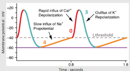
-Ca2+ channels switch off at max depolarisation-Activation of voltage-gated K+ channels occurs
-K+ leaves causing repolarisation
-
What is Phase 0? (Atrial & ventricular muscle action potentials)
Rapid depolarisation
-
What happens in Phase 0? (3) (Atrial & ventricular muscle action potentials)

-Atrial and Ventricular muscle receives depolarisation stimulus from SA node causing:
-Voltage-gated Na+ channels open, Na+ influx.-Voltage-gated Ca2+ channels start to open very slowly
-
What is Phase 1? (Atrial & ventricular muscle action potentials)
Early Repolarisation
-
What happens in phase 1? (1) (Atrial & ventricular muscle action potentials)

-Na+ channels close cells beginning to repolarise
-
What is phase 2? (Atrial & ventricular muscle action potentials)
Plateau phase
-
What happens in phase 2? (2) (Atrial & ventricular muscle action potentials)

-Voltage gated calcium channels fully open - Ca2+ influx halts the repolarisation
-Voltage-gated K+ channels start to open slowly
-
What is phase 3? (Atrial & ventricular muscle action potentials)
Rapid repolarisation
-
What happens in phase 3? (Atrial & ventricular muscle action potentials)
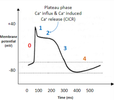
-Ca2+ channels close & K+ channel open fully so K+ efflux
-
What is phase 4? (Atrial & ventricular muscle action potentials)
Resting phase
-
What happens in phase 4? (2) (Atrial & ventricular muscle action potentials)

-Stable - Na+/K+ pump (– 3Na+ out & 2K+ in)-Membrane slightly impermeable to Na+ slightly permeable to K+
-
Heart muscle cells are also known as?
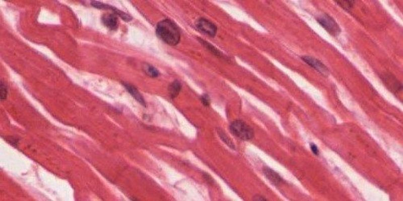
Cardiomyocytes
-
Cardiomyocytes are connected through....? Which allows what to happen? (2)
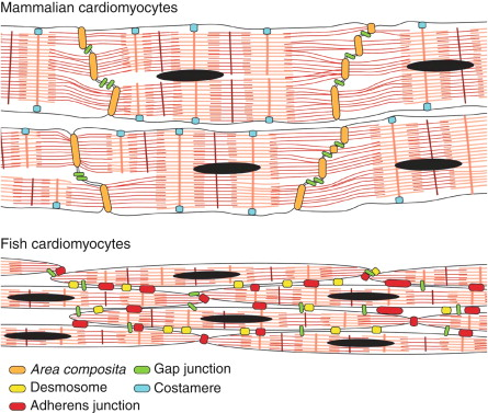
Gap junction-rich intercalated discs
-allows an action potential event in one cardiomyocyte to induce action potential events in adjacent cells with a slight time delay
-This allows a wave of depolarisation to pass through the cardiac muscle in a co-ordinated way causing contraction of atria or ventricles
-

What happens in step 1? (Electrical conduction through the heart)
-Electrical activity generated in the sinoatrial node (SA node) spreads out via gap junctions into atria
-
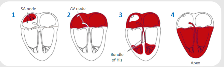
What happens in step 2? (Electrical conduction through the heart)
-At the atrioventricular node (AV node) conduction is delayed to allow correct filling of ventricles
-

What happens in step 3? (Electrical conduction through the heart)
-Conduction occurs rapidly through bundle of His into ventricles
-

What happens in step 4? (Electrical conduction through the heart)
-Conduction through Purkinje fibres spreads quickly throughout the ventricles
-
On an ECG graph what does the X and Y acid represent?
-X axis represents time
-Y axis represents amplitude of the depolarisation mV
-
What does the P wave represent?

Atrial depolarisation
-
What does the PR segment represent?

AV nodal delay
-
What does the QRS complex represent?

Ventricular depolarisation contraction (atria repolarising simultaneously)
-
What does the ST segment represent?

Ventricles contracting and emptying
-
What does the T wave represent?

Ventricular repolarisation
-
What does the TP interval represent?
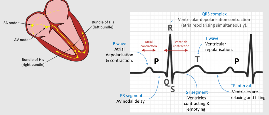
Ventricles are repolarising and filling
-
Electrical activity is converted into what type of contraction? This creates what?
-Myocardial contraction
-This creates pressure changes within chambers of the heart
-
Low pressure in the ventricles allows what valves to open? And as a result what chamber(s) fill with blood?
-Mitral and Tricuspid valve
-Ventricles fill with blood
-
What is isovolumetric contraction?
Process of using energy to contract, while not actively pumping any blood

