-
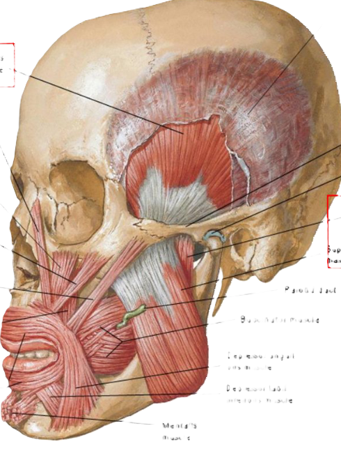
Temporal fascia
Temporalis
muscle
Levator labii
superioris
alaeque nasi
muscle
Levator labii
superioris
muscle
Zygomaticus
minor muscle
Zygomatic arch
Articular disc of
temporomandibular
joint
Deep part of
masseter muscle
Superficial part of
masseter muscle
Zygomaticus
major muscle
Levator
anguli
oris
muscle
Parotid duct
Buccinator muscle
Orbicularis
oris muscle
Depressor anguli
oris muscle
Depressor labii
inferioris muscle
Mentalis muscle
-
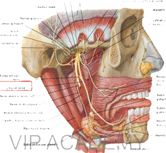
Anterior division
Posterior division
Foramen ovale
Temporal fascia and temporalis muscle
Posterior deep temporal nerve
Anterior deep temporal nerve
Masseteric nen
Lateral pterygoid nerve and muscl
Nervus spinosus
Foramen spinosum
Middle meningeal artery
Auriculotemporal
neve
Chorda tympani
nerve
Lingual nerve
Inferior alveolar nerve (cut)
Nerve to mylohyoid
Medial pterygoid muscle (cut)
Digastric muscle (posterior belly)
Stylohyoid muscle
Buccal nerve and buccinato muscle (cut)
Submandibul ganglion
Sublingual gland
Mylohyoid
muscle (cut)
Mental nerve
V interio aiveolar nerve (cul)
selly)
-
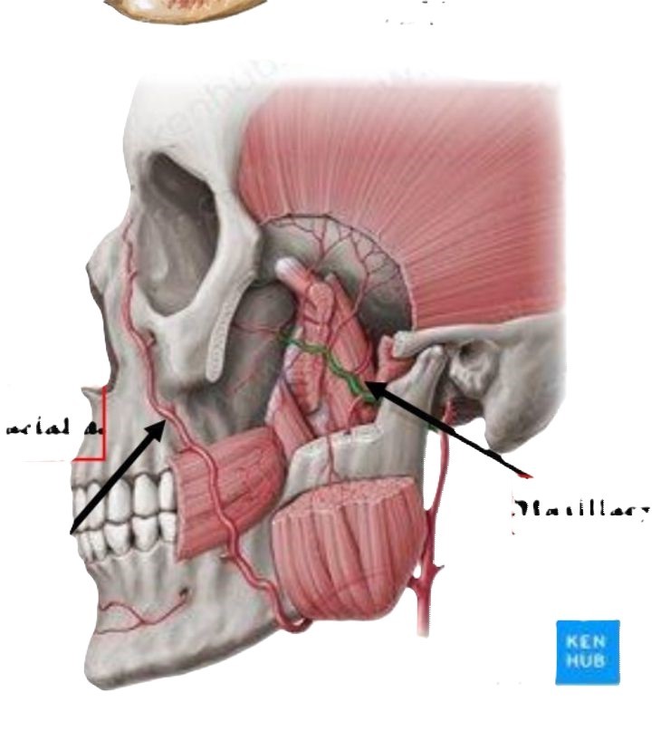
Facial a.
Maxillary a.
-
Digastrie museles
Hyoid bone
Muscular triangle
Midline of the neck
Omohyoid muscle
Sternocleidomastoid muscle
-
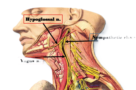
Hypoglossal n.
Sympathetie chain
Vagus n.
-
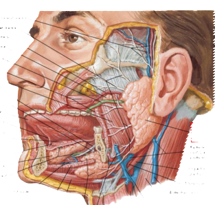
Salivary Glands
Superficial temporal artery and vein and
auriculotemporal nerve
Branches of facial nerve
Transverse facial artery
Accessory parotid gland
Parotid duct
Buccinator muscle (cut)
Masseter muscle
Lingual nerve
Submandibular ganglion
Tongue
Frenulum of tongue
Sublingual fold with openings
of sublingual ducts
Sublingual caruncle with opening
of submandibular duct
Sublingual gland
Submandibular duct
Parotid gland
Retromandibular vein (anterior and posterior branches)
Digastric muscle (posterior belly)
Stylohyoid muscle
External jugular vein
Sternocleidomastoid muscle
Common facial vein
Sublingual artery and vein
Mylohyoid muscle (cut)
Digastric muscle (anterior belly)
Submandibular gland
Facial artery and vein
Hyoid bone
Internal jugular vein
External carotid artery
-
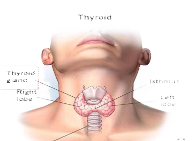
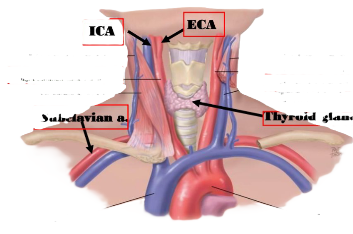
Thyroid
Thyroid gland
Right lobe
Isthmus
Left lobe
Trachea
-
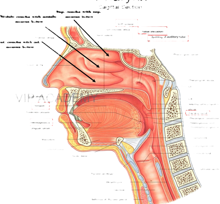
Pharynx
Sagittal Section
Sup. concha with sup. meatus below
Middle concha with middle meatus below
Soft palate
Sphenoidal sinus
Hard palate
Tubal elevation
Opening of auditory tube
Frontal sinus
Inf. concha with inf. meatus below
Pharyngeal tonsil
Nasal cavity
Pharyngeal recess
CI
Tongue-
Foramen cecum
Genioglossus
Lingual tonsil
Mandible
Geniohyoid
CIl
Salpingopharyngeal fold
Uvula
Palatine tonsil
Pharyngeal constrictors
Palatoglossal arch
Palatopharyngeal arch
Epiglottis
Laryngeal inlet
Vocal fold
Hyoid bone
Cricoid cartilage
Thyroid cartilage
Esophagus
Trachea
Isthmus of thyroid gland
-
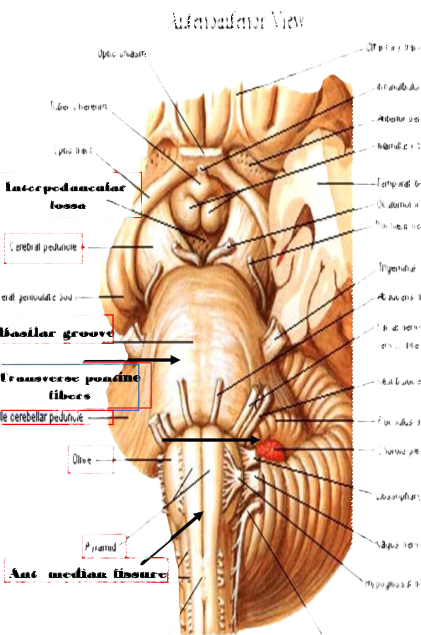
Brainstem
Anterioiferior View
Optic chiasm
Tuber cinereum
Optic tract
Interpeduncular
fossa
Cerebral peduncle-
Lateral geniculate body-
Basilar groove
Transverse pontine
fibers
iddle cerebellar peduncle
Olive-
Pyramid
Ant. median fissure
Olfactory tract
-Infundibulum (pituitary stalk
Anterior perforated substanc
⁃ Mamilary bodies
⁃ Temporal lobe (out surface)
Oculomotor nerve (Il)
-Trochlear nerve (IV)
Trigeminal nerve (V)
Abducens nerve (M)
⁃ Facial nerve (VII) and nervus intermedius
⁃ Vestibulocochlear nerve (MI
• Flocculus of cerebellum
Choroid plexus of 4h ventri
-Glossopharyngeal nerve (IX)
*Vagus nerve (1)
•Hypoglossal nerve (XII)
Pyramidal decussation
Accessory nerve (XI)
-
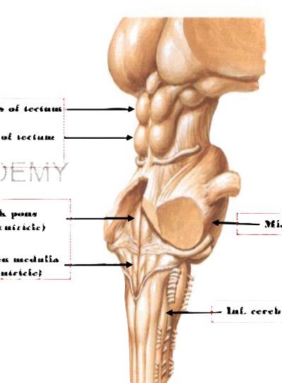
Neuroanatomy
Brainstem
Posterolateral View
Sup. colliculus of tectum
Inf. colliculus of tectum
P ACADEMY
Back pons
(4 ventricle)
Back open medulla -
(4* ventriele)
— Middle cerebellar pedunele
- Inf. cerebellar peduncle
-
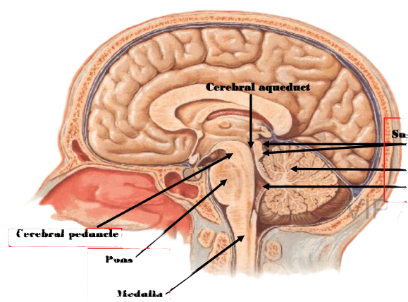
Brainstem, cerebellum & 4% ventriele
Sagittal Section - Medial View
Cerebral aqueduet
Sup. & inf. colliculi of tectumm
Cerebellum
4th ventriele
Cerebral peduncle
Pons
Medulla•
-
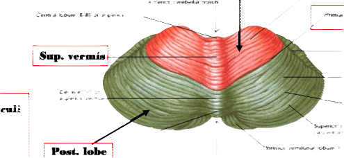
Cerebellun
Superior Suface
Anterior lobe
Anterior cerebellar notch
Central lobule (-Ill] of superior
Quadrangular lobule [H IV.
Primary fissure
Sup. vermis
Horizontal
Simple lobule [H
Declive (VI] of superior vermis-
- Postlunate
Superior semilunar
(anseriform) lobule [H VII A]
Inferior semilunar lobule [H VII
Post. lobe
Posterior cerebellar
-
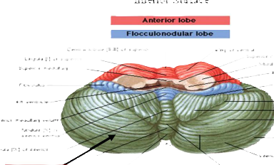
Cerebellum
Inferior Suface
Anterior lobe
Flocculonodular lobe
Central lobule (II-Ill) of superior
Lingula [1) of superior
Superior medullary
Wing of central
Superior cerebellar peduncle
Middle cerebellar
- Inferior cerebellar peduncle
Posterolateral
Flocculus-
4th ventricle
Retrotonsillar fissure
-Tonsil
Biventral lobule [H
Inferior medullary velur
Nodule [X] of inferior vermis
Uvula [IX) of inferior
Horizontal fissure
• Inferior semilunar lobule [H VII|
Post. lobe
Secondary (postpyramidal)
Posterior cerebellar
Posterior lobe
-
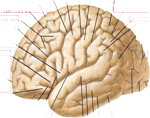
Cerebrum
Lateral View
Central sulcus (Rolando)
Precentral gyrus
Postcentral gyrus
Postcentral sulcus
Precentral sulcus
Superior parietal lobule
Superior frontal gyrus
Superior frontal sulcus
Middle frontal gyrus
Inferior frontal sulcus
Intraparietal sulcus
Inferior parietal lobule
inferior frontal gyrus
IP A
Frontal pole
occipital
pole
Posterior ramus of lateral sulcus (Sylvius)
Temporal pole
Superior temporal gyrus
Superior temporal sulcus
Middle temporal gyrus
Inferior temporal sulcus
Inferior temporal gyrus
-
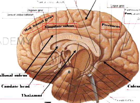
Cerebrum - Hemisphere with Brainstem Excised
Medial View
Paracentral lobule
Splenium of corpus callosum
Trunk of corpus callosum
Lingual gyrus
Cingulate gyrus,
Genu of corpus callosum
Med. frontal syrus
dingulate sulens.
Parietooccipital sulcus
PostC alcarine sulcus
Cuneus
Preeneus
Callosal suleus
Caudate head
Calcarine suleus
Thalamus
Uncus
*Parahippocampal gyrus
Collateral sulcus
Medial occipitotemporal gyrus
Occipitotemporal sulcus
Lateral occipitotemporal gyrus
-
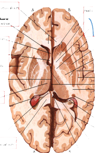
Horizontal Sections through Cerebrum
Genu of corpus callosum
Ant. horn
Lateral ventricle,
Head of caudate nucleus
Internal capsule
Septum pellucidum .-
Insula (island of Reil)
lentiform nucleus
Thalamus -
*Uccipital (posterior) hom of lateral ventricle
Splenium of corpus callosum
-
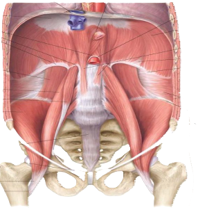
Caval foramen (transmitting inferior vena cava and right phrenic nerve at 18)
Esophageal hiatus
(transmitting esophagus and vagal trunks at T10)
Central tendon
Arouate ligaments:
Median Media!
Lateral
Aortic hiatus (transmitting aorta and thoracic duct at T12)
Diaphragm
Right and left crura
Quadratus lumborum muscle
Lumbocostal
triangle
12th rib
Psoas major muscle
Psoas minor muscie*
Iliacus muscie
External abdominal oblique muscle
Internal abdominal oblique muscle
Transversus abdominis muscie
liac crost
Anterior superior
Inguinal ligament
Anterior longitudinal ligament
Lacunar ligament
Ilopsoas tendon
Lesser trochanter
-
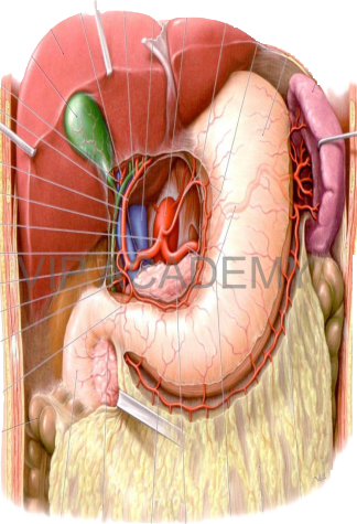
Proper hepatic artery, left branch
Proper hepatie artery, right branch
Inferior vena cava
Liver
Abdominal aorta
Cut edge of
lesser omentum
Left gastric artery
Stomach
Gall bladder
Spleen
Cystic artery
Cut edge of free border of lesser omentum
Proper hepatic artery
Portal vein
Celiac trunk
Common hepatic artery
Common bile duct
Right gastric artery
Superior pancreatico-duodenal artery (posterior branch)
Gastroduodenal artery
Right colic flexure
2nd part of duodenum
Right gastro-epiploic artery
Superior pancreaticoduodenal artery
Pancreas
(anterior branch)
Splenic artery
Left gastro-epiploic artery
Greater omentum
-
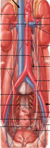
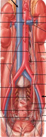
Arteries of Ureters and Urinary Bladder
NC
Abdominal aorta
Superior mesenterio artery-
⁃ Inferior suprarenal artery
⁃ Renal artery and vein
Ovarian (testicular) artery-
Inferior mesenteric artery (out)-
Ureteric branches from ovarian and common iliac
⁃ Ureterio branch from renal artery
⁃ Ureter
⁃ Psoas major muscle
⁃ Ureteric branch from aorta
Common iliac artery
nternal ilac artery
Superior gluteal artery-
Inferior gluteal and internal pudendal arteries
Obturator artery -
Vaginal artery-
External iliac A.
— Middle rectal artery
Uterine artery
⁃ Inferior epigastric artery
⁃ Inferior vesical artery and ureteric brai
Ureteric branch from superior vesical artery
Superior vesical arteries
-
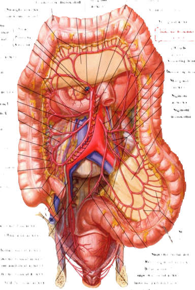
Middle colic artery
Transverse mesocolon
Marginal artery
Straight arteries (arteriae rectae)
Inferior pancreatico-duodenal artery
Marginal artery
Stem
Posterior
Anterior
Right colic artery
Ileocolic artery
Colic branch
Teal branch.
Marginal artery
Anterior caecal artery
Posterior caecal artery
Appendicular artery
Superior mesenteric artery
Ist jejunal artery
Jejunal and ileal arteries
Marginal artery
Inferior mesenterie
artery
Left colic artery
-
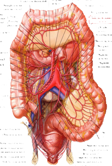
Ascending branch
Descending branch
Marginal artery
Sigmoid arteries
Sigmoid
mesocolon
Internal iliac artery
Obturator artery
Median sacral artery
Superior vesical artery (from umbilical artery)
Inferior vesical artery
Middle rectal artery
Branch of superior rectal artery
Straight arteries
Superior rectal artery
Rectosigmoid arteries
Bifurcation of superior rectal artery
Internal pudendal artery in pudendal canal
Inferior rectal artery
-
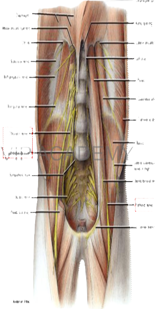
Daphragm
Medial arcuale Igament
12h tb
Subcostal nene
lichypogas tic rese
Rongunal neve
Obturalor nene
Lntest
Sympathelic bunk
Sciatic nerve
Psoas (cut end)
Anterior View
Escphagol opering
Aorte opening
Lateral arcuate lgarnent
Laft cus
-
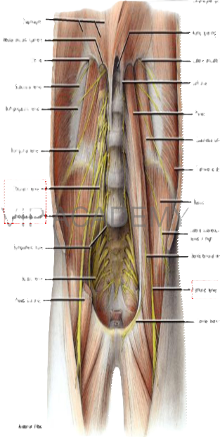
Quadratus lumbarm
Transverse abdominal
lacus
Laleral culaneous
neve of trigh
Genitofemoral nene
Femoral nerve
Genital branch
-
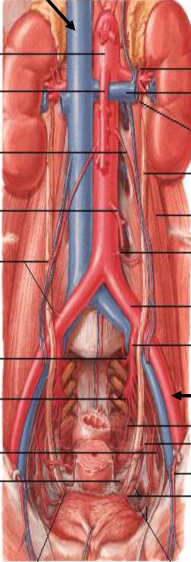
Arteries of Ureters and Urinary Bladder
NC
Abdominal aorta
Superior mesenterio artery-
⁃ Inferior suprarenal artery
⁃ Renal artery and vein
Ovarian (testicular) artery-
Inferior mesenteric artery (out)-
Ureteric branches from ovarian and common iliac
⁃ Ureterio branch from renal artery
⁃ Ureter
⁃ Psoas major muscle
⁃ Ureteric branch from aorta
-
Common iliac artery
nternal ilac artery
Superior gluteal artery-
Inferior gluteal and internal pudendal arteries
Obturator artery -
Vaginal artery-
External iliac A.
— Middle rectal artery
Uterine artery
⁃ Inferior epigastric artery
⁃ Inferior vesical artery and ureteric brai
Ureteric branch from superior vesical artery
Superior vesical arteries
-
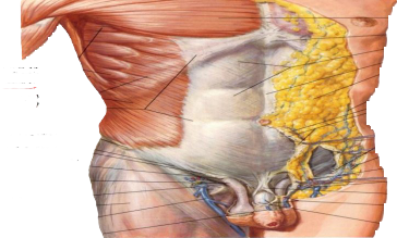
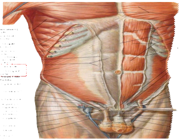
Serratus anterior
muscle
Latissimus dorsi
Fleshy
External oblique
Aponeurotie
Anterior superior
Pectoralis major muscle
Xiphoid process
Rectus sheath
Linca alba
Superficial fascia of the abdomen
Thoracoepigastric vein
Camper's fascis Searpa's fascia
Inguinal ligament
Intererural fibers
Superficial inguinal ring
External spermatic fascia over spermatic cord
Cribriform fascia
Fascia lata
Great saphenous vein
Superficial dorsal vein
Attachment of Scarpa's fascia to fascia lata
Superficial circumflex
Superficial epigastric
Superficial external pudendal vessels Fundiform ligament
Superficial fascia of penis and scrotum
and deep dorsal vein
of penis
-
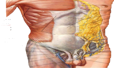
Thoracoepigastric vein
Camper's fascis Searpa's fascia
Inguinal ligament
Intererural fibers
Superficial inguinal ring
External spermatic fascia over spermatic cord
Cribriform fascia
Fascia lata
Great saphenous vein
Superficial dorsal vein
Attachment of Scarpa's fascia to fascia lata
Superficial circumflex
Superficial epigastric
Superficial external pudendal vessels Fundiform ligament
Superficial fascia of penis and scrotum
and deep dorsal vein
of penis
Latissimus dorsi
muscle
Serratus anterior
muscle
External oblique muscle (cut)
External intercostal
muscles
External oblique
aponeurosis (cut edge) Rectus sheath
Internal obliqueness
-
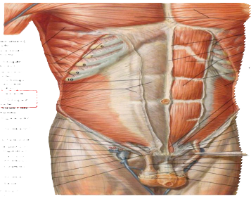
Anterior superior iline spine Inguinal ligament
Cremasteric muscle
Conjoint tendon Reflected part of inguinal ligament Femoral vein
(in femoral sheath) Saphenous opening
Cremasteric muscle Fascia lata
Great saphenous
vein
Pectoralis major
muscle
Anterior wall of
rectus sheath (cut) Linea alba
Rectus abdominis
muscle
External oblique muscle (cut)
Tendinous intersection Internal oblique
muscle Pyramidalis muscle Conjoint tendon
Inguinal ligament Anterior superior iliac spine
External oblique aponeurosis (cut) Pectineal ligament
Lacunar ligament
Reflected part of inguinal ligament
Pubic tubercle Suspensory ligament of penis
Cremasteric muscle and fascia
Deep fascia
of penis
External spermatic
fascia (cut) Superficial fascia of penis and scrotum
-
Superior epigastric
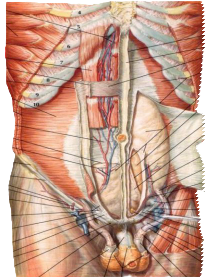
vessels
Serratus anterior
muscle
External oblique musele (cut).
Rectus abdominis
muscle.
External oblique aponeurosis (cut).
Internal oblique aponeurosis (cut)
Transversus abdomini
muscle
Internal oblique muscle
(cut)
Posterior wall of rectus sheath
Arcuate line.
Inferior epigastric vessels-
Anterior superior iliac spine
Inguinal ligament (Poupart's ligament) Superficial circumflex iliac artery
Superficial epigastric
artery
Superficial external-
pudendal artery
Conjoint tendon
Pectineal ligament (Cooper's ligament)-
Lacunar ligament
(Gimbernat's ligament)
Reflected part of inguinal ligament
Fascia lata
Pubic tubercle
-
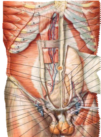
Pubic tubercle
Cremasteric muscle and fascia
External spermatic fascia (cut) Deep fascia of penis
Superficial fascia of penis and scrotum (dartos fascia)
Anterior wall of rectus sheath (cut)
Linea alba
Anterior wall of rectus sheath Transversus abdominis muscle (cut)
Fascia transversalis
Peritoneum and extraperitoneal fatty tissue
Medial umbilical ligament
Umbilical prevesical
fascia
Arcuate line
Inferior epigastric artery and vein Site of deep inguinal ring (origin of internal spermatic fascia)
Cremasteric and pubic branches of inferior epigastric artery Femoral sheath (containing femoral artery and vein) Inguinal ligament (Poupart's ligament) Lacunar ligament (Gimbernat's ligament) Pectineal ligament (Cooper's ligament)
Fat in retropubic space
Pectineal fascia
Sartorius muscle
Internal spermatic fascia
Cremasteric muscle and fascia
External spermatic fascia
-
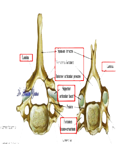
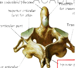
Dens (odontoid process)
Superior articular facet for atlas
Interarticular part
Posterior articular facet for transverse ligament of atlas
Transverse process
• Spinous process
Inferior articular process
Posterior View
Axis (Second Cervical Vertebra)
-
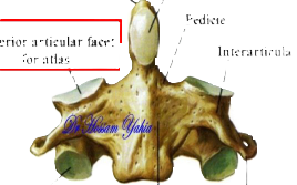
Dens (odontoid process)
Superior articular facet for atlas
Interarticular part
Posterior articular facet for transverse ligament of atlas
Transverse process
• Spinous process
Inferior articular process
Posterior View
Axis (Second Cervical Vertebra)
-
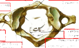
Anterior tubercle
Anterior arch
Articular facet for dens
Lateral mass
Transverse process
Tubercle for transverse
ligament of atlas
Foramen transversarium
Superior articular facet (for occipital condyle)
- Vertebral foramen
Posterior arch
- Posterior tubercle
Groove for vertebral artery
Atlas Vertebra: superior view
-
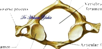
Anterior tubercle
Anterior arch
Articular facet for dens
Lateral mass
Transverse process
Tubercle for transverse
ligament of atlas
Foramen transversarium
Superior articular facet (for occipital condyle)
- Vertebral foramen
Posterior arch
- Posterior tubercle
Groove for vertebral artery
Atlas Vertebra: superior view
-
Anterior tubercle
Anterior arch
Articular facet for dens
Lateral mass
Transverse process
Tubercle for transverse
ligament of atlas
Foramen transversarium
Superior articular facet (for occipital condyle)
- Vertebral foramen
Posterior arch
- Posterior tubercle
Groove for vertebral artery
Atlas Vertebra: superior view
-
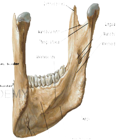
Coronoid process
Mandibular notch
Ptergoid fovea
Mylohyoid line
Lingula
Mandibular foramen
Mylohyoid groove
rder
Angle
Submandibular fossa
Sublingual fossa
' Digastric fossa
Mental spines (Genial tubercles)
Mandible; inner surface
-
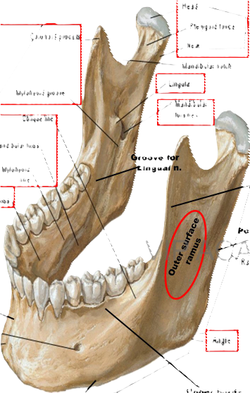
Goronoid proess
Inner
Surfa ce
Head
Pterygoid fovea
Neck
Mandibular notch/Lingula
Mandibular foramen
Mylohyoid groove
Oblique line
Submandibular fossa,
Mylohyoid
line
Groove for
Lingual n.
Ant. l
Sublingual fossa
Outer surface ramus
Post. bord
Ramus
Symphysis menti
Mental foramen
Mental protuberance
Angle
Upper border (alveolar margin)
Lower border
Body
-
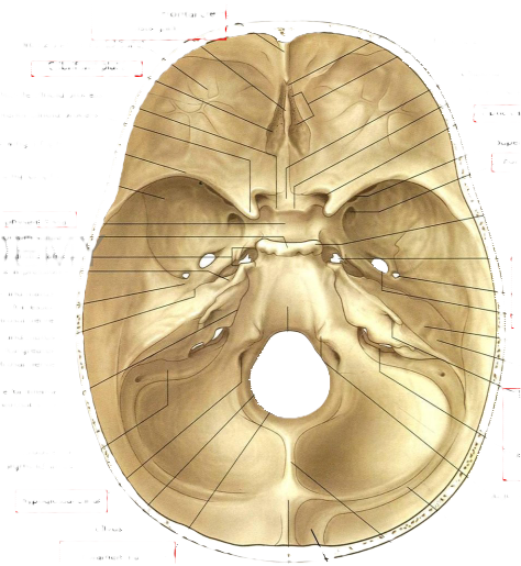
Norma Basalis Interna
Frontal crest
Crista galli
Orbital part (of frontal bone)
Cribriform plate -
Middle clinoid process
Anterior clinoid process
Lesser wing (of sphenoid)
Greater wing (of sphenoid)
Foramen caecum
Foramina of cribriform plate
Chiasmatic sulcus
Tuberculum sellae
Optic canal
-
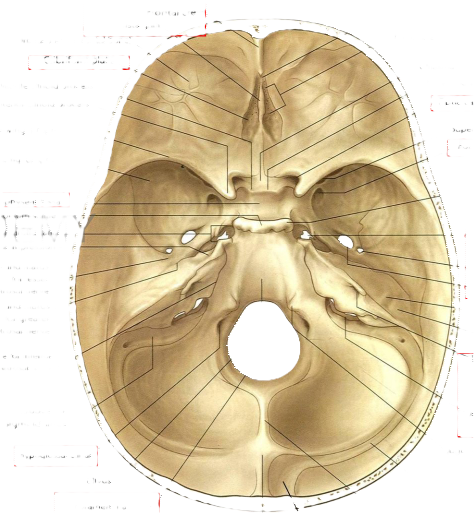
Superior orbital fissure
- Foramen rotundum
Posterior clinoid process
Hypophyseal fossa
Dorsum sellae
opening of carotid canal
Trigeminal impression
Groove and hiatus
for lesser petrosal nerve
Groove and hiatus
for greater petrosal nerve
Groove for inferior petrosal sinus
Groove for sigmoid sinus
Hypoglossal canal
Clivus
Foramen magnum
Foramen lacerum
Foramen spinosum
Foramen ovale
Arcuate eminence
- Tegmen tympani
Internal acoustic meatus
Jugular foramen
Jugular tubercle
Groove for transverse sinus
Internal occipital crest
Internal occipital protuberance
Groove for superior sagittal sinus
-
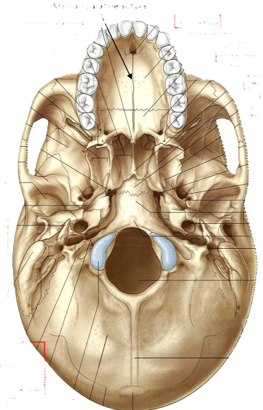
Norma Basalis Externa
Median palatine suture
Posterior nasal spine
Posterior nasal aperture (choana)
Alveolar arch.
Pterygoid
Pterygoid fossa
Greater wing (of sphenoid bone)
Opening of pterygoid canal
Articular tubercle
Mandibular fossa
Groove for auditory tube
Styloid process
-
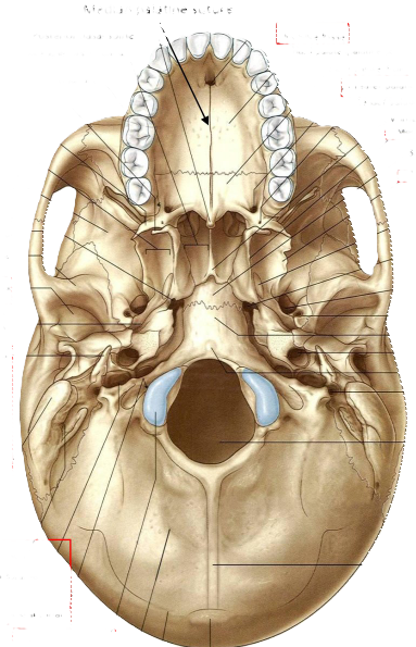
Incisive fossa
Hard palate (palatine process of maxilla)
, Hard palate (horizontal plate of palatine bone)
Greater palatine foramen
Lesser palatine foramen
Vomer
Medial plate of pterygoid process
-Body of sphenoid
Lateral plate of pterygoid process
- Scaphoid fossa
Foramen lacerum
Foramen ovale
Foramen spinosum
Basilar part of occipital bone
Carotid canal
Pharyngeal tubercle
Stylomastoid foramen
Foramen magnum
Mastoid process
Mastoid notch
Jugular foramen /
Hypoglossal canal
Occipital condyle
Inferior nuchal line
Superior nuchal line
External occipital protuberance
External occipital crest
-
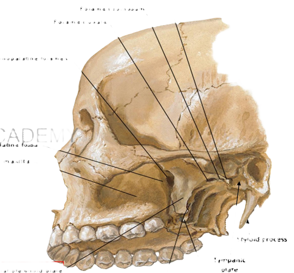
Infratemporal Fossa Exposed
Lateral View
Spine of sphenoid bone
Foramen spinosum
Foramen ovale
Sphenopalatine foramen
Pterygopalatine fossa
Back of maxilla
Styloid process
Lateral pterygoid plate
ma lateralis
Tympanic plate
Pterygoid hamulus
Medial pterygoid plate
-
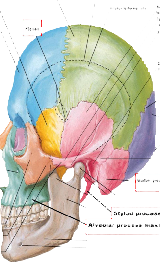
Greater wing of sphenoid
Frontal bone
Parietal bone
Temporal fossa
Superior temporal line
Inferior temporal line
Pterion
Squamous part of temporal bone
Zygomatic process of temporal bone
Articular tubercle
External acoustic /meatus
Lambdoid suture
Nasal bone
Occipital bone
External occipital protuberance
Infraorbital foramen
Suprameatal
Mastoid process
Stylod process
Alveolar process maxilla
zygomatic bone
Temporal process... of zygomatic bone
Zygomatic arch
-
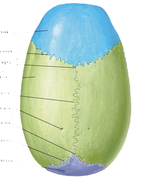
Norma verticalis
Frontal bone
Coronal suture
Bregma
Paretal bone
Sagittal suture
Parietal foramen
Lambda
Lambdoid suture
Occipital bone
-
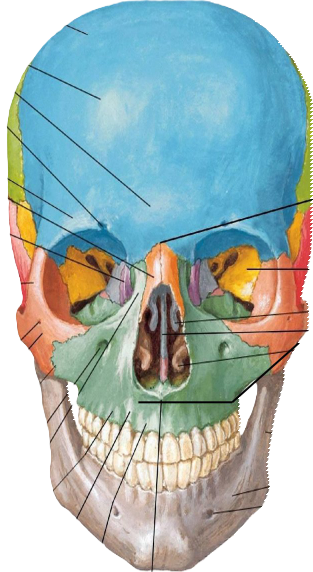
Norma frontalis
Frontal bone
Frontal eminence
Glabella
Supraorbital notch (foramen)
Superciliary arch
Nasal bone
Nasion
Lacrimal bone
Frontal process of zygomatic bone
Orbit
VI Midate nasal concha DE
Nasal septum
Inferior nasal concha
Zygomaticofacial
foramen
Zygomatic bone
Zygomatic process of maxilla
Ramus
Body
Mental foramen
Infraorbital foramen
of maxilla
Frontal process of maxilla
Canine fossa
Canine eminence
Anterior nasal spine
Incisive fossa
-
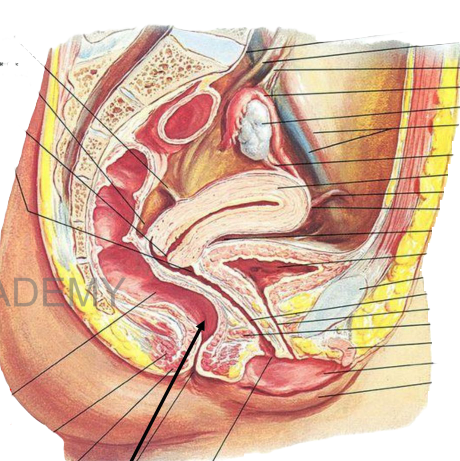
SACRO-UTERINE
LIGAMENT
POSTERIOR
CUL-DE-SAC
OF DOUGLAS
CERVIX
FORNIX OF VAGINA.
HEACADE
SACRAL PROMONTORY
URETER
INFUNDIBULOPELVIC
LIGAMENT
• FALLOPIAN TUBE
OVARY
EXTERNAL ILIAC
VESSELS
⁃ OVARIAN LIGAMENT CORPUS OF UTERUS ROUND LIGAMENT FUNDUS OF UTERUS ANTERIOR CUL-DE-SAC BLADDER SYMPHYSIS PUBIS VAGINA
⁃ URETHRA UROGENITAL DIAPHRAGM CRUS CLITORIS LABIUM MINUS LABIUM MAJUS
RECTUM
LEVATOR ANI
MUSCLE
EXTERNAL ANAL
SPHINCTER
Anal canal ORGINAL
Sagittal section female pelvis
-
Kidney
Superior pole
— Medial margin
Lateral margin
- Renal artery
Renal vein
Renal pelvis
Anterior View
Interior pole
Ureter
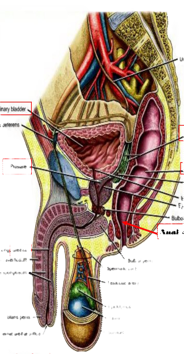
Testis & spermatic cord
Spermatic cord
Testis
Ureter
Uninary bladder
Ductus deferens
Seminal vesicle
Prostate
⁃ Rectum ampulla)
⁃ Ampulla of ductus defere
Internal trethral orifice
Ejaculatory duct
Bulbourethral gland
Anal canal
Spong
urethra
Corpus cavemosum
Corpus spongiosum
Bulb of penis
Spermatic cord
•Testicular artery
Glans penis
External urethal crifice
Epididymus
Testis
Scrotum
Sagittal section male pelvis
-
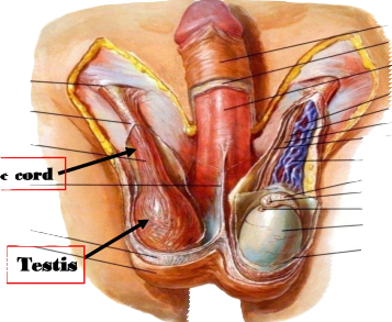
Testis & spermatic cord
Spermatic cord
Testis
-
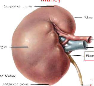
Kidney
Superior pole
— Medial margin
Lateral margin
- Renal artery
Renal vein
Renal pelvis
Anterior View
Interior pole
Ureter
-
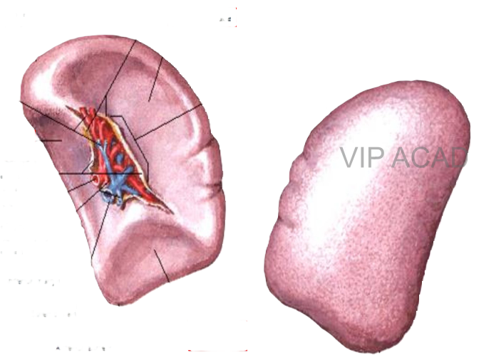
Spleen
Visceral and Diaphragmatic Surfaces
Posterior extremity
Gastrosplenic ligament
Gastric surface
Superior margin
Posterior extremity
Hilus
short astric ressels
Rena surface
Tenorenal)
gament
Splenic artery
eft gastroepiploid
gastroomental)
Inferior margin
Inferior margin
Splenio vein
Colic surface
Anterior extremity
Anterior extremity
Micrarsl curfara
Nianhranmatin curfaro
-
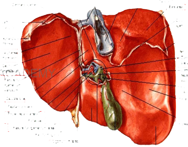
Surfaces and Bed of Liver
Visceral Surface
Hepatic veins
Caudate process,
Inferior vena cava
, Suprarenal impression
Coronary ligament,
Bare area
Left triangular
Appendix-fibrosa
Esophagea mpression
Gastric-impression
Fissure for-gamentum
Kenosum
Caudate lobe
Papillary prooess
Proper hepatic artery
Portal vein
Fissure for ligamentum teres
Falciform ligament
-gamentum teres Quadrate lobe Porta hepatis
Coronary ligament
Right triangular ligament
⁃ Common bile duct
• Common hepatio duct
⁃ Cystic duct
⁃ - Renal impression
*Duodenal impression
Gallbladder
Colic impression
-
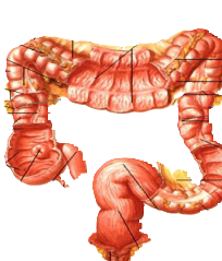
ransverse colon
Transverse mesocoion
Right colic (hepat
Greater omentum (cut away)
Epiploic appendices
Semdunar foids
Left colic (splenic) flexure
Omental tenia
Haustra
Peritoneum (cut away)
Omental tenia (exposed by hook)
Free tenia (tenia libera,
Ascending colon
• Haustra
Peritoneum (cut away)
Descending colon
Mesocolic tenia
(exposed by hook)
Ceoum
Free tenia (tenia ibera)
• Sigmoid mesocolon
leum
Vermiform appendoe
Sigmoid colon
Rectosigmoid junction (teniae spread out and unite to form longitudinal muscle layer)
Levator ani musc
External anal sphincter muscle
Rectum
-
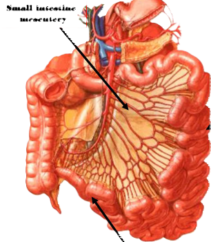
Small intestine mesentery
Jejunum
Ileum
Mucosa and Musculature of Large Intestine
-
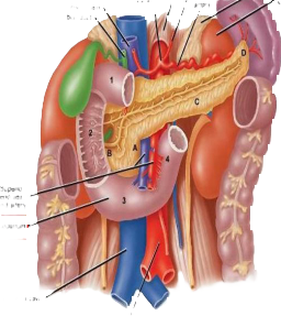
Celiac trunk
Left gastric artery
Diaphragm
Splenic artery
Portal vein
Bile duct
Spleen
• Left suprarenal gland
Left kidney
Vertebral levels
Superior mesenteric vein and artery
Duodenum
Inferior vena cava
Aorta
-
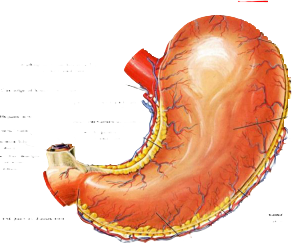
Fundus of stomach
Esophagus
Short gastric vessels
Esophageal branches of left gastric artery and vein
Cut edge of lesser omentum
Left gastric artery and vein
Cut edge of gastrosplenie ligament
Body of stomach
Hepatic artery
Portal vein
Common bile duct
Right free margin of lesser omentum
Lesser curvature of stomach
Right gastric artery and vein
Left gastroepiploie vessels
First part of duodenum
Greater curvature of stomach
Cut edge of greater omentum
Annular groove between pylorus and duodenum
Right gastroepiploie vessels
Pyloric antrum

