 Matthew Burn
-
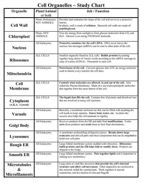
Functions of Cell Organelles
-
Functions of Plasma membrane
In all living cells, the plasma membrane functions as the boundary and is selectively permeable, by allowing the entry and exit of certain selective substances. Along with these, the plasma membrane also acts as a connecting system by providing a connection between the cell and its environment.
-
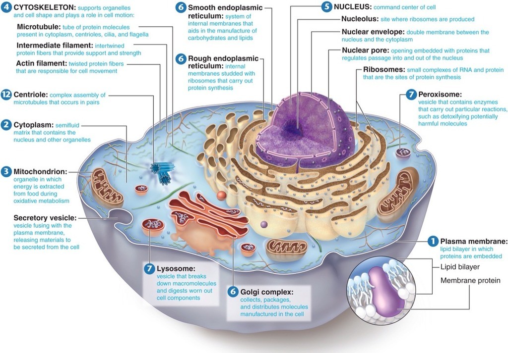
Cell Structure and Function
-
Endocytosis vs. Exocytosis
Endocytosis is the process of capturing a substance or particle from outside the cell by engulfing it with the cell membrane, and bringing it into the cell.
Exocytosis describes the process of vesicles fusing with the plasma membrane and releasing their contents to the outside of the cell.
Both endocytosis and exocytosis are active transport processes.
-
Na+/K+ Pump
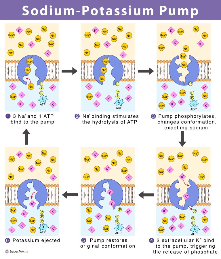
In general, the inside of a cell has a higher concentration of potassium (K+, plus, end superscript) and a lower concentration of sodium than the extracellular fluid around it.
If sodium ions are outside of a cell, they will tend to move into the cell based on both their concentration gradient (the lower concentration of Na+ in the cell) and the voltage across the membrane (the more negative charge on the inside of the membrane).
Because K is positive, the voltage across the membrane will encourage its movement into the cell, but its concentration gradient will tend to drive it out of the cell (towards the region of lower concentration). The final concentrations of K on the two sides of the membrane will be a balance between these opposing forces.
The combination of concentration gradient and voltage that affects an ion’s movement is called the electrochemical gradient.
-
Ionic and Covalent Bonds
1. An ionic bond essentially donates an electron to the other atom participating in the bond, while electrons in a covalent bond are shared equally between the atoms.
2. The only pure covalent bonds occur between identical atoms. Usually, there is some polarity (polar covalent bond) in which the electrons are shared, but spend more time with one atom than the other.
3. Ionic bonds form between a metal and a nonmetal. Covalent bonds form between two nonmetals.
-
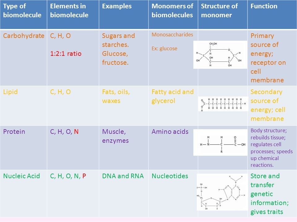
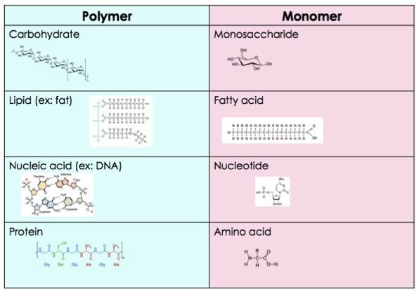
Components of biomolecules
1. Functional Groups react and Name of the Bond
a.) Carbs - 2 Hydroxyl Groups (-OH), called Glycosidic Bond/Linkage
b.) Lipids - Hydroxyl Groups on glycerol and the hydroxyl component of the carboxyl group on the fatty acid, called Ester Bond/Linkage
c.) Proteins - between hydroxyl component of the carboxyl group on one amino acid and the amine group on the other amino acid, called Peptide bond.
-
Micromolecules and macromolecules
-
% Percent Formula and Example
% = grams of solute/100 mL solution.
ex: 5% glucose solution
% = 5g glucose/100 mL
1. d. Ex: Calculate the % solute of a solution containing 4g fructose in 100 mL solution .
i. 4g/100 mL solution= 4%
2. Calculate the % solute of a solution containing 6g MgBr2 in 25 mL solution.
i. 6g/25 mL X 4/4= 24/100= 24%
-
Molarity (M) and Molality (m)
Molal/molality (m) = moles solute per kg solvent
1. Density H2O=1
A. 1g/1mL or 8 lb/1 gal
B. 1kg/1L (1kg= 1000g and 1L= 1000 mL)
Molar/molarity (M) = moles solute per 1L solution
3 Things to Know:
1. NaCl= 58.5 g/mole
B. Monosaccharides (180 g/mole)
C. Disaccharides (342 g/mole)
-
Osmosis and Diffusion
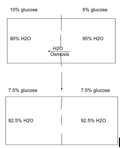
1. Diffusion - moves down concentration gradient, from high to low concentration.
2. Osmosis - diffusion of water
i.) Semipermeable: permeable to water but impermeable to the solute.
ii.) Semipermeable must have for osmosis to occur, so that water can move in through it but glucose can not. Water moves towards higher solute concentration, or water moves down it's own concentration gradient.
-
Tonicity Comparison (Isotonic, hypertonic, hypotonic)
1. Isotonic - no net movement
2. Hypertonic - cells shrink (Crenation) due to more water coming out than going in.
3. Hypotonic - more water moves in then out; cells will burst (lysis), in RBC its hemolysis
-
Positive vs. Negative Feedback
1. Negative feedback - system produces a product that reaches a set point, which will turn off system.
2. Positive feedback - system produces a product that reaches set point, will stimulate system. ex: milk production
-
Blood Sugar and Homeostasis
1. Alpha Cells - Glucagon: increase blood sugar; glycogenolysis
2. Beta Cells - Insulin: decrease blood sugar; glycogenesis
3. Thyroxine - increases blood sugar; glycogenolysis
4. Cortisone - increases blood sugar; glycogenolysis
5. Epinephrine - increases blood sugar; glycogenolysis (rapid increase)
-
G-words
1. Glycogenolysis - split glycogen, raises blood sugar
2. Glycogenesis - formation of glycogen; decreases blood sugar
3. Gluconeogenesis - creation of new sugar; burn something other than carbs and convert amino acids to glucose.
4. Glycolysis - split glucose into two molecules of pyruvate
-
Osteoblasts vs. Osteoclasts
1. Osteoblast - increase bone mass/density. Takes Ca+ out of blood into bone.
i.) Stimulated by testosterone and estrogen, Thyroxine, Growth Hormone.
ii.) Abuse of cortisone will inhibit osteoblasts; decreasing bone density leading to stress/pathological fractures.
2. Osteoclast - decrease bone mass/density. Takes Ca+ out of bone into blood.
i.) Stimulated by Parathyroid Hormone (PTH).
ii.) Inhibited by calcitonin; stops the decrease in bone mass.
-
Integumentary System Function
1. Body Temp Regulation
i.) Sweating
ii.) Vasomotor Control - control of blood vessels
a.) Dilate = Hot, Constrict = Cold
2. Protection - serves as first line of defense, pH of skin is not conductive to growth of microorganisms.
3. Perception - feeling/tactile/texture
4. Excretion and Secretion
i.) Excretion - leaves body (sweat, oil)
ii.) Secretion - glands release something but stays in body. eg: melanin, goblet cells (mucus secretion)
5. Synthesis
i.) Sweat glands (Sudoriferous)
ii.) Oil Glands (Sebaceous)
iii.) Mammary glands
-
Epidermis
5 layers; called epidermal layers
1. Stratum Coreneum (extremly thick)
2. Stratum Lucidium (layer missing in thin layer, very thin)
3. Stratum Granulosum (very thin)
4. Stratum Spinosum (very thick)
5. Stratum Basale (Very thin)
-
Layers of skin
1. Epidermis
2. Dermis
i.) Top (20-35%) Papillary region (loose CT, elastic CT) composed of dermal papillae and parallel pegs, and Meissner's corpuscles.
ii.) Bottom (65-80%) Reticular region - (dense irregular CT, collagen and elastic fibers), adipocytes, hair follicles and associated structures.
3. Hypodermis - (loose CT and adipose CT) made of Pacinian corpuscles and sudoriferous glands.
-
Functions of Skeletal System
1. Movement- muscles pull bones to move
2. Support - how you are standing
3. Storage - Ca+ and Triglycerides (fats)
4. Production- hemopoiesis blood made in bone
5. Protection (x2) - brain inside skull, ribs protect organs and WBC immunity.
-
Vertebral Column
1. Curves
a.) Cervical - Secondary
b.) Thoracic - Primary
c.) Lumbar - Secondary
d.) Pelvic - Primary
2. Number of vertebrae - 32-34
3. Types of Vertebrae
a.) Cervical- 7
b.) Thoracic- 12
c.) Lumbar - 5
d.) Sacral - 5
e.) Coccygeal - 3-5
-
Endochondral Ossification
Occurs until fully grown. (Hyaline cartilage -> Long bone)
1. Blood vessels grow through perichondrium.
2. Blood brings Ca+ into cartilage and becomes primary ossification center. (cells burst changing pH)
3. Repeat steps 1-2
4. Ossification spreads from each epiphysis toward the diaphysis and from the diaphysis toward the epiphysis.
5. When done growing, epiphyseal plate ossifies and becomes the epiphyseal line.
-
Intramembranous Ossification
Occurs until you DIE. (Fibrous membrane -> Flat Bone)
1. fibrous membranes, starts during embryo genesis, membrane is being replaced by bone. At birth ossification is not complete to pass birth canal and brain grows faster than the skull.
-
Disorders of the vertebral column
1. All disorders associated with Ca+ or Vit. D deficiency.
2. Lordosis - lumbar curvature exaggerated. Obesity, gestation lordosis
3. Kyphosis - thoracic cavity exaggerated. Part of aging, compress more anteriorly, bends forward.
4. Scoliosis - lateral bending of vertebral column, associated with leg length discrepancy. Can interfere with lungs ability to expand and contract.
5. Spina Bifida- bifurcated/forked spinous process of the vertebral column. variable expression, cause paraplegia. caused by too little folic acid or too much retinoic acid (vit A).

