What is the general structure of blood vessels that are NOT capillaries?
- tunica externa (CT)
- tunica media (smooth muscles, electic fibers (CT))
- tunica intima/interna (endothelium (simple squamous epithelium), continues with endocardium)
- lumen (contains blood, not a layer)
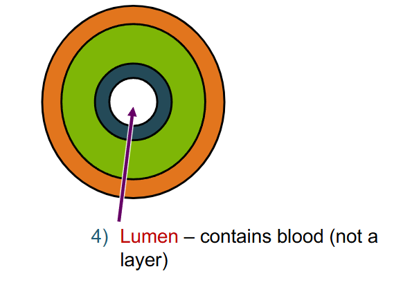
Name layers of this vein (not capillary)
- orange: tunica externa
- dark green: tunica media
- light green: tunica intima/interna
What are the blood vessel types? (following path from heart and back to heart)
- arteries
- arterioles
- capillaries
- venules
- veins
What are arteries? what do they do?
- carry blood AWAY from heart (doesnt refer to oxygenenated or deoxygenated blood)
- 2 types: elastic arteries and muscular arteries
What are elastic arteries?
- large conducting arteries exiting the heart
- elastic CT in all 3 layers
- largest arteries (near heart)\
- e.g. aorta
What are muscular arteries?
- smaller distributing arteries
- a lot of smooth muscle
- most arteries
- e.g. coronary artery
What are arterioles?
- little arteries
- regulate blood flow and blood pressure
what are capillaries?
- ONLY layer is tunica intima endothelium (one cell layer) plus a basement membrane
- allow exchange of gases and nutrients
- most have gaps between cells that allow exchange of fluid and solutes with the interstitial fluid
What are venules?
- collect blood from capillaries
- intima (endothelium) with thin media and externa layers
What are veins?
- carry blood INTO heart (doesnt refer to oxygenenated or deoxygenated blood)
- large lumen
- can have one way valves to prevent backflow of blood
- thin media, more CT but less smooth muscle than arteries
Whats included the cardiovascular system? What does it transport?
- heart, blood vessels, blood
- transports gases, nutrients, hormones, wastes, and heat
- protects from disease and fluid loss (clotting)
Where is the heart?
cavity called mediastinum (between lungs in thoracic cavity)
What makes up the structure of the heart?
Coverings (pericardium), heart wall, four chambers, septa, cardiac (fibrous) skeleton, valves, and cardiac muscle cells
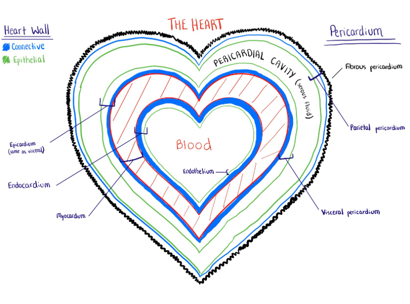
What is the pericardium and what are its layers?
- double-walled sac surrounding the heart
- 3 layers: fibrous pericardium
serous pericardium (made of)
parietal pericardium
visceral pericardium
- between parietal and visceral is the pericardial cavity (serous fluid)
What is the fibrous pericardium?
- outermost layer of pericardium
- anchors to surrounding structures (diaphragm, vessels like aorta, vena cava etc.)
- dense irregular connective tissue
What is the parietal pericardium?
- 2 layers, epithelial and connective
- connected to fibrous pericardium
- pericardial sac (outer layer of the pericardial cavity)
What is the visceral pericardium?
- also called epicardium
- 2 layers, epithelial and connective
- fused to heart surface (part of heart wall)
What are the layers of the heart wall?
- epicardium (visceral pericardium), simple squamous epithelium and CT
- myocardium (cardiac muscle) arranged in spiral/circular pattern & reinforced with CT
- endocardium (simple squamous epithelium & CT) the epitheliums named endothelium and lines all inner surfaces of heart and all blood vessels
Name the layers surrounding the heart
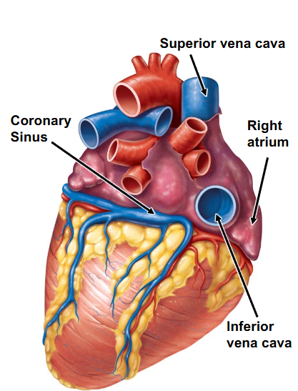
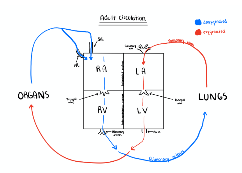
What vessels are associated with the right atrium of the heart?
- 3 major veins carry deoxygenated blood into the chamber
inferior vena cava (carries in blood from the body below heart)
superior vena cava (carries blood from the body above heart)
coronary sinus (carries blood from myocardium)
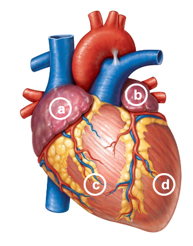
a. right atrium
b. left atrium
c. right ventricle
c. left ventricle
What vessels are associated with the right atrium of the heart (location/image)?
What vessels are associated with the left atrium?
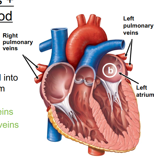
- 4 veins carry oxygenated blood from lungs into left atrium
( 2 left pulmonary veins and 2 right pulmonary veins)
What vessels are associated with the right ventricle?
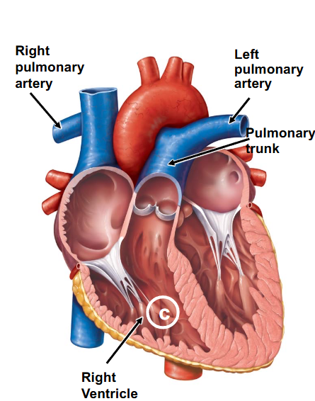
- 1 artery (the pulmonary trunk) exists then divides to form 2 pulmonary arteries (left and right) which carry deoxygenated blood towards lungs
What vessels are associated with the left ventricle?
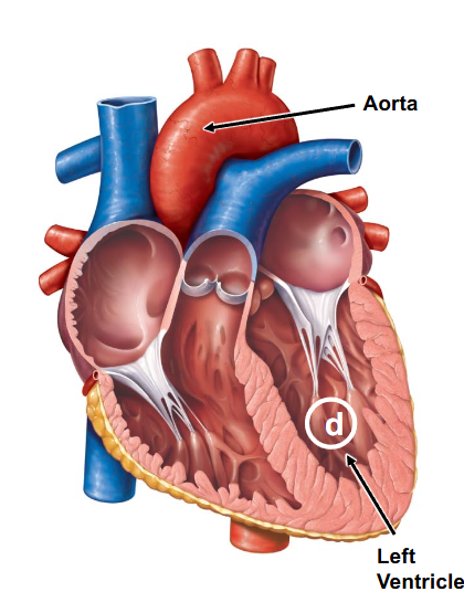
- 1 artery (the aorta) exists and carries oxygenated blood to all organ systems (this has thick walls to squeeze all blood out)
What is the order of blood chambers and where they go?
What are the two types of Septa?
- interatrial septum (separates atria)
- interventricular septum (separates ventricles)
What is the cardiac (fibrous) skeleton?
- fibrous connective tissue separating atria and ventricles
- forms solid rings around heart valves, base of aorta and pulmonary trunk, to provide structural support for these structures and hold them in place
What does the cardiac (fibrous) skeleton do?
- provides firm attachment point for cardiac muscles
- provides electrical insulation (prevents simultaneous contraction of atria and ventricles)
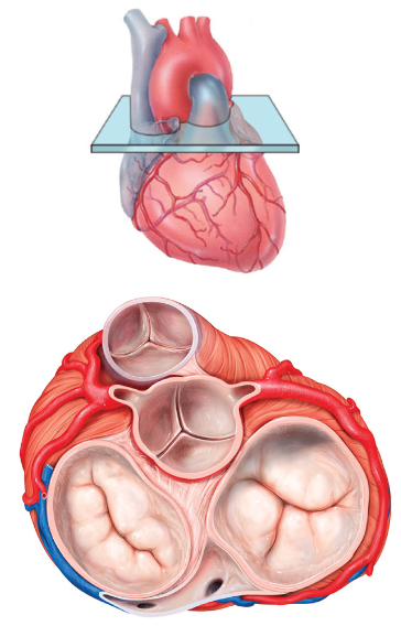
What are the types of valves?
- Atrioventricular (AV) valves, (bicuspid and tricuspid valves)
- semilunar valves (aortic and pulmonary)
What are and what are the two types of semilunar valves?
- 3 cup-like cusps each
- aortic (separates left ventricle and aorta)
- pulmonary (separates right ventricle and pulmonary trunk)
Where is the bicuspid (mitral) valve?
- between left atrium and left ventricle
- has 2 sheet-like cusps composed of CT
Where is the tricuspid valve?
- between right atrium and right ventricle (bc its always right to try)
- has 3 sheet-like cusps composed of CT
What are chordae tendineae?
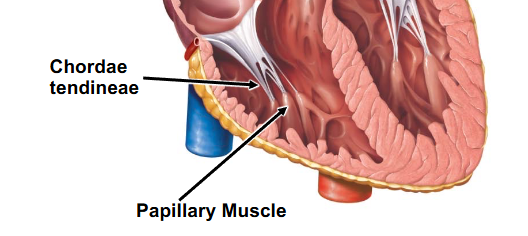
- strings of connective tissue that attach to atrioventricular valve cusps to papillary muscles that project from the ventricular myocardium
- prevents eversion of cusps
in cardiac muscle cells what cell types are there?
- contractile cells (forms majority of myocardium)
- conduction system cells (forms rest of myocardium)
How are contractile cells different and similar to skeletal muscles?
- similar: striated (myofibrils with sarcomeres), has sarcoplasmic reticulum & T-tubules
- different: branched (myofibrils with sarcomeres), uninucleated, intercalated discs (region where two fibers meet) with anchoring and gap junctions (to contract at same time)
What do conduction system cells do?
- modified cardiac muscle cells to produce and conduct electrical impulses
- they do NOT contract
- have many gap junctions that help electrical signals spread very quickly
What are the parts of conduction system cells?
sinoatrial (SA) node, atrioventricular (AV) node, Atrioventricular bundle (bundle of his), atrioventricular (AV) bundle branches, purkinje fibers
What is a sinoatrial (SA) node? Where is the atrioventricular (AV node)?
- in right atrium at base of superior vena cava
- generates impulses the fastest (sets pace)
AV: base of right atrium
what is the atrioventricular bundle (bundle of his)? What do atrioventricular bundle branches do?
- superior part of the interventricular septum
- electrically connects atria to ventricles
bundle branches: carry impulse to apex (bottom tip) of heart
What are purkinje fibers?
- terminal fibers in ventricles (not found in atria) that carry signals from apex upward to all parts of the ventricle