What are the components of the skin? (3)
Skin
Hair
Nails
What percentage of body weight does the skin make up? (1)
Approximately 15%
What is the integumentary system? (2)
It includes the skin, hair, and nails.
It is the largest organ system in the body.
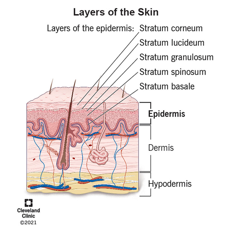
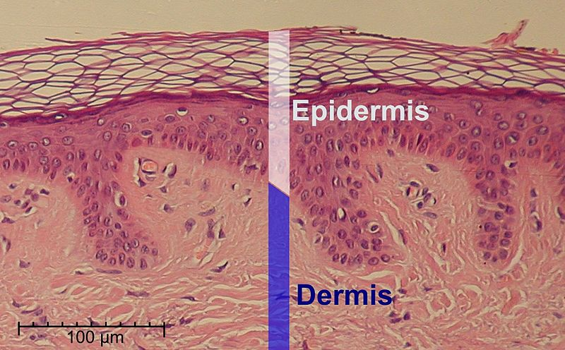
What are the three main layers of the skin? (3)
Epidermis
Dermis
Hypodermis
What structures are found in the skin? (4)
Hair
Glands
Nails
Sensory receptors
What are the main functions of the skin? (5)
Acts as a barrier against dehydration, infection, injury/abrasion, and solar radiation.
Thermoregulation.
Sensation.
Repair of damaged tissues.
Vitamin D production.
What is the epidermis? (4)
The most superficial layer of the skin.
It gives our skin its color.
Made up of keratinized stratified squamous epithelium.
Contains 4 distinct cell types and 5 layers.
What are the functions of the epidermis? (3)
Protects the skin from the external environment.
Contributes to skin color.
Acts as a site for Vitamin D production.
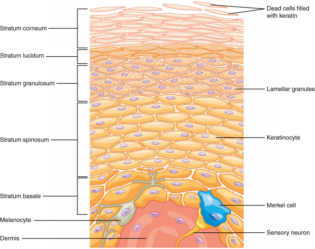
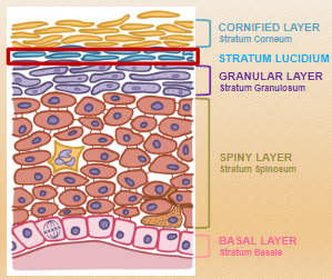
What are the five layers of the epidermis, starting from the surface? (5)

Cornified layer (Stratum Corneum)
Stratum Lucidum (only present in thick skin, e.g., palms and soles)
Granular layer (Stratum Granulosum)
Spiny layer (Stratum Spinosum)
Basal layer (Stratum Basale) --------------------------------------------------------------------------------------------------------------"Come, Let's Get Sun Burned"
Come = Corneum (Cornified layer)
Let's = Lucidum (only in thick skin)
Get = Granulosum (Granular layer)
Sun = Spinosum (Spiny layer)
Burned = Basale (Basal layer)
Which layer is only present in thick skin, such as on the palms and soles? (1)
Stratum Lucidum
What is the basal layer (stratum basale) of the epidermis? (4)
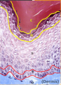
The deepest layer of the epidermis, attached to the dermis.
Contains stem cells that constantly proliferate (many mitotic nuclei are seen).
Dynamic layer: Daughter cells move upward (distally) through the epidermis, differentiating as they go.
10-25% of the cells in this layer are melanocytes.
How long does it take for epidermal cells to move from the basal layer to the outer surface? (1)

Approximately 20-50 days.
What is the spiny layer (stratum spinosum) of the epidermis? (2)

Consists of keratinocytes connected by numerous desmosomes, visible as “spines” between cells.
Strong desmosomal bonds hold the epidermis together.
What is the granular layer (stratum granulosum) of the epidermis? (4)
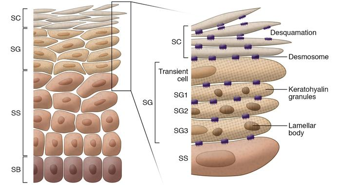
Consists of 1-4 layers of cells containing keratohyalin granules (precursors to keratin).
Contains lamellar bodies filled with lipids (visible under TEM).
Cells are differentiating to form the outermost layer.
Plays a key role in skin barrier formation.
What is the cornified layer (stratum corneum) of the epidermis? (4)
The outermost protective layer of the epidermis.
Cells are keratinized (cornified), with cytoplasm full of tough keratin derived from keratohyalin granules.
Cells are flattened and have lost their nuclei, forming a dense, protective barrier.
Nonpolar lipids between cells (from lamellar bodies) make the layer waterproof.
What are melanocytes and their function in the epidermis? (2)
Specialized cells responsible for producing melanin, the pigment that gives skin its color.
Protects underlying tissues from UV radiation by absorbing harmful rays.
What are Langerhans cells and their function in the epidermis? (2)
Immune cells that function as antigen-presenting cells (APCs).
Detect and present foreign substances to the immune system, aiding in skin defense.
What are Merkel cells and their function in the epidermis? (2)
Specialized mechanoreceptor cells associated with sensory nerve endings.
Involved in detecting light touch and contributing to the sensation of touch.
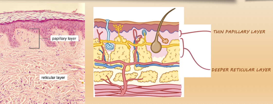
What is the dermis, and what is it made of? (3)
The dermis is the layer beneath the epidermis, composed of connective tissue.
Characterized by an interconnected mesh of elastin and collagen fibers.
These fibers are produced by dermal fibroblasts, providing strength, elasticity, and resilience to the skin.
What type of connective tissue is found in the dermis and what are its key characteristics? (3)
The dermis is made of dense irregular connective tissue.
Contains an interconnected mesh of elastin fibers, providing skin elasticity.
Rich in blood vessels, sensory receptors, and glands.
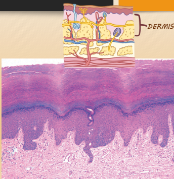
What is the function of the dermal-epidermal border? (2)
The dermal-epidermal border is wavy to resist shear forces, such as rubbing sideways.
Helps to strengthen the connection between the dermis and epidermis.
What are dermal papillae and rete ridges? (4)
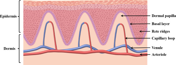
Dermal papillae are finger-like protrusions of dermal connective tissue into the epidermal layer.
Rete ridges are extensions of the epidermis into the dermal layer.
These structures interlock the dermis and epidermis for better mechanical stability.
More prominent in thick skin, such as on the hands and feet, forming fingerprints.
What does the Verhoeff-Van Gieson stain highlight in the dermis? (2)
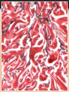
The Verhoeff-Van Gieson stain is used to stain collagen fibers.
In this stain, collagen fibers appear red.
What is the effect of UV light on the dermis? (2)
UV light leads to the loss of elastic fibers in the dermis.
This results in the formation of wrinkles and a loss of skin elasticity.
What is the structure and function of the hypodermis? (3)

The hypodermis is made up of well-vascularized, loose areolar connective tissue and adipose tissue.
It functions as a mode of fat storage.
The hypodermis provides insulation and cushioning for the integument.
What is the composition and function of the hypodermis? (4)
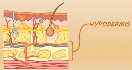
The hypodermis is composed of fat, containing glands, hair follicles, nerves, and blood vessels.
It is often the thickest layer of skin, with thickness varying based on age, body site, and nutrition.
The hypodermis provides insulation, cushioning, and energy storage.
It is the site for hypodermic needles during subcutaneous injections.
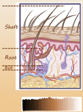
What are the components of a hair strand? (3)
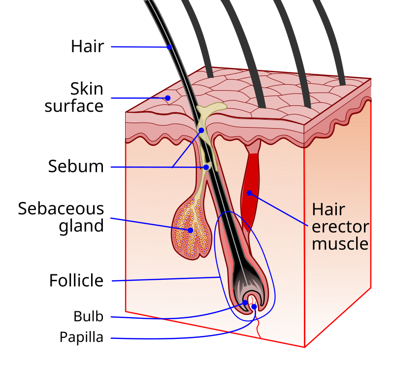
The hair strand is composed of the shaft, root, and bulb.
The bulb sits in a pouch-like structure called the hair follicle.
The hair follicle is made of epidermal tissue and extends into the dermis.
What structures are associated with the hair follicle? (4)
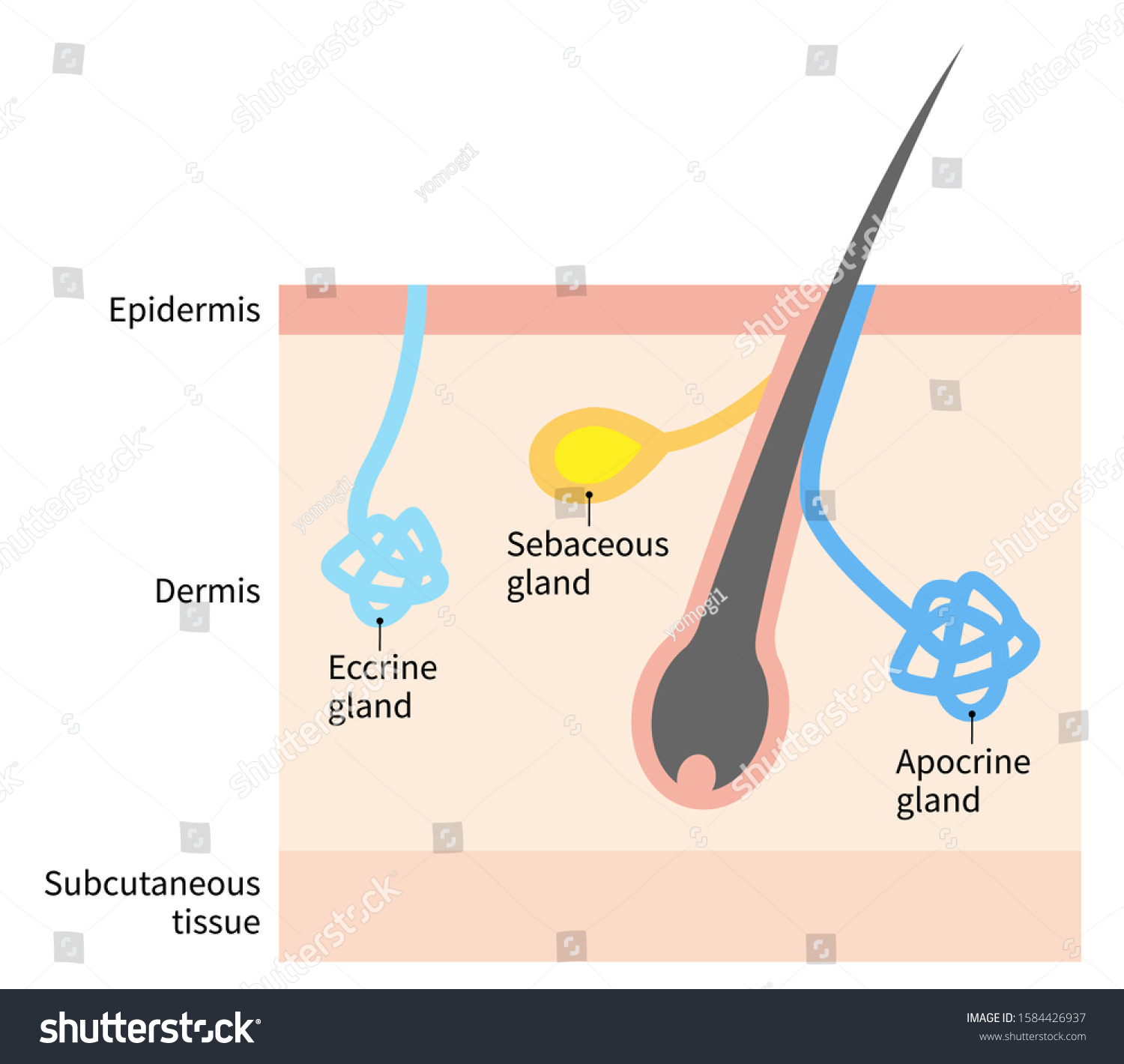
The hair follicle interacts with other structures like apocrine glands, sebaceous glands, the arrector pili muscle, and nerve receptors.
It is involved in various functions like hair growth and sensation.
Where is hair found on the skin? (1)
Hair is found on nearly every part of the skin except for the palms, soles, and lips.
What is the hair bulb and its role in hair growth? (3)

The hair bulb contains the hair matrix, which is the active site of hair growth and pigmentation.
The bulb contains two types of cells: follicular keratinocytes and melanocytes.
Follicular keratinocytes produce hard keratin, which forms the hair shaft.
What happens to follicular keratinocytes during hair formation? (2)

Follicular keratinocytes are filled with hard keratin and flatten out as they are pushed up the follicle.
This process leads to the formation of the hair root and shaft.
What causes hair loss and baldness? (2)

Follicular keratinocytes at the bulb replicate a limited number of times, after which the hair follicle stops growing and the hair falls out.
This process contributes to baldness.
Why does hair turn white as people age? (1)
Over time, melanocytes in the hair bulb stop producing melanin, which causes hair to turn white.
What is the function of eccrine sweat glands? (2)
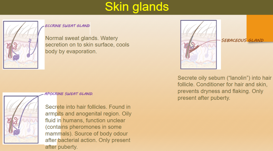
Eccrine sweat glands secrete watery sweat onto the skin surface.
This helps cool the body by evaporation.
What is the function and secretion of sebaceous glands? (3)

Sebaceous glands secrete oily sebum (also known as "lanolin") into hair follicles.
The sebum acts as a conditioner for hair and skin, preventing dryness and flaking.
Sebaceous glands are present after puberty.
What is the function of apocrine sweat glands? (3)

Apocrine sweat glands secrete an oily fluid into hair follicles.
These glands are found in the armpits and anogenital region.
The function of apocrine glands is unclear in humans but can contain pheromones in some mammals and are a source of body odor after bacterial action.
When do sebaceous and apocrine sweat glands become active? (1)

Both sebaceous and apocrine sweat glands are only present and become active after puberty.
What are nail folds and their function? (2)

Nail folds are areas where the skin seals off the edges of the nail.
They help protect the nail from external factors.
What is the eponychium and its role? (3)
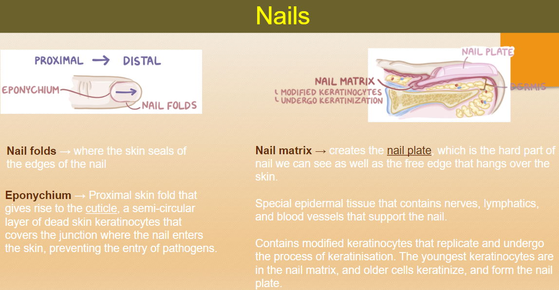
The eponychium is the proximal skin fold that gives rise to the cuticle.
It forms a semi-circular layer of dead skin keratinocytes.
The eponychium covers the junction where the nail enters the skin, preventing the entry of pathogens.
What is the nail matrix and its function? (4)

The nail matrix is responsible for creating the nail plate, the hard part of the nail we can see.
It also creates the free edge of the nail that hangs over the skin.
The matrix contains modified keratinocytes that replicate and undergo keratinization.
The youngest keratinocytes are in the nail matrix, with older cells keratinizing to form the nail plate.
What is contained within the nail matrix? (3)

The nail matrix contains nerves, lymphatics, and blood vessels.
These structures support the nail's growth and function.
It also contains modified keratinocytes that undergo keratinization to form the nail plate.
What are free nerve endings and their functions? (3)

Free nerve endings are sensory receptors found in the skin.
They detect nociception (pain), thermal sensation (temperature), and light touch.
These receptors are important for responding to environmental stimuli like temperature changes and pain.
What are Pacinian corpuscles and their function? (2)

Pacinian corpuscles are encapsulated sensory receptors.
They are rapidly adapting and detect vibration or brief touch.
What are Merkel's discs and their function? (2)
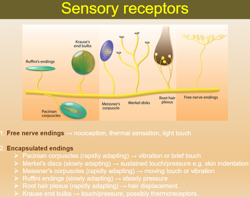
Merkel's discs are encapsulated sensory receptors.
They are slowly adapting and detect sustained touch or pressure, such as skin indentation.
What are Meissner's corpuscles and their function? (2)

Meissner's corpuscles are encapsulated sensory receptors.
They are rapidly adapting and detect moving touch or vibration.
What are Ruffini endings and their function? (2)

Ruffini endings are encapsulated sensory receptors.
They are slowly adapting and detect steady pressure.
What are root hair plexuses and their function? (2)

Root hair plexuses are rapidly adapting sensory receptors.
They detect hair displacement, sensing when hair moves.
What are Krause end bulbs and their function? (2)

Krause end bulbs are sensory receptors that detect touch and pressure.
They may also function as thermoreceptors.
What is the skin's function in preventing dehydration and which skin layer is involved? (2)
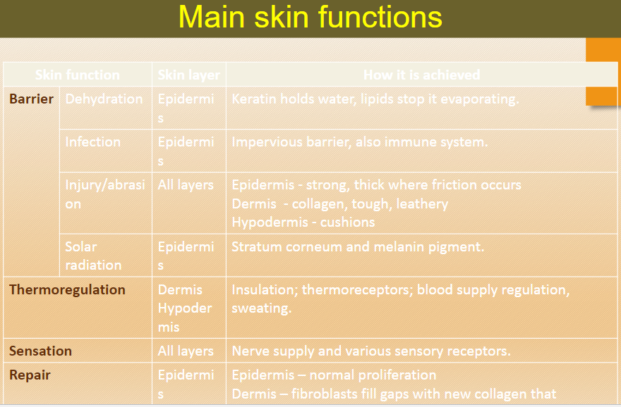
The skin prevents dehydration by using keratin to hold water and lipids to stop evaporation.
This function is achieved through the epidermis.
How does the skin protect against infection and which skin layer is involved? (2)

The skin provides a barrier that is impervious to pathogens and also involves the immune system.
This function is achieved through the epidermis.
How does the skin protect against injury and abrasion, and which layers are involved? (3)

The epidermis is strong and thick where friction occurs.
The dermis contains collagen, making the skin tough and leathery.
The hypodermis cushions and absorbs shock.
How does the skin protect against solar radiation and which skin layer is involved? (2)

The stratum corneum and melanin pigment protect against solar radiation by absorbing and blocking UV light.
This function is achieved through the epidermis.
How does the skin regulate thermoregulation and which skin layers are involved? (3)

The dermis and hypodermis help with thermoregulation through insulation, thermoreceptors, blood supply regulation, and sweating.
How does the skin sense stimuli and which skin layers are involved? (2)

The skin detects sensory information through nerve supply and various sensory receptors located in all layers of the skin.
How does the skin repair itself and which skin layers are involved? (2)

The epidermis repairs itself through normal cell proliferation.
The dermis fills gaps with new collagen using fibroblasts to promote healing.
What is the integument and what does it consist of? (2)
The integument is the skin and its appendages.
It includes hair, nails, and glands.
What are the three main layers of the skin? (3)
Epidermis: The outer layer of the skin, made of stratified, squamous, keratinized epithelium.
Dermis: The second layer, composed of fibrous, dense connective tissue.
Hypodermis (Subcutis): The third layer, consisting of fatty connective tissue.
What is the cutis and what layers does it consist of? (2)
The cutis (also called cutaneous) consists of the epidermis and the dermis.
What is keratin and what is its role in the skin? (2)
Keratin is a protein made inside keratinocytes.
It is the main component of hair, hooves, horns, nails, and the outer epidermis, giving strength and hardness.
What are keratinocytes and what is their function? (2)
Keratinocytes are epithelial cells.
They are the main cells that compose the epidermis and produce keratin.
What are melanocytes and what do they produce? (2)
Melanocytes are cells found in the epidermis.
They produce melanin, the pigment responsible for the color of the skin, hair, and eyes.
What is collagen and where is it found? (2)
Collagen is a strong, fibrous extracellular protein.
It is the main component of the dermis and is also found in leather.
What are fibroblasts and what do they do? (2)
Fibroblasts are the main cell type in the dermis and other soft connective tissues.
They generate the extracellular matrix, including collagen.
What is the function of the sebaceous gland? (2)
The sebaceous gland produces sebum, an oily substance that conditions the skin and hair.
What is the function of the eccrine sweat gland? (1)
The eccrine sweat gland produces normal sweat to regulate body temperature.
What is the function of the apocrine sweat gland and where is it found? (2)
The apocrine sweat gland produces a different type of sweat found in areas like the armpits.
This gland becomes active after puberty.