What does the somatosensory system convey? (4)
Touch
Proprioception
Heat and cold
Pain and itch
What are the types of nerves connecting the CNS to the body? (2)
Spinal nerves: 31 pairs
Cranial nerves
What is the structure of a peripheral nerve? (3)
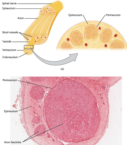
Bundle of axons ensheathed in connective tissue
Epineurium: Connective tissue covering the entire nerve
Perineurium: Connective tissue surrounding axon bundles (fascicles)
What are dorsal root ganglion cells and their role? (1)
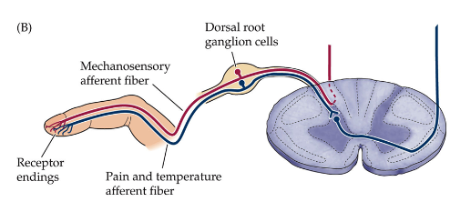
They are the sensory receptors of the somatosensory system.
What are the two subsystems of somatosensory fibres, and their functions? (2)

Large fibres:
Large diameter, myelinated, fast conduction
Responsible for tactile and proprioceptive sensations
Small fibres:
Small diameter, thinly myelinated or unmyelinated, medium or slow conduction
Responsible for temperature, pain, itch, and crude touch
How does afferent fibre type affect sensation? (2)
Specificity: Different fibres are sensitive to specific stimuli.
Example: Mechanosensitive fibres are insensitive to thermal stimulation.
Thermosensitive fibres: Respond to warming or cooling.
What is an example of thermosensitive fibre activity? (2)
Cooling the skin from 34°C to 26°C activates cold receptors.
Warming back to 34°C deactivates them.
What are the afferent types and their roles in the somatosensory system? (4)
A-α afferents:
Large diameter, myelinated, the fastest conducting (≤100 m/s)
Associated with proprioception (e.g., muscle spindles)
A-β afferents:
Large diameter, myelinated, 2nd fastest conducting (30-70 m/s)
Responsible for discriminative touch
What are the tactile afferent receptors and their classification? (4)
Superficial receptors:
Meissner’s corpuscles
Merkel’s discs
Deep receptors:
Ruffini corpuscles
Pacinian corpuscles
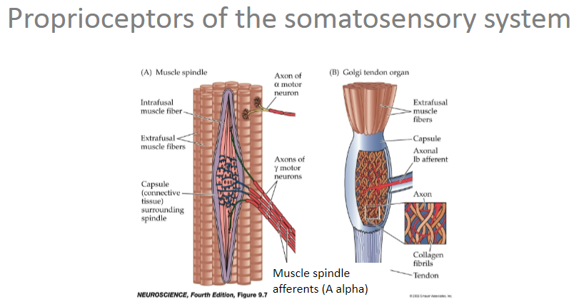
Picture demonstrating Proprioceptors of the somatosensory system:
What happens during biceps contraction? (2)

Muscle fibres shorten, decreasing the angle of the elbow joint (flexion)
Triceps is lengthened
What happens during triceps contraction? (2)
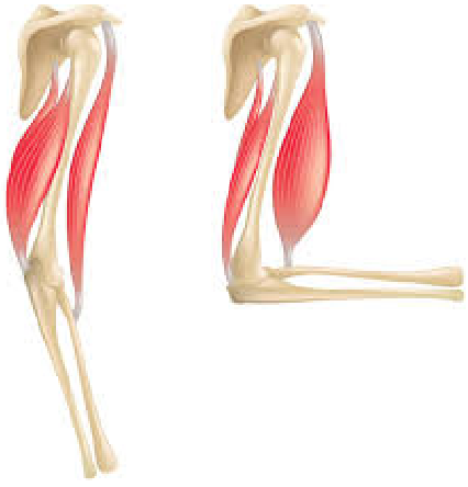
Muscle fibres contract, increasing the angle of the elbow joint (extension)
Biceps is lengthened
What are the characteristics of A-α afferents in proprioception? (3)
Large diameter
Myelinated
Fastest conduction (≤100 m/s)
What are the characteristics of A-β afferents in tactile afferents (discriminative touch)? (4)
Large diameter
Myelinated
2nd fastest conduction (30-70 m/s)
Specialized sensory endings in skin
What are free nerve endings responsible for, and what are their fibre types? (3)
Responsible for low-resolution tactile, temperature, and pain
Fibre types:
A delta fibres: Small diameter, thinly myelinated, moderate conduction velocity (≤30 m/s)
C fibres: Small diameter, unmyelinated, slow conducting (≤1 m/s)
Picture demonstrating the Cutaneous receptors of the somatosensorysystem:
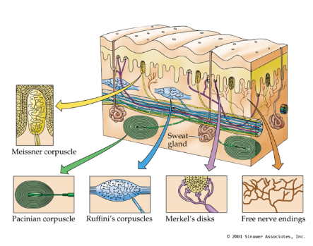
Which mechanosensory afferent classes have small receptive fields? Where are they located? (3)
Merkel's disks
Meissner’s corpuscles
Located at the dermis/epidermis border
Which mechanosensory afferent classes have large receptive fields? Where are they located? (3)
Ruffini’s corpuscles
Pacinian corpuscles
Located in the dermis
Which mechanosensory afferent classes are slowly adapting, and what stimuli do they respond to? (2)
Merkel's disks (small receptive fields): Pressure, light touch
Ruffini’s corpuscles (large receptive fields): Skin stretch
Which mechanosensory afferent classes are rapidly adapting, and what stimuli do they respond to? (2)
Meissner’s corpuscles (small receptive fields): Vibration (low frequency), light touch
Pacinian corpuscles (large receptive fields): Vibration (high frequency)
What are the two major central pathways of the somatosensory system, and what do they mediate? (2)
Dorsal column – medial lemniscal system (DCML): Mediates discriminative touch, vibration, proprioception
Spinothalamic tract (STT) (also known as the anterolateral system): Mediates coarse touch, temperature, pain
Which afferent fibres provide input to the dorsal column – medial lemniscal system (DCML)? (2)
A-β fibres
A-α fibres
Which afferent fibres provide input to the spinothalamic tract (STT)? (2)
A-δ fibres
C fibres
What is the route map of the dorsal column – medial lemniscal system (DCML)? (5)
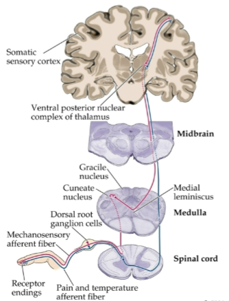
Primary afferents enter the spinal cord and ascend ipsilaterally to the brain stem.
First synapse occurs in the dorsal column nuclei of the medulla.
Second-order neurons cross over (decussate) and ascend contralaterally to the thalamus.
Second synapse takes place in the ventral posterior (VP) nuclei of the thalamus.
Third-order neurons project to the primary somatosensory cortex.
What is the route map of the spinothalamic tract (STT)? (5)
Primary afferents enter the spinal cord and terminate in the dorsal horn.
First synapse occurs in the dorsal horn of the spinal cord segment.
Second-order neurons cross over (decussate) and ascend the spinal cord contralaterally to the thalamus.
Second synapse takes place in the ventral posterior (VP) nuclei and medial thalamic nuclei of the thalamus.
Third-order neurons project to the primary somatosensory cortex.
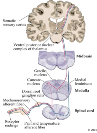
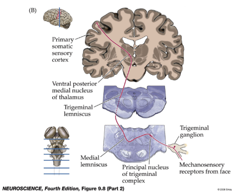
What is the route map of the somatosensory system (body)? (5)
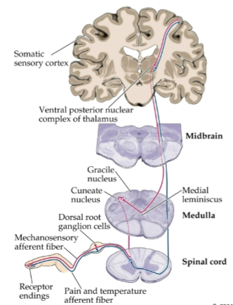
Primary afferents enter the spinal cord and ascend ipsilaterally to the brain stem.
First synapse occurs in the dorsal column nuclei of the medulla.
Second-order neurons cross over (decussate) and ascend contralaterally to the thalamus.
Second synapse takes place in the ventral posterior (VP) nuclei of the thalamus.
Third-order neurons project to the primary somatosensory cortex.
Picture demonstrating Central pathways of the somatosensory system: face and head:
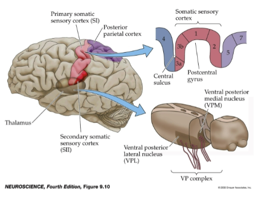
Picture demonstrating Cortical representation of tactile information:
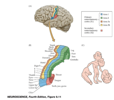
Picture demonstrating Somatotopic maps:
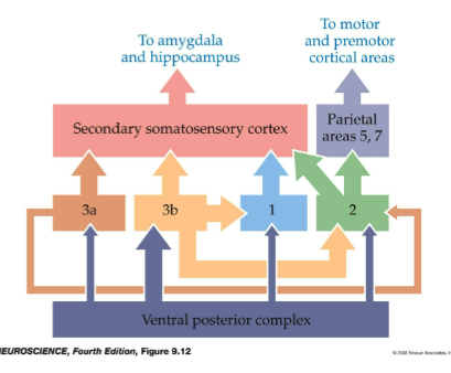
Picture demonstrating Cortical connections:
What are the key features of regional variation in cortical cytoarchitecture? (4)
Same basic cell types organized in layers across different areas of the cortex.
Regional differences are identified by variations in layer thickness, cell size, and density (cytoarchitectural differences).
Brodmann defined and numbered over 50 areas in the human cortex based on subtle cytoarchitectural differences.
Brodmann areas 1, 2, and 3 are associated with the somatosensory cortex.
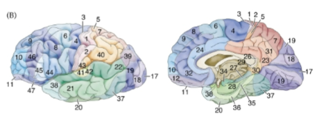
Picture demonstrating Brodmann areas:
What are A-alpha afferents (Ia) and their functions? (2)
Axons associated with muscle spindles, sensory structures within voluntary muscles.
Important for reflex control of movement and our sense of limb position.
What are A-beta afferents and their functions? (3)
Axons associated with cutaneous receptors.
Responsible for our sense of fine touch, pressure, and vibration.
What are A-delta afferents and their functions? (3)
Axons associated with free nerve endings in the skin.
May function as mechanosensory receptors (crude, poorly localized touch).
Sensitive to temperature (painful and non-painful) and noxious stimuli (potentially damaging, painful).
What are C-fibres and their characteristics? (4)
Thin, unmyelinated axons associated with free nerve endings in the skin.
Slow conducting.
Sensitive to noxious and non-noxious stimuli (chemosensory, mechanosensory, temperature).
Often multimodal in function.
What are dorsal root ganglia and their role in the somatosensory system? (2)
Ganglia located along spinal nerves, just outside the spinal cord.
Contain the cell bodies of primary afferents of the somatosensory system.