Function
-Gas exchange: Supplies oxygen (O2) and removes carbon dioxide (CO2)
Respiratory System Processes
-Ventilation
-External Respiration
-Transport
-Internal Respiration
Ventilation
Movement of air in and out of the lungs
External Respiration
Exchange of gases between the lungs and the blood
Transport
Circulation of gases from the lungs, to cells, and back to the lungs
Internal Respiration
Exchange of gases between the blood and cells of the body
Upper Respiratory System
-Outside air enters the nasal cavity through the nostrils (nares) of the nose.
Nasal Cavity
-Hollow space above the roof of the mouth lined with respiratory mucosa to warm air and trap particles
-Nasal conchae: bony projections of the nasal septum to increase surface area.
Nasal Cavity
-Paranasal sinues
-----Hollow air-filled spaces lined with mucosa surrounding the nasal cavity
-----Produce mucus, resonate sound, and lighten the skull
Nasal Cavity pt 2
-Lacrimal (tear) glands drain into nasal cavity via tear ducts
-Eustachian tube links the nasopharynx to the middle ear
Nasal Cavity pt 3
-Palate: roof of the mouth
--Separates the nasal and oral cavities
--Hard palate is anterior
--Soft palate is posterior
Pharynx
-Air travels from the nasal cavity to the pharynx ("throat")
-Common passageway for both food and air
Pharynx pt 2
-Three parts:
----Nasopharynx: Posterior to the nasal cavity
---Oropharynx: Posterior to the oral cavity
---Laryngopharynx: Posterior to the larynx
Pharynx pt 3
-Tonsils
---Made of lymphatic tissue to defend against pathogens
---Adenoids: Single pharyngeal tonsil
---Palatine tonsils: Rear of oropharynx
---Lingual tonsils: Base of tongue
Larynx
-Cartilage structure connecting the pharynx and trachea
-Thyroid cartilage: Large upper portion referred to as the "Adam's Apple"
-Cricoid cartilage: Inferior to the thyroid cartilage
Larynx pt 2
-Contains the vocal cords ("voice box").
---Glottis: opening between vocal folds
---Epiglottis: flap of elastic cartilage that covers the glottis when swallowing.
Lower Respiratory System
--The trachea and bronchi constitute the lower respiratory conducting tract.
--Actual gas exchange occurs in the respiratory unit
Trachea
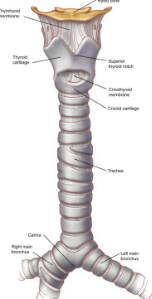
Tube that connects the larynx to the primary bronchi
-Lined w/ pseudostratified ciliated columnar epithelium to help trap particles and send them back up to the throat.
-Held open by C-shaped rings of cartilage
--Rings can be felt anteriorly
--Opening of "C" is posterior so food can move down the esophagus
Bronchioles
-Trachea divides into smaller and smaller branches
-Bronchioles: Are the smallest conducting airways
---Contain ciliated epithelium and smooth muscle
---Constrict w/ asthma
-Bronchioles lead to alveoli
Alveoli
-Functional unit of the respiratory system
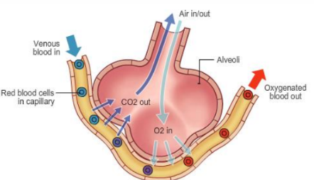
-Exchange of gases occurs here
-Compromised of simple squamous epithelium
-Lined w/ surfactant to help maintain surface tension
-Surrounded by capillaries
Lungs
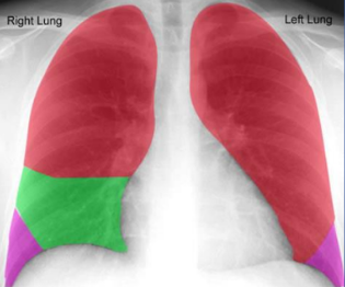
-Comprise most of thoracic cavity
-Mediastinum comprises the rest of the thoracic cavity
---Apex is superior (near the clavicle)
---Base is inferior resting upon the diaphragm
---Left lung is comprised of two lobes in order to make room for heart
---The right lung is comprised of three lobes
Respiratory Surface
-Large surface area (500-800 sq ft)
----Equivalent to one side of a tennis court
-Plasma membranes must be moist
-O2 and CO2 must first dissolve to diffuse across the simple squamous epithelial membrane.
Pleural Space
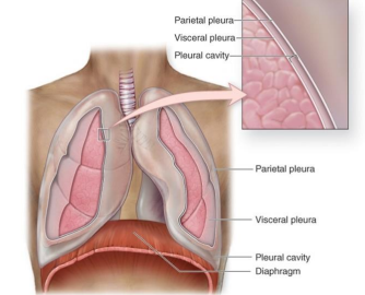
-Lungs are lined w/ visceral pleura
-Cavity is lined w/ parietal pleura
-There is high surface tension between the visceral and parietal pleura which enables the lungs to expand
Respiratory Volumes
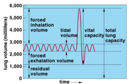
Measurement of pulmonary function
Respiratory Volumes
-Total lung capacity
-Tidal volume
-Vital capacity
-Forced inhalation volume
-Forced exhalation volume
-Residual volume
-Dead space air
Total Lung Capacity
Maximum amount of air that the lungs are able to hold (~6 liters)
Tidal Volume
Volume air that moves in and out w/ each breath (~1/2 liter)
Vital Capacity
Maximum volume of air that can be forced in and out of the lungs in a single breath (~4.5 liters)
Forced Inhalation Volume
(Inspiratory Reserve Volume): Amount of air that can be inhaled beyond the tidal volume (~3 liters).
Forced Exhalation Volume
(Expiatory Reserve Volume): Amount of air that can be exhaled beyond the tidal volume (~1.5 liters)
Residual Volume
Air that always remains inside the lungs (so they don't collapse) (~1.3 liters)
Dead Space Air
Air in the passageways that do not reach alveoli (~30%)
-To ensure that air reaches alveoli it is best to breath slowly and deeply.
Breathing Control Centers
The regulation of the respiratory rate is controlled by the brain stem
-Medulla oblongata controls basic breathing function
-Pons adds rhythm and control
Breathing Control Centers pt 2
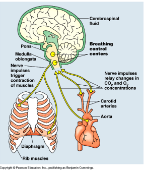
-The brain stem is mostly sensitive to levels of CO2 (not levels of oxygen) for the regulation of the respiratory rate.
-The medulla oblongata and pons monitor the carbon dioxide and pH levels in the blood and cerebrospinal fluid.
-Normal blood pH is 7.4
Acidotic
-Carbonic acid levels are increased and the blood becomes ACIDOTIC (pH <7.35)
----Cause: Hypoventilation
----Condition: Emphysema
----Response: Respiratory rate increases (hyperventilation) to rid the body of excess CO2
Alkalotic
-Carbonic acid levels are reduced and the blood becomes ALKALOTIC (pH > 7.45)
----Cause: Hyperventilation
----Condition: Anxiety
----Response: Respiratory rate decreases (hypoventilation) to increase the levels of CO2
Gas Exchange and Transport
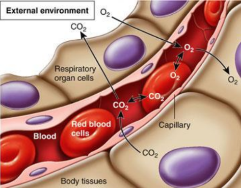
External Respiration
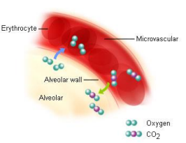
-Gases are exchanged along their concentration gradients between the air in the alveoli and the blood in the capillaries
---Oxygen diffuses from the alveoli and into the blood (loading)
----Condition dioxide diffuses from the blood and into the alveoli (unloading)
Internal Respiration
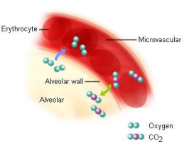
-Gases are exchanged along their concentration gradients between the blood in the capillaries and the cells in the body.
----Oxygen diffuses from the blood and into the cells (loading)
----Carbon dioxide diffuses from the cells and into the blood (unloading)
Transport
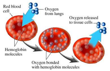
-Transport of respiratory gases
----Oxygen is carried from the lungs to the tissues
----Carbon dioxide is carried from the tissues to the lungs
Oxygen Transport
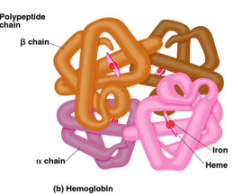
-Oxygen combines with the iron portion of hemoglobin in the red blood cells (RBC)
-Hemoglobin (HgB): iron containing pigment in RBC
-Each HgB molecule can carry 4 oxygen molecules; there are about 250,000 of HgB in a RBC; therefore, each RBC carries about 1,000,000 oxygen molecules.
Carbon Dioxide Transport
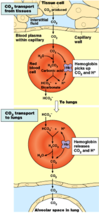
-Most carbon dioxide is transported in the blood plasma as bicarbonate ion (HCO3-)
-When oxygen enters the blood stream from the alveoli, it is converted through a RBC to (HCO3-) and then enters the plasma for transport.
-When the blood returns to the lungs, (HCO3-) is converted back to CO2 through the RBC and enters the alveoli to be exhaled.
Anatomy of Respiratory System
-Nostrils (Nares)
-Nasal Cavity
-Pharynx (throat)
-Larynx (Glottis)
-Trachea (windpipe)
-Bronchi
-Bronchioles
-Alveoli
All the "tubes" are just conducting pathways to alveoli- purify, humidify, warm the air before it gets to the respiratory surface.
Trachea
-Trachea divides into right and left primary bronchi for each lung
-Primary bronchi divide into secondary bronchi for each lobe of each lung
-Secondary bronchi divide into smaller tertiary bronchi
-Bronchioles are smallest conducting airways
----Carbon ciliated epithelium and smooth muscle
----Constrict w/ asthma
-Bronchioles lead to alveoli
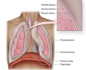
Lungs
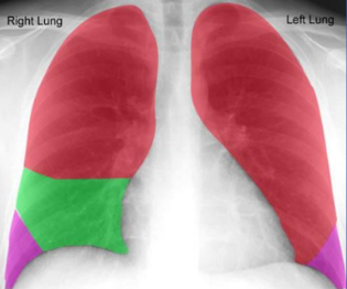
-Comprise most of thoracic cavity (except mediastinum)
-Apex is close to clavicle while the base is at the diaphragm
-2 lobes in left lung (make room for heart), 3 in right
-Enclosed by double sac filled w/ fluid
-----Hollow surface tension between the visceral pleura (next to lung) and parietal pleura (next to body wall).
Pulmonary Ventilation
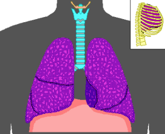
Ventilation ("breathing"):
-Movement of air IN and OUT of the lungs
Gas Exchange- Concentrations
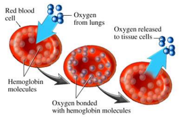
-In lungs (external respiration): oxygen concentration in alveoli is high while oxygen concentration in blood is low; so, oxygen diffuses into blood (loading)
----Response: CO2 is simultaneously unloading at this time (from blood into alveoli) due to its concentrations.
Gas Exchange - Concentrations pt 2
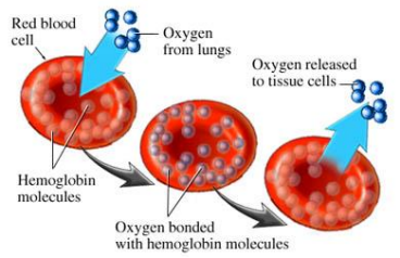
-In tissues (internal respiration): oxygen concentration in cells is low while oxygen concentration in blood is high; so, oxygen diffuses out of blood capillaries and into tissues (unloading).
---Note: CO2 is simultaneously loading at this time (from tissues into blood) due to its; concentrations.
Ultimately Supports Cellular Respiration
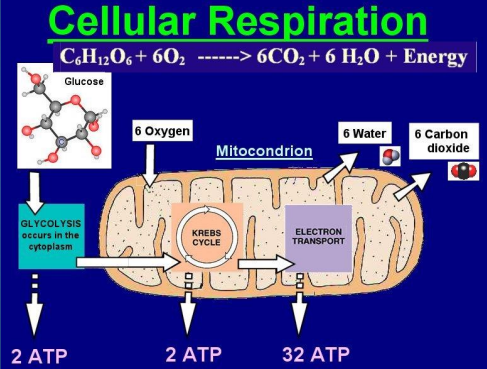
Oxygen Transport
-Hemoglobin (HgB): iron containing pigment in RBC.
-There are four subunits of the hemoglobin molecule
-Each subunit has heme group cofactor w/ iron in the center.
-Oxygen combines w/ the iron portion of hemoglobin.
-Each HgB molecule can carry 4 oxygen molecules; there are about 250,000 of HgB in a RBC; therefore, each RBC carries about 1,000,000 oxygen molecules.
What is the name for the entrance to the nasal cavity?
Nares
Name the structures (2) that separate the nasal and oral cavities.
Hard palate and soft palate
Name three ways that the nasal cavity prepares inhaled air for gas exchange.
1.) Warm
2.) Moisten
3.) Filter
What structure separates the nasal cavity into two sides?
Nasal septum
What structures give the nasal cavity increased surface area?
Nasal conchae
What covers these structures
Mucous membranes
Name two paranasal sinuses and give three functions of sinuses.
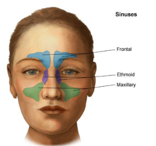
-Frontal Sinus
-Maxillary Sinus
Produce mucus, lighten skull, resonate sound
What is the atomic name for the throat?
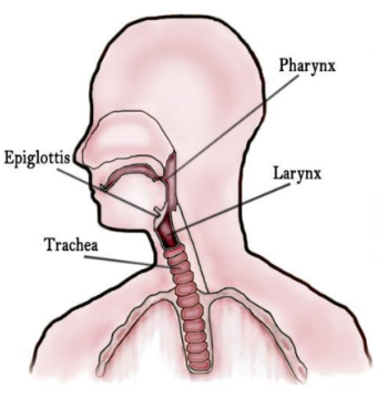
Pharynx
Name the two cartilages that make up the larynx.
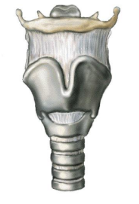
-Thyroid cartilage
-Cricoid cartilage
Name and describe the three sections of the throat.
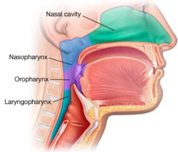
-Nasopharynx
-Oropharynx
-Laryngopharynx
Name the structure that covers the vocal cords when swallowing
Epiglottis
Name the space between the vocal cords.
Glottis
What type of wave is sound?
Longitudinal
What is the technique used to dislodge objects stuck in the trachea?
Heimlich maneuver
Name the tissue that, lines the throat
Stratified squamous epithelium
Name the tissue that comprises the larynx
Hyaline cartilage
Name the tissue that comprises the epiglottis
Elastic cartilage
Name the tissue that comprises the vocal cords
Elastic cartilage
Name the tissue that covers the vocal cords
Mucous membranes
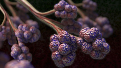
What is the functional unit of the respiratory system?
Alveoli
Name the type of tissues that comprises the lining of the trachea
Pseudostratified ciliated columnar epithelium
Name the type of tissue that comprises the rings of the trachea
Hyaline cartilage
Describe what is meant by conducting passageway.
Portions of the respiratory system that allow air to travel from outside the body to the alveoli, gas exchange does not occur here
What part of the lower respiratory system is affected by asthma and what makes these tubes so susceptible?
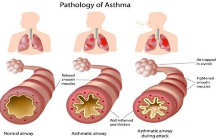
Bronchioles are most affected because they are made of only smooth muscle without cartilage rings for support.
What is the function of the respiratory system?
Exchange gases
What is the name for the type of breathing mammals do?
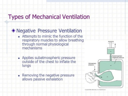
Negative pressure breathing
Hpoventilation may be caused by______ and causes a (n) _______ in levels of CO2, which gives a pH reading that is more _________.
-Emphysema
-Increase
-Acidotic
Name and describe the four processes involved in the function of the respiratory system,
Ventilation- breathing
External respiration- exchange of gases between lungs and capillaries
Transportation- movement of gases throughout the body
Internal respiration- exchange of gases between cells and capillaries
Name the portion of the brain that controls respiratory: rate and rhythm
1.) Medulla Oblongata
2.) Pons
Name the part of blood that carries oxygen
Red blood cell (RBC)
Name the part of the blood that carries CO2.
Plasma
What is the name of the form that CO2 is converted to in order to travel throughout the body?
Bicarbonate ion
What is the name for air that remains in the respiratory system in order to hold open the passageways?
Residual volume
Describe "dead-space" air
Air moved in and out of the respiratory system that does not get exchanged
Describe three things that help to allow air to enter the lungs
-Rib elevation
-Diaphragm contraction
-Open trachea
-Decrease in thoracic pressure
-Increase in thoracic volume
Describe the difference between total lung volume and vital capacity.
Total lung volume is the full amount of air that the respiratory system can hold.
Vital capacity is the maximum amount of air that can be inhaled and exhaled.
Name five measurements from a pulmonary function test.
-Tidal volume
-Forced inhalation (IRV)
-Forced exhalation (ERV)
-Vital capacity
-Residual volume
Name the non-respiratory movement where a deep breath is taken, the glottis is closed, and air is forced against the glottis clearing the upper respiratory passageway.
Sneeze
Name the membranes associated with the lungs
Visceral pleura
Parietal pleura
What is the force called that allows the lungs to "stick" to the ribs as they expand?
Surface tension
What type of membranes line the lungs and lung cavity?
Serous membranes
What happens if the membranes are separated by air or fluid?
Collapsed lung
Identify if the following conditions affect the Upper Respiratory System, or the Lower Respiratory System:
1. Emphysema
2. Tonsilitis
3. Otitis media
4. Pneumonia
5. Laryngitis
1. Lower
2. Upper
3. Upper
4. Lower
5. Upper
True or false- There is no "safe" way to smoke.
True
T or F -Smoking affects the heart
True
T or F- Chewing tobacco is a safe alternative to smoking
False
T or F - Wherever nicotine touches cells it can cause cancer
True