What are the two main types of cells in the human brain? (2)
Neurons
Glial cells
What is the "reticular theory," and who proposed it? (2)
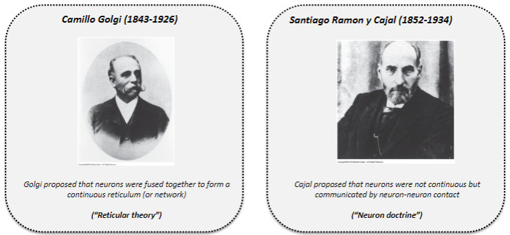
Proposed by Camillo Golgi (1843–1926).
Theory states that neurons are fused together to form a continuous network (reticulum).
What is the "neuron doctrine," and who proposed it? (2)

Proposed by Santiago Ramón y Cajal (1852–1934).
Doctrine states that neurons are not continuous but communicate via neuron-neuron contact.
How can neural tissue be visualized? (2)

Sectioning: A microtome is used to cut slices from a block of embedded brain tissue.
Fixation: Brain tissue is fixed for preservation and then embedded (e.g., in paraffin or frozen).
What is the Golgi stain, and what does it stain? (2)

Comprised of a silver chromate solution.
Stains neurons and their projections.
What is the Nissl stain, and what does it stain? (2)
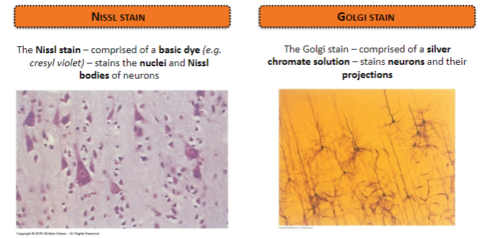
Comprised of a basic dye (e.g., cresyl violet).
Stains the nuclei and Nissl bodies of neurons.
What evidence definitively supported the neuron doctrine? (2)

The resolving power of the electron microscope.
Neurons were shown to be discrete, individual units.
What is the limit of resolution for standard light microscopy and how does it relate to neuron spacing? (2)

Resolution limit: 0.1 μm.
Neuron spacing: Approximately 0.02 μm (or 20 nm), below the resolution of light microscopy.
How does electron microscopy improve our understanding of neuron structure? (2)

Resolution limit: 0.1 nm, allowing visualization of structures smaller than 20 nm.
Provides insights into the fine structure of neurons.
What did Cajal's neural circuitry drawings demonstrate? (1)

Neurons are discrete and not continuous, supporting the neuron doctrine.
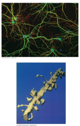
What are "Brainbow" mice, and how are they visualized? (2)

Definition: Genetically engineered mice where individual neurons or glial cells are labeled with fluorescent proteins.
Visualization techniques: Fluorescence microscopy and genetic manipulation (e.g., Cre-Lox system).
What brain regions can be studied using "Brainbow" mice? (2)
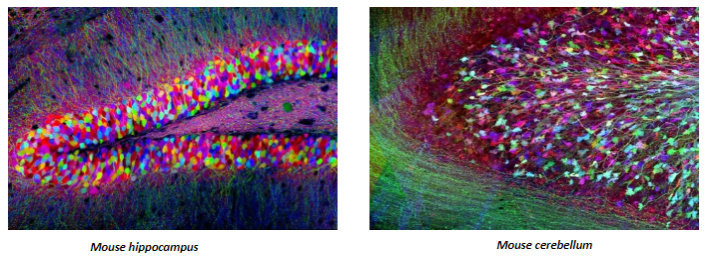
Mouse hippocampus.
Mouse cerebellum.
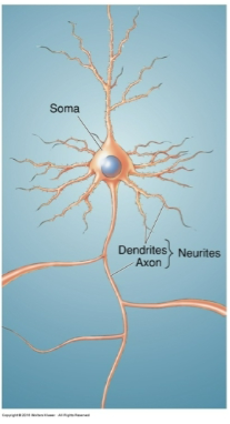
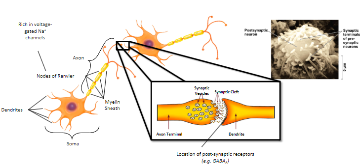
What are the main components of a prototypical neuron? (3)

Cell body (soma).
Axon.
Dendrites.
What is the function of neurons? (2)
Information processing cells within the nervous system.
Specialized for the conduction and transmission of electrical and chemical signals.
What organelles are found in the cell body (soma) of a neuron? (5)
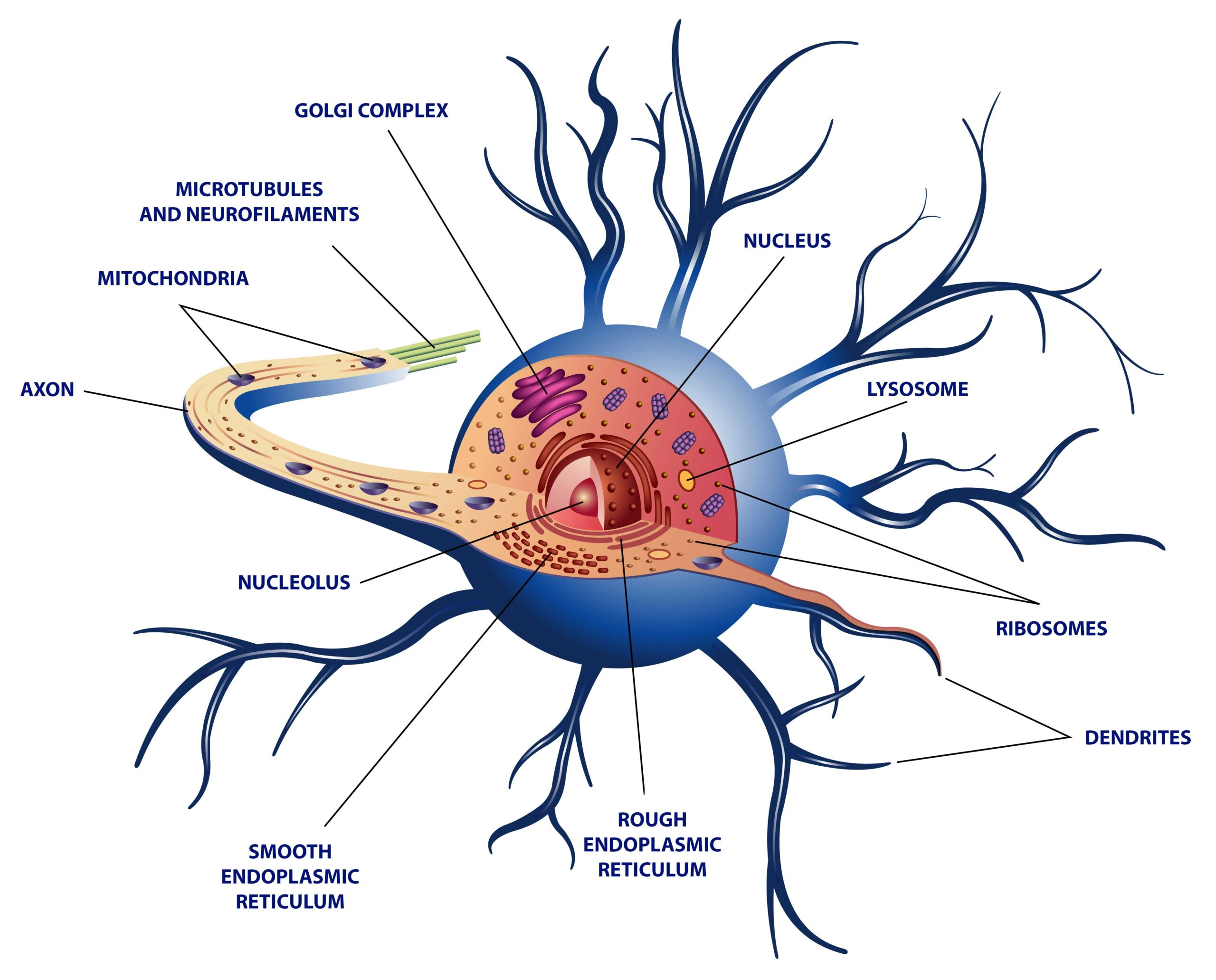
Nucleus.
Rough endoplasmic reticulum (RER).
Smooth endoplasmic reticulum (SER).
Golgi apparatus.
Mitochondrion.
What is the cytoskeleton of a neuron, and what are its components? (4)

The cytoskeleton is the internal ‘scaffolding’ that gives a neuron its shape.
Comprised of:
Microtubules.
Microfilaments.
Neurofilaments.
What are microtubules, and where are they located in neurons? (2)
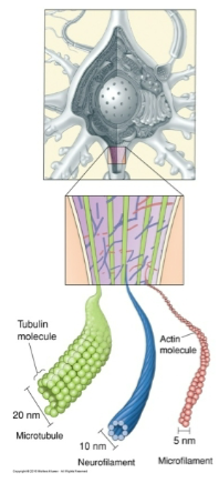
Microtubules are polymers of the protein tubulin.
Located in axons and dendrites, important for axoplasmic transport.
What are microfilaments, and where are they abundant in neurons? (2)

Microfilaments are polymers of the protein actin.
Found throughout the neuron but particularly abundant in axons and dendrites.
What are neurofilaments, and why are they significant? (3)

Neurofilaments are a type of intermediate filament.
Abundant in axons, important for regulating axonal shape, through crosslinks.
Promising biomarker for neurodegenerative disorders (e.g., Alzheimer’s).
What is the structure of an axon and its key regions? (4)
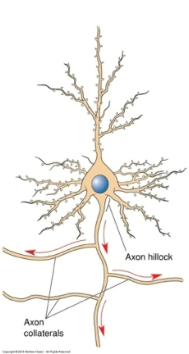
Axon Hillock: Tapers away from the soma to form the initial segment of the axon.
Axon 'Proper': The main portion of the axon, which can branch to form axon collaterals and recurrent collaterals.
Axon Terminal: The site where the axon comes into contact with other neurons at a synapse.
What is myelin, and what is its function? (2)

Myelin is a membranous sheath that wraps around and insulates axons.
It speeds up the conduction of nerve impulses (action potentials).
What are the Nodes of Ranvier, and what is their role? (2)

Gaps in the myelin sheath along the axon.
Highly enriched in voltage-gated Na+ ion channels, facilitating faster transmission of action potentials.
Which cells are responsible for myelinating axons? (1)
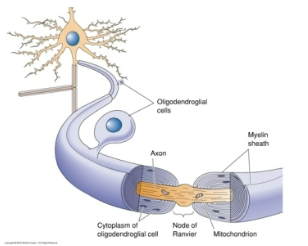
Glial cells are responsible for myelinating axons.
What are dendrites and their function? (2)

Dendrites are highly specialized neuronal projections that receive synaptic inputs from other neurons.
They are integral to transmitting signals from other neurons to the cell body
What is a dendritic tree? (1)
The dendrites of a single neuron collectively form the dendritic tree.
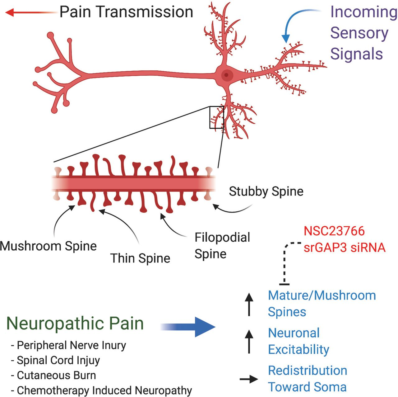
What are dendritic spines, and what is their role? (2)

Dendritic spines are small sacs of membrane that protrude from the dendrites of some cells.
They receive synaptic input and play a crucial role in synaptic signaling.
How does dendritic spine structure relate to synaptic activity? (1)

The structure of dendritic spines is sensitive to the type and amount of synaptic activity, which can influence their formation and function.
What conditions have been associated with abnormal dendritic spine numbers? (2)

Alzheimer’s disease.
Schizophrenia.
What is the fundamental function of neurotransmission? (1)
Neurotransmission is the fundamental process that drives information transfer between neurons and their targets.
How can neurons be classified based on the number of projections? (3)
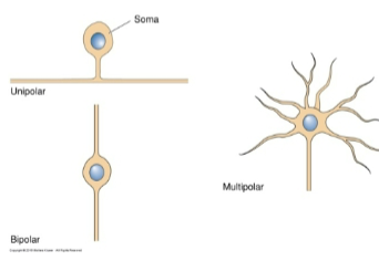
Neurons can be classified by the total number of projections (or neurites), which includes: Unipolar, Bipolar and Multipolar
How can neurons be classified by their dendritic trees and spines? (1)
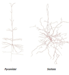
Neurons can be classified by their dendritic trees and dendritic spines into categories such as stellate and pyramidal neurons.
How can neurons be classified based on their connections? (1)

Neurons can be classified based on their connections into sensory neurons, motor neurons, and interneurons.
How can neurons be classified by axon length? (1)
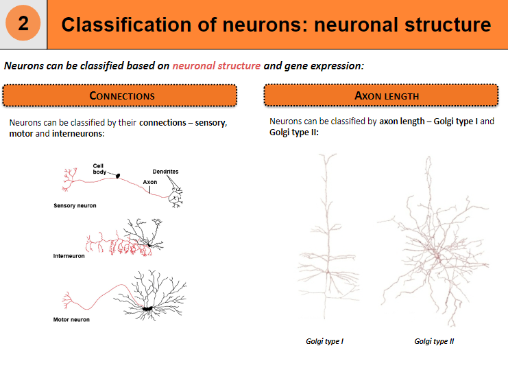
Neurons can be classified by axon length into Golgi type I and Golgi type II neurons.
What is the difference between Golgi type I and Golgi type II neurons? (2)

Golgi type I neurons have long axons that extend over long distances, such as motor neurons.
Golgi type II neurons have short axons, typically confined to local areas, such as interneurons
How can neurons be classified based on gene expression? (1)
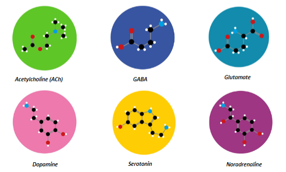
Neurons can be classified by the neurotransmitter they use, which results from the differential expression of proteins involved in neurotransmitter synthesis, storage, and release.
What are some neurotransmitters that neurons can use for classification based on gene expression? (6)

GABA
Dopamine
Serotonin
Acetylcholine (ACh)
Noradrenaline
Glutamate
What are glial cells, and what is their primary function? (1)
Glial cells are the “support cells” within the nervous system that assist neurons in various ways, including structural support, nutrient supply, and waste removal.
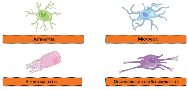
How are glial cells classified? (4)

Glial cells can be classified into four categories based on structure and function:
Oligodendrocytes/Schwann cells
Astrocytes
Microglia
Ependymal cells
What are astrocytes, and what is their shape? (1)

Astrocytes are star-shaped glial cells in the brain.
What is the primary function of astrocytes in the brain? (2)
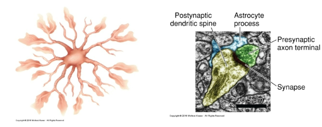
Astrocytes regulate the extracellular environment of the brain by enclosing synaptic junctions and removing neurotransmitters from the synaptic cleft.
How numerous are astrocytes in the human brain? (1)

Astrocytes are the most numerous type of glial cell in the human brain.
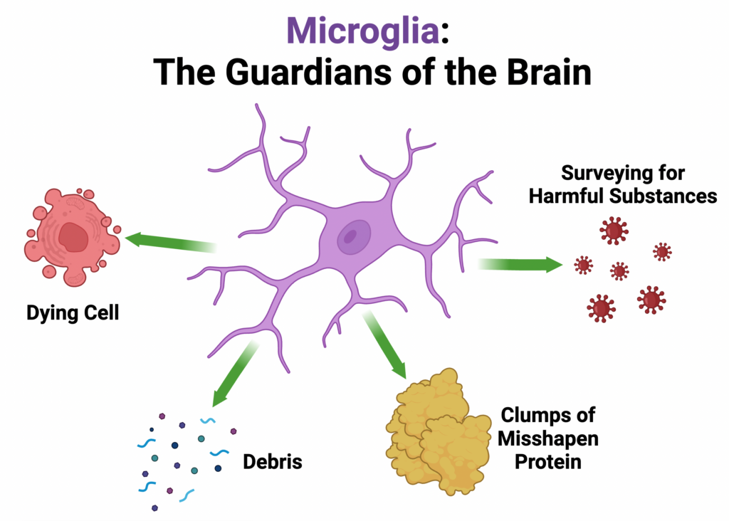
What are microglia, and what is their function? (2)

Microglia are glial cells that function as phagocytes within the nervous system to remove neuronal and glial debris.
What percentage of total CNS cells do microglia account for, and where are they distributed? (2)
Microglia account for 5-15% of total CNS cell number, depending on the anatomical region, and are distributed throughout the brain and spinal cord.
What roles do microglia play in the nervous system? (3)

Phagocytosis of neuronal and glial debris (e.g., at injury sites).
Synaptic connection remodeling.
Directing neuronal migration during brain development.
What are ependymal cells, and where are they located? (2)

Ependymal cells are a type of glial cell that provide the lining of the ventricular system in both the brain and spinal cord.
What is the function of ependymal cells? (3)
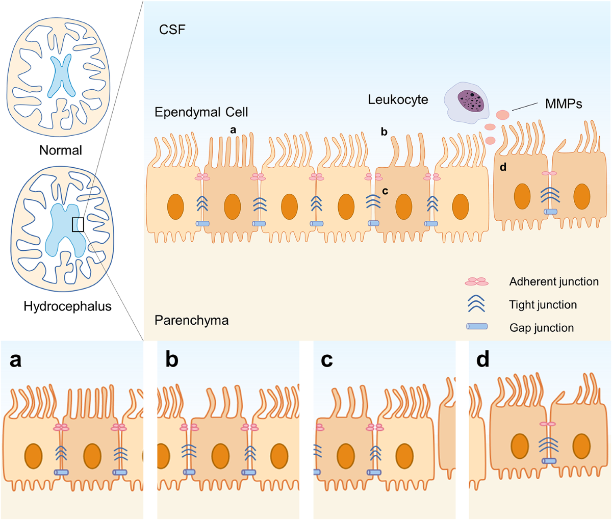
They act as a physical barrier separating brain tissue from cerebrospinal fluid (CSF).
They regulate osmotic balance of cerebrospinal fluid.
They assist in the flow of cerebrospinal fluid and direct cell migration during brain development.
What condition is linked to deficits in ependymal cell function? (1)

Hydrocephalus.
What are oligodendrocytes and Schwann cells, and what is their function? (2)

Oligodendrocytes and Schwann cells are glial cells that provide myelin, a membranous sheath around axons.
Myelin helps speed up nerve signal transmission.
How do oligodendrocytes and Schwann cells differ in myelination? (2)

Oligodendrocytes myelinate several axons in the central nervous system (CNS).
Schwann cells myelinate a single axon in the peripheral nervous system (PNS).
Where are oligodendrocytes and Schwann cells located? (2)
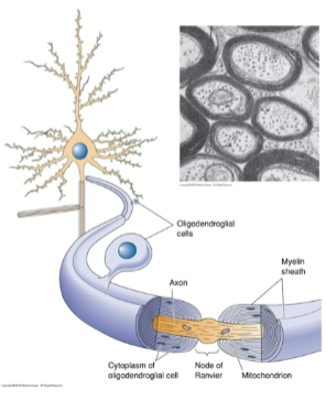
Oligodendrocytes are located in the central nervous system (CNS).
Schwann cells are located in the peripheral nervous system (PNS).
Compare and contrast the function of oligodendrocytes and Schwann cells (5)
Oligodendrocytes myelinate multiple axons in the CNS, Schwann cells myelinate one in the PNS.
Oligodendrocytes are in the CNS, Schwann cells are in the PNS.
Oligodendrocytes have multiple processes; Schwann cells wrap around one axon.
Both increase signal conduction speed via myelin.
Schwann cells aid PNS regeneration; oligodendrocytes have limited CNS regeneration.
What is anterograde transport? (1)
A cellular process responsible for moving substances from the cell body to distal parts of a cell.
What is an aspinous neuron? (1)
A neuron lacking dendritic spines.
What is an astrocyte? (1)
A star-shaped glial cell of the central nervous system.
What is an axon? (1)
The long thread-like part of a neuron that conducts impulses from the cell body to other cells.
What is an axon collateral? (1)
Branches off a neuron’s main axon.
What is the axon hillock? (1)
The site in the soma where membrane potentials are summed before being transmitted to the axon.
What is an axon terminal (bouton)? (1)
The enlarged, club-shaped endings where axons form synaptic connections with other neurons or effector cells.
What is axoplasmic transport? (1)
A cellular process responsible for the movement of substances from the soma down the axon.
What is a bipolar neuron? (1)
A neuron with two neurites extending from the soma.
What is the cell body? (1)
The nucleus-containing part of a cell.
What is cytoplasm? (1)
The material or protoplasm within a living cell, excluding the nucleus.
What is the cytoskeleton? (1)
A microscopic network of protein filaments and tubules in the cytoplasm, giving cells shape and coherence.
What is cytosol? (1)
The aqueous component of the cytoplasm, within which organelles and particles are suspended.
What is a dendrite? (1)
A short, branched extension of a neuron that transmits impulses received from other cells.
What is a dendritic tree? (1)
The structure formed by multiple dendrites from the same neuron.
What is a dendritic spine? (1)
A small membranous protrusion from a neuron’s dendrite that typically receives input from a single axon at the synapse
What is a glial cell? (1)
Supporting cells of the nervous system; includes astrocytes, oligodendrocytes, and Schwann cells.
What is an interneuron? (1)
A neuron that transmits impulses between other neurons.
What are microglia? (1)
Immune cells in the CNS that function as macrophages, removing debris.
What is a terminal bouton? (1)
The specialized presynaptic terminal at the end of an axon.