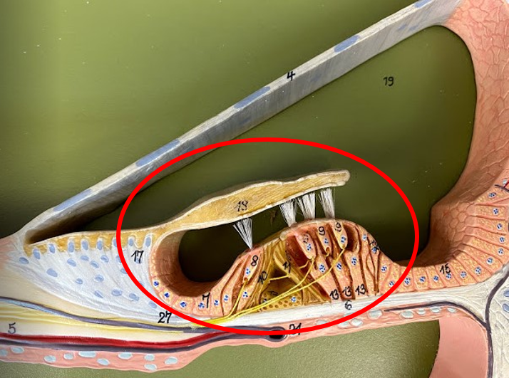
Organ of Corti / Spiral Organ
- part of the Cochlear Model
- includes
- the Basilar Membrane
- the Hair Cells
- the Tectorial Membrane
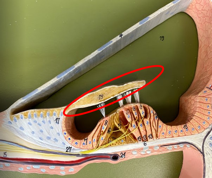
Tectorial Membrane
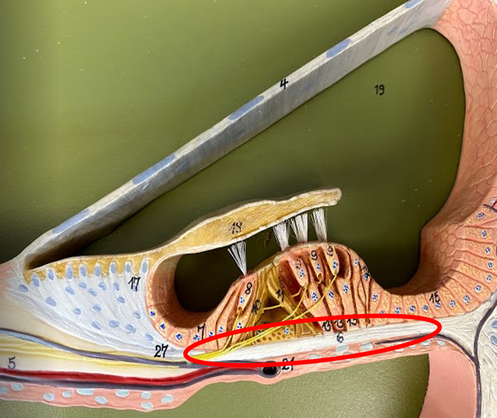
Basilar Membrane
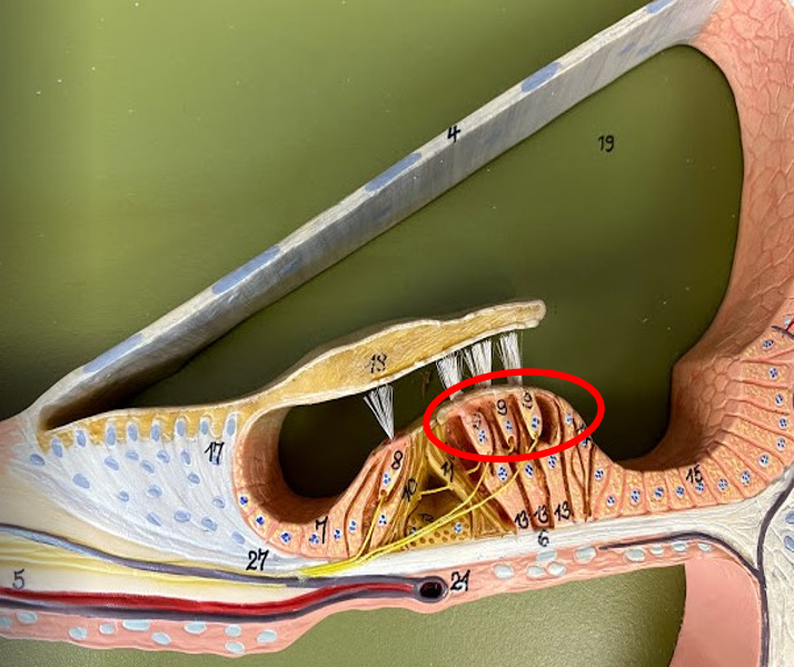
Hair Cells
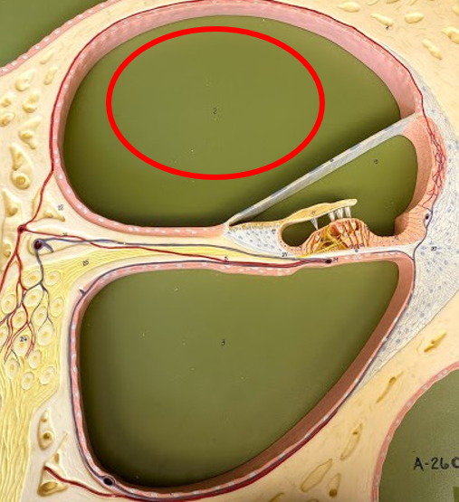
Vestibular Duct
- filled with Perilymph fluid (outer fluid)
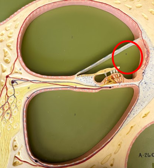
Cochlear Duct
- filled with Endolymph fluid (inner fluid)
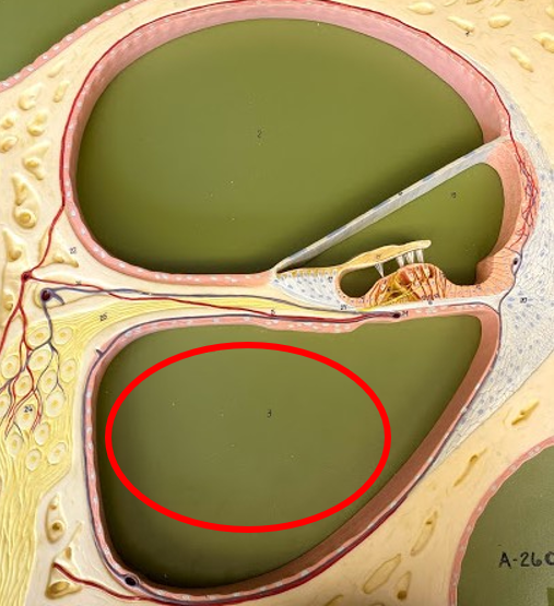
Tympanic Duct
- filled with Perilymph fluid (outer fluid)
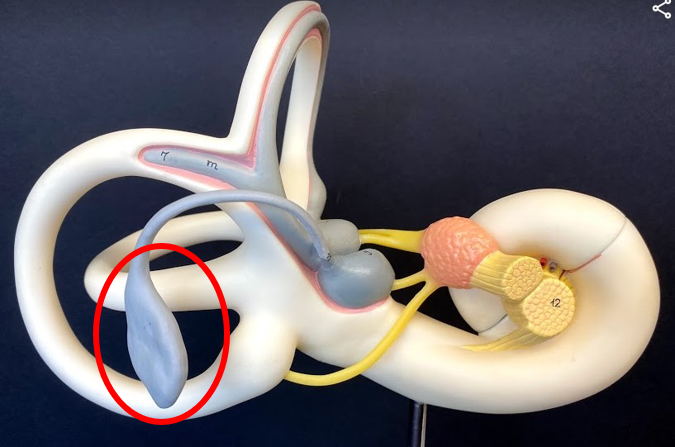
Endolymph Sac
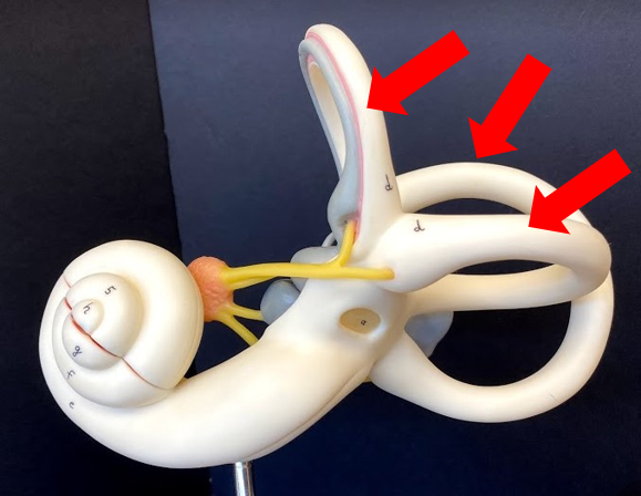
Semicircular Canals
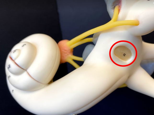
Oval Window
- where the Stapes is attached and vibrates the oval window membrane.
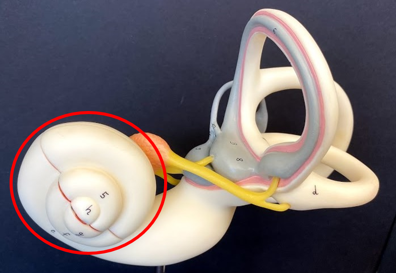
Cochlea
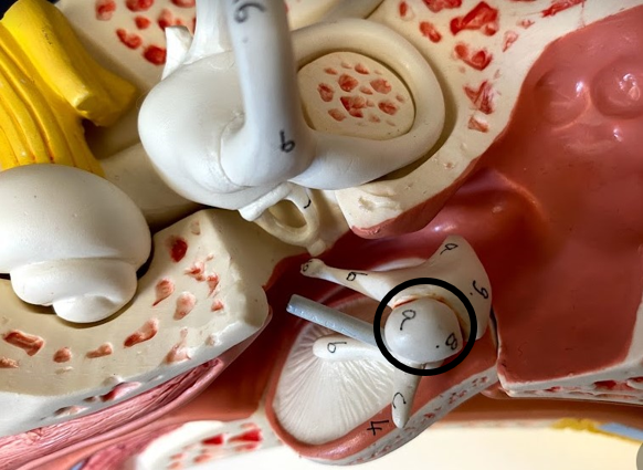
Malleus
- first auditory ossicle
- attached to the tympanic
membrane
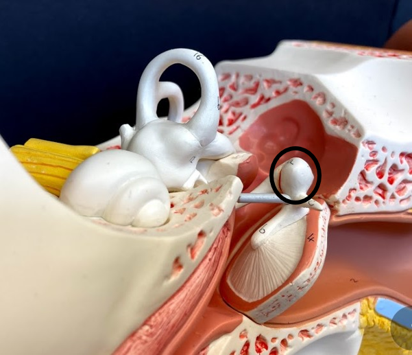
Malleus
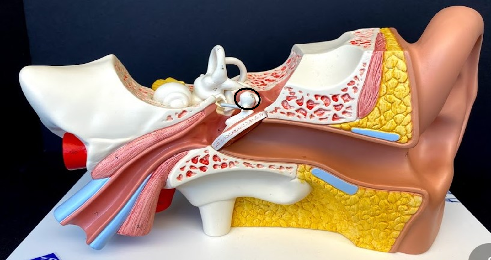
Malleus
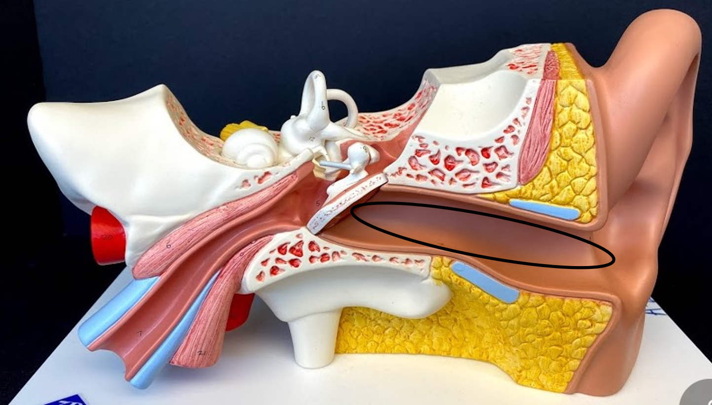
External Auditory Canal
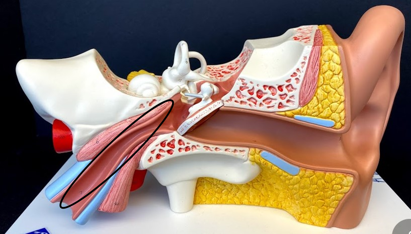
Auditory Tube/ Pharyngotympanic Tube/ Eustachian Tube
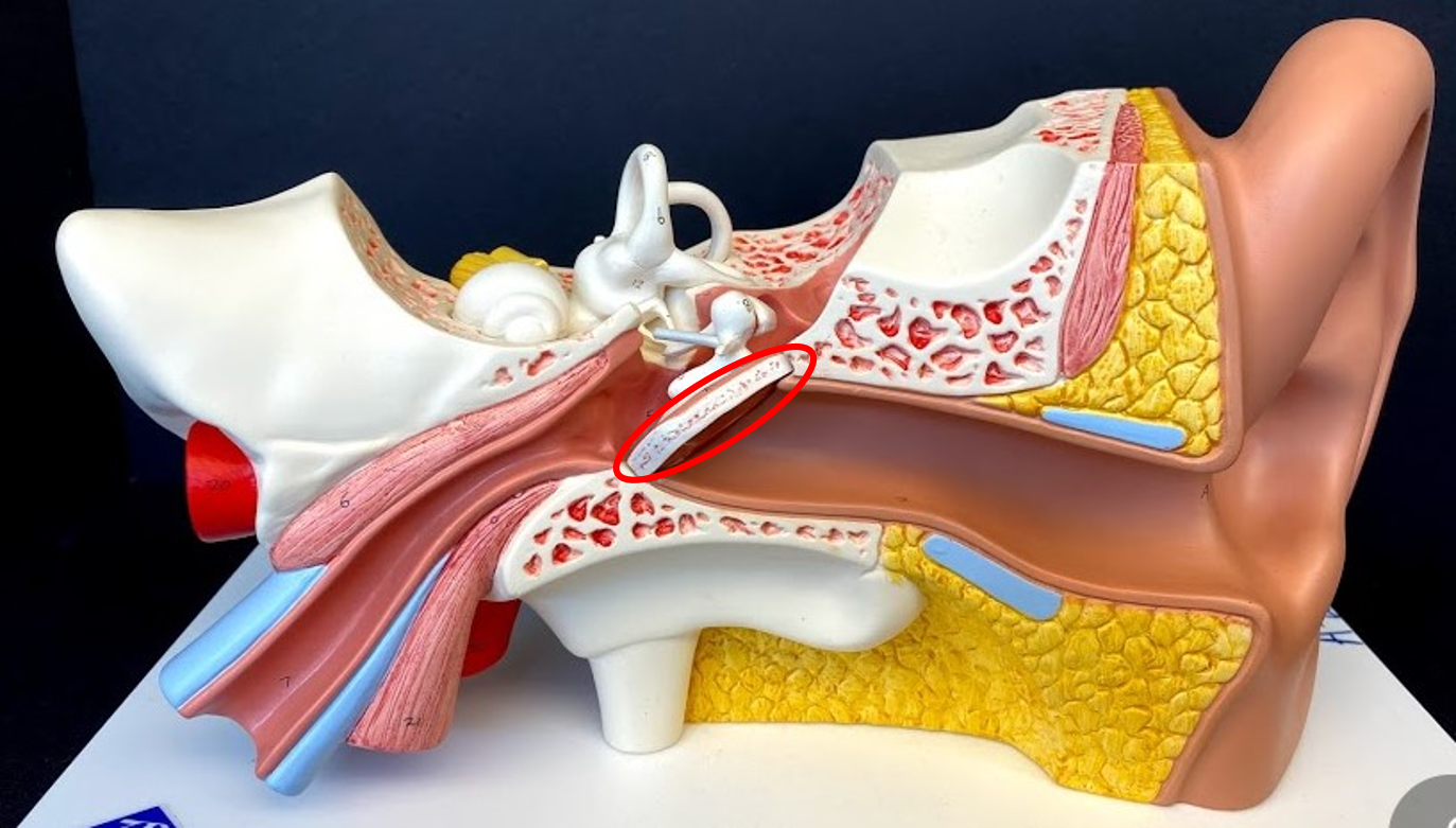
Tympanic Membrane
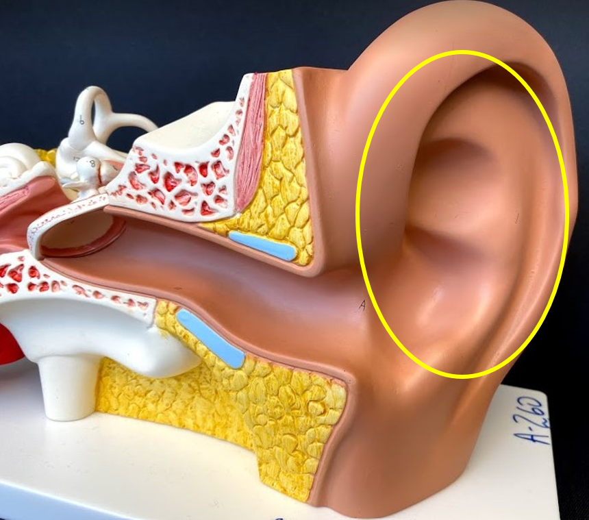
Auricle/ Pinna
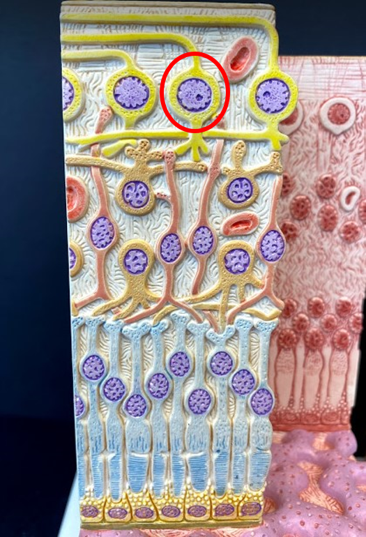
Ganglion Cells
- form Optic Nerve II
- get stimulated by the bipolar
cells which are signaled by the
photoreceptors
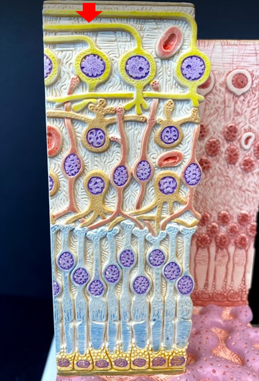
Optic Nerve II
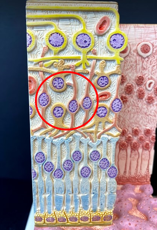
Bipolar Cells
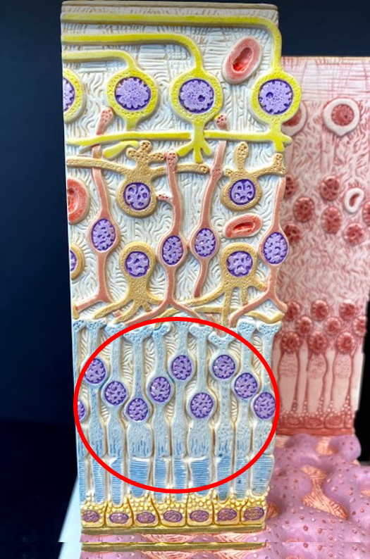
Photoreceptors
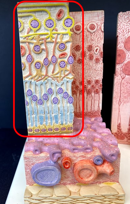
Retina layer
- inner/ deepest part of the eye
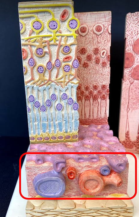
Choroid layer
- middle part of the eye
- with all of the blood vessels
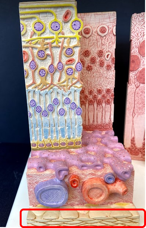
Sclera
- outer part of the eye
- the white part
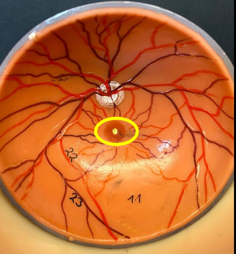
Fovea Centralis
- within the Macula Lutea
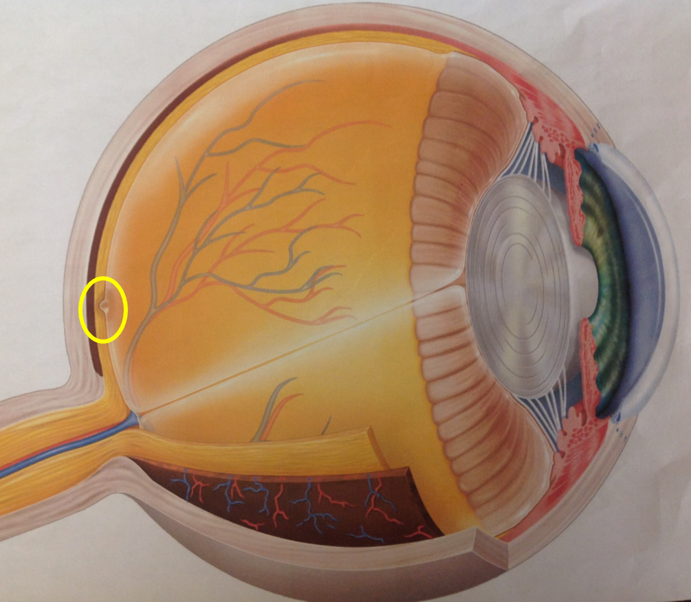
Fovea Centralis
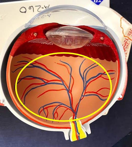
Posterior Cavity
- filled with Vitreous Humor
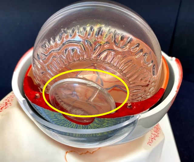
Lens
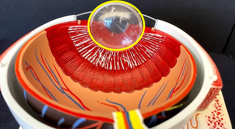
Lens
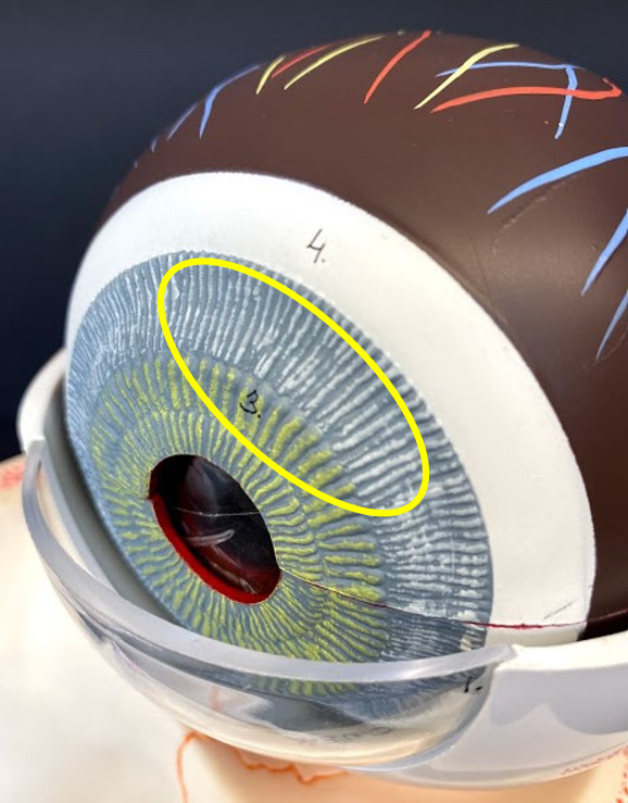
Iris
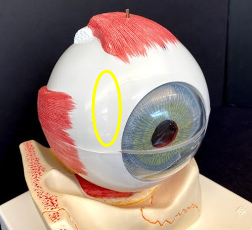
Sclera
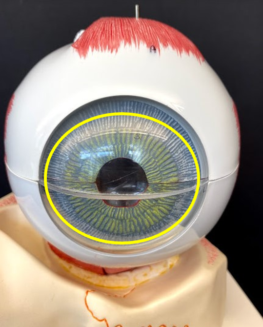
Cornea
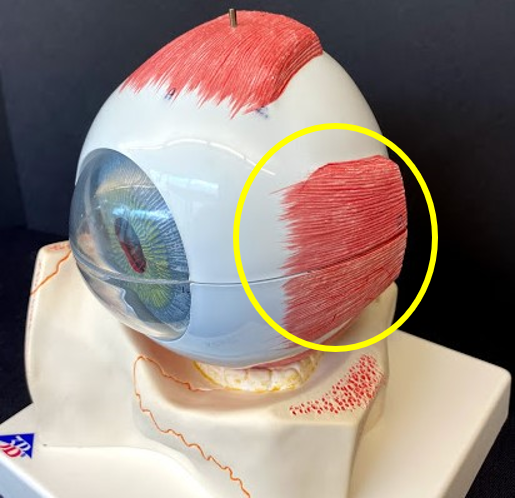
Lateral Rectus
- pulls the eye laterally
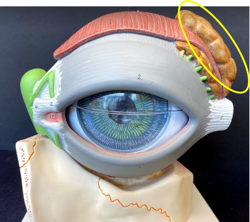
Lacrimal Gland
- makes tears
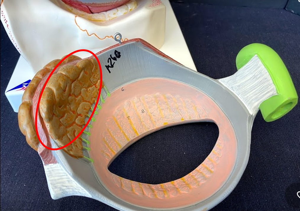
Lacrimal Gland
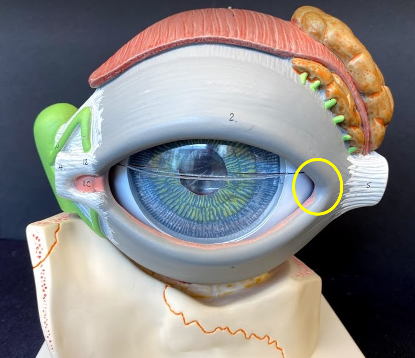
Lateral Canthus/ Lateral Commissure