gapeworm
advertisement
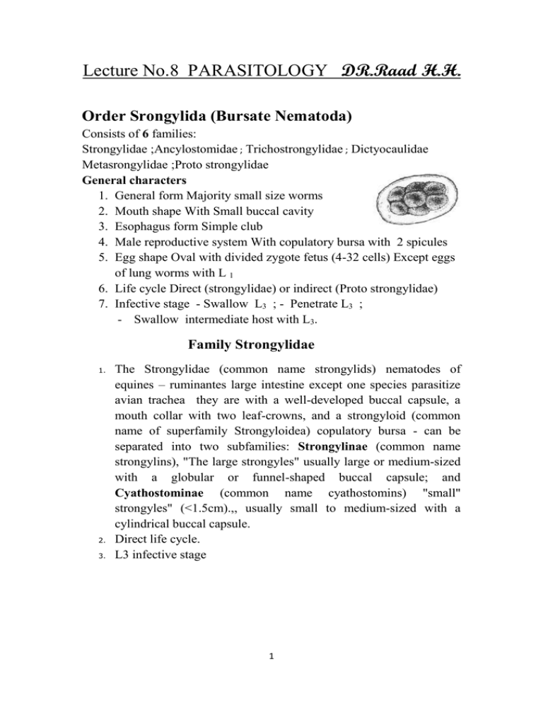
Lecture No.8 PARASITOLOGY DR.Raad H.H. Order Srongylida (Bursate Nematoda) Consists of 6 families: Strongylidae ;Ancylostomidae ; Trichostrongylidae ; Dictyocaulidae Metasrongylidae ;Proto strongylidae General characters 1. General form Majority small size worms 2. Mouth shape With Small buccal cavity 3. Esophagus form Simple club 4. Male reproductive system With copulatory bursa with 2 spicules 5. Egg shape Oval with divided zygote fetus (4-32 cells) Except eggs of lung worms with L 1 6. Life cycle Direct (strongylidae) or indirect (Proto strongylidae) 7. Infective stage - Swallow L3 ; - Penetrate L3 ; - Swallow intermediate host with L3. Family Strongylidae 1. 2. 3. The Strongylidae (common name strongylids) nematodes of equines – ruminantes large intestine except one species parasitize avian trachea they are with a well-developed buccal capsule, a mouth collar with two leaf-crowns, and a strongyloid (common name of superfamily Strongyloidea) copulatory bursa - can be separated into two subfamilies: Strongylinae (common name strongylins), "The large strongyles" usually large or medium-sized with a globular or funnel-shaped buccal capsule; and Cyathostominae (common name cyathostomins) "small" strongyles" (<1.5cm).,, usually small to medium-sized with a cylindrical buccal capsule. Direct life cycle. L3 infective stage 1 Genus Strongylus Characterized by : 1. Species members infects equine cecum and colon. 2. Red or brown colored short small size called Blood Worms, Red Worms. 3. usually with large or medium-sized globular or funnel-shaped buccal capsule with teeth except S. S. edentatus There are three major species of the large strongyles: Strongylus vulgaris S. edentatus S. equinus Note the large buccal capsule and the leaf crowns (C) at the anterior end. There is a dorsal "gutter" (D) with a bifid dorsal tooth (B) at its base and two smaller ventral teeth (A) at the base of the buccal capsule. 2 Species S. equinus S. edentatus Strongylus vulgaris Length up to 5 cm up to 4 cm up to 2.5 cm in No teeth Pair of Ear shape dorsal teeth Larvae penetrate the intestine and migrate via the portal vein to the liver, where moulting occurs. After 9 weeks, the L4 wander in the peritoneum, causing the formation of nodules. Migrating to the large intestine, the strongyle larvae form nodules in the gut wall, which they rupture to enter the colon lumen. The third-stage larvae penetrate the intestinal mucosa, where they moult to L4 in several days. Penetrating nearby blood vessels, the larvae wander through the arteries for 2 weeks before reaching the anterior mesenteric artery, where they remain for 4 months. After moulting to immature adults, S. vulgaris returns via the arteries to the large intestine and burrows into the lumen. cecum and colon and ileoceco- anterior mesenteric artery . About 7 months after the original infection eggs begin to pass in the faeces. S. vulgaris is the most pathogenic; roughening of the arterial walls, leading for Clotting , Thrombosis. Aneurisms, Verminous arteritis; aneurisms may burst, causing death. The majority of Verminous Colic in horses has been attributed to the lesions caused by the migratory stages . Pair of teeth Triangular shape in base of buccal cavity plus one dorsal divided large tooth Infective stage After burrowing into the submucosa and moulting to L3 and life L4, these parasites migrate cycle to the liver where they wander for 6-7 weeks. Emerging from the liver, they molt to immature adults in various abdominal organs, then return to the large intestine. Common in spring , winter season. Teeth no. Site infection cecum and colon cecum and colon adult maturation period Pathogenecity About 9 months after infection mature adults lay eggs. cause liver damage , hepatitis and peritonitis About 10 months after infection mature adults lay eggs. cause liver damage , hepatitis and peritonitis 3 Genus Trichonema Characterized by: 1. includes more than 35 spp. 2. Buccal cavity without teeth. 3. Spp. moderate length ;male 1cm; female 2 cm. 4. Habitats equine cecum and colon. 5. Life cycle similar to Strongyles and larvae may tend to capsulated about 2 months in colon mucosa leading to sever inflammations . Genus Triodontophorus Characterized by 1. Spp. moderate length ;male 1cm; female 2 cm. 2. Habitats equine cecum and colon. 3. Buccal cavity at base with 3 pairs of teeth. 4 Strongylidae Worms Importance In Equine ( Srongylosis ): 1. Adult large strongyles are "plug feeders," which feed by ingesting plugs of mucosa and capillaries. Heavy feeding of this type produces intestinal damage, anaemia, fluid loss into the intestine, and blood protein loss due to sucking blood and intestinal tissue by its large buccal cavity and its digestive and anticoagulant enzymatic secretions . 2. Young age horse (Foals) more susceptible and highly prevalent infection (80-95% ) due to that worms cause severe intestinal mucosa irritations and bleeding leading to death. 3. Large age horse more resist in spite of harboring highly burden of these worms and called "carrier hosts" although Older horses are often observed to have arterial lesions and lameness without a history of specific signs. 4. The acute signs associated with S. vulgaris are due to migrating larvae and are seen during the first few weeks after infection. The severity of these is related to the number of larvae ingested and undoubtedly to the age (particularly yearlings) and previous experience of the host. marked increase in temperature, loss of appetite, rapid loss of body weight, depression and recumbency , abdominal distress, constipation, or occasionally, intermittent diarrhea with foul smell. Most animals died 14 to22 days after infection. 5. Larval migration inside intestinal mucosa form inflamed infiltrated nodules then fibrosis. 6. The most pathogenic spp. Is S.vulgaris due to induction of sudden infection onset symptoms expressed by colic due to clots and thrombosis in ileo-ceco- anterior mesenteric artery and bursting leading to death due to internal hemorrhage. Diagnosis : 1. 2. 3. 4. Symptoms. Fecal ; dung ,flotation exam. for eggs. Larval identification by Fecal culture. Rectal exam. To explore ileo-ceco- anterior mesenteric artery aneurysms. 5. P.M. particularly equine cecum & colon. 5 Control : 1. 2. 3. 4. 5. 6. Mare breeding away from foals. Overcrowding must be avoided. Feed exposure to sun light to kill larvae. Avoid early morning or before sunset grazing. Deworming application to animals. Treatment with an effective anthelmintic is necessary. In particular, since foals are very susceptible, brood mares should be treated and moved to clean pastures. 7. Removal of faeces & manure . 8. Ploughing to break up dense ground cover. 9. Frequent faecal checks will aid in Strongylus control. Genus Chabertia 1. Chabertia spp. members are commonly called the large-mouthed bowel worm. In domestic animals, its predilection site is the colon of sheep and goats and it is occasionally seen in cattle. 2. It has a worldwide distribution but it tends to be more common in temperate areas of the world. 3. Chabertia is one of the easiest ruminant nematodes to identify because of its size (1-2cm long), location (colon) and its prominent, curved, bell-shaped buccal capsule which lacks teeth. 4. Buccal cavity provide with 2 raws of chitinos teeth called Cuticular elements .male coupulatory spicules large . Chabertia ovina Head - both sexes Tail 6 5. L3 infective stage . 6. The life cycle is direct with the preparasitic phase similar to the Trichostrongyles. After ingestion by the final host, L3s exsheath in the small intestine, penetrate the mucosa and molt to L4s. These emerge, gather in the cecum, molt to immature adults (L5s) and pass on the colon to mature. The prepatent period is approximately 6 weeks. 7. Pathogenic effects are caused by the feeding adults which become attached to the mucosa and draw a plug of mucosa into the buccal capsule which is then digested. This results in area of mucosal ulceration and local hemorrhage with protein loss into the gut through these lesions. In heavy infections, the feeding effects of 200-300 adult worms results in a colon that is edematous and thickened with local areas of hemorrhage where the worms were attached. Some reports claim that larvae and immature adults are blood suckers 8. Adults of the large-mouth bowel worm e.g. Chabertia ovina, are ~12 mm long and bent ventrally at the anterior end. There is a typical direct life cycle. The larvae penetrate the mucosa of the small intestine shortly after ingestion and later emerge and pass to the colon. The prepatent period is ~7 wk. Larvae and adults may cause small hemorrhages with edema in the colon and passage of feces coated with mucus ; Infected sheep pass soft faeces and brown mucus containing flecks of blood . Clinical chabertiasis is seldom, if ever, seen in cattle. 9. A specific diagnosis is not usually possible in live animals for the reasons mentioned above. At necropsy the worms are readily identified from their location, size and shape of the buccal capsule. 1. DIAGNOSIS Eggs in faeces are similar to those of other nematodes. Worms identified at P.M. or from enema sample. EGG C. ovina Oesphagostomum venulosum 7 Genus Oesphagostomum 1. Members of this genus are known as the "nodular worms" because they are associated with nodule formation in the intestines of their hosts. They are common parasites of ruminants, pigs, primates and rodents colon . The species found in domestic animals are often of pathogenic importance 2. Adults are 1-2cm long. The buccal capsule is relatively shallow and the head end is distinctive because of the cephalic inflations of the cuticle with Leaf crowns plus cephalic alae . Males have a bursa and the egg passed by the female worms is a strongyle-type egg. Note the small buccal capsule and the leaf crowns (C) at the anterior end. There is a dorsal "gutter" (D) with a bifid dorsal tooth (B) at its base and two smaller ventral teeth (A) at the base of the buccal capsule. 3. Clinically Young animals suffer from the effects of adult worms, whereas in older animals, the effect of the nodules is more important. Infection causes anorexia; severe, constant, dark, persistent, fetid diarrhea; weight loss; and death. In older, resistant animals, the nodules surrounding the larvae become caseated and calcified, thus decreasing the motility of the intestine. Stenosis or intussusception occasionally occurs. Nodules can be palpated per rectum, and the worms and nodules can be seen readily at necropsy. 4. Diagnosis as in Chabertia ovina 8 5. P.M. look for nodules in large intestine of sheep. Syngamus trachea 1. This is the gapeworm of poultry, found in the trachea of chickens, turkeys, guinea fowl and many species of wild birds. It is of particular importance in farm-raised pheasants. Although Syngamus is found throughout the world, conversion of domestic poultry raising from outdoors to indoors has significantly reduced its prevalence in the United States. 2. The most distinctive feature of this nematode is that males and females are joined together in a state of permanent copulation forming a Y shape as seen in the image on the right. Females are much larger (up to 20mm long) than males (up to 6mm long). 3. Buccal cavity with 6-10 small teeth. 4. The life cycle is complicated in both its preparasitic and parasitic phases. In the preparasitic phase L3s develop inside the eggs at which time they may hatch. Earthworms play an important role in the life cycle, serving as transport (paratenic) hosts. Larvae have been shown to remain viable for more than three years encapsulated in earthworm muscles. Other invertebrates may also serve as paratenic hosts and these include terrestrial snails and slugs as well as the larvae of Musca domestica (the common house fly) and Lucilia sericata (the greenbottle fly responsible for cutaneous myiasis). The 9 parasitic phase involves substantial migration in the definitive host to reach the predilection site. 5. Pathogenesis Young birds are most severely affected with migration of larvae and adults through the lungs causing a severe pneumonia. Lymphoid nodules form at the point of attachment of the worms in the bronchi and trachea. Adult worms appear also to be blood suckers. Worms in the bronchi and trachea provoke a hemorrhagic tracheitis and bronchitis with formation of large quantities of mucus, plugging the air passages and, in severe cases, causing asphyxiation. 6. Epidemiology Earthworm transport hosts are important factors in the transmission of Syngamus trachea where poultry and game birds are reared on soil. The longevity of L3s in earthworms (up to 3 years) is particularly important in perpetuating the infection from year to year. Wild birds may serve as reservoirs of infection and have been implicated as the sources of infections in outbreaks on game-bird farms as well as poultry farms. Wild reservoir hosts may include pheasants, ruffed grouse, partridges, turkeys, magpies, meadowlarks, robins, grackles, jays, jackdaws, rooks, starlings and crows. There is also evidence to suggest that strains of Syngamus trachea from wild bird reservoir hosts may be more infective for domestic birds if they first pass through an earthworm transport host rather than by direct infections via ingestion of L3s or eggs containing L3s. 7. Clinical signs Blockage of the bronchi and trachea with worms and mucus will cause infected birds to gasp for air. They stretch out their necks, open their mouths and gasp for air producing a hissing noise as they do so. This "gaping" posture has given rise to the common term "gapeworm" to describe Syngamus trachea. These clinical signs first appear approximately 1-2 weeks after infection. Severely affected birds, particularly young ones, will deteriorate rapidly; they stop drinking and become anorexic. At this stage, death is the usual outcome. Adult birds are usually less severely affected and may only show an occasional cough or even no obvious clinical signs. 8. Diagnosis 10 i. ii. iii. A diagnosis is usually made on the basis of the classical clinical signs of "gaping". Subclinical infections with few worms may be confirmed at necropsy by finding copulating worms in the trachea and also by finding the characteristic eggs in the feces by Fecal exam. of infected birds. for eggs which resemble Capillaria eggs 9. Some control or reduction in infection density (worms/bird) is achieved by alternating the use of range pens every other year and/or using a pen for only one brood each year. Tilling the soil in the pens at the end of the growing season helps to reduce the residual infection. Treating the soil to eliminate earthworms, snails and slugs is possible but the cost is usually prohibitive. battery breeding system up to 3months of age . offal's disposal; avoiding nearby turkey breeding due to its higher suscptability . 10.Treatment of Gapeworms are best by administering a wormer at fifteen to thirty day intervals or including a drug at low levels continuously beginning fifteen days after birds are placed in the infected pens. One drug that is effective for eliminating gapeworms is fenbendazole . 11.Recently genus name found as Mammomonogamus ( Syngamus) in the litreture. 11
