Powerpoint template for scientific posters (Swarthmore
advertisement
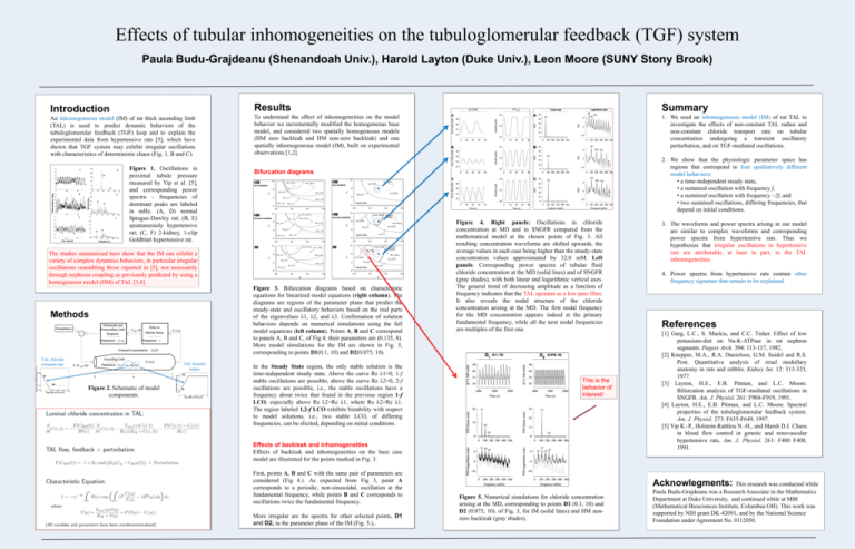
Effects of tubular inhomogeneities on the tubuloglomerular feedback (TGF) system Paula Budu-Grajdeanu (Shenandoah Univ.), Harold Layton (Duke Univ.), Leon Moore (SUNY Stony Brook) Introduction Results Summary An inhomogeneous model (IM) of rat thick ascending limb (TAL) is used to predict dynamic behaviors of the tubuloglomerular feedback (TGF) loop and to explain the experimental data from hypertensive rats [5], which have shown that TGF system may exhibit irregular oscillations with characteristics of deterministic chaos (Fig. 1, B and C). To understand the effect of inhomogeneities on the model behavior we incrementally modified the homogeneous base model, and considered two spatially homogeneous models (HM zero backleak and HM non-zero backleak) and one spatially inhomogeneous model (IM), built on experimental observations [1,2]. 1. We used an inhomogeneous model (IM) of rat TAL to investigate the effects of non-constant TAL radius and non-constant chloride transport rate on tubular concentration undergoing a transient oscillatory perturbation, and on TGF-mediated oscillations. Figure 1. Oscillations in proximal tubule pressure measured by Yip et al. [5], and corresponding power spectra - frequencies of dominant peaks are labeled in mHz. (A, D) normal Sprague-Dawley rat; (B, E) spontaneously hypertensive rat; (C, F) 2-kidney, 1-clip Goldblatt hypertensive rat. Bifurcation diagrams The studies summarized here show that the IM can exhibit a variety of complex dynamics behaviors, in particular irregular oscillations resembling those reported in [5], not necessarily through nephrons coupling as previously predicted by using a homogeneous model (HM) of TAL [3,4]. Figure 3. Bifurcation diagrams based on characteristic equations for linearized model equations (right column). The diagrams are regions of the parameter plane that predict the steady-state and oscillatory behaviors based on the real parts of the eigenvalues λ1, λ2, and λ3. Confirmation of solution behaviors depends on numerical simulations using the full model equations (left column). Points A, B and C correspond to panels A, B and C, of Fig.4; their parameters are (0.135, 8). More model simulations for the IM are shown in Fig. 5, corresponding to points D1(0.1, 10) and D2(0.075, 10). Methods TAL chloride transport rate TAL luminal radius Figure 2. Schematic of model components. 2. We show that the physiologic parameter space has regions that correspond to four qualitatively different model behaviors: • a time-independent steady state, • a sustained oscillation with frequency f, • a sustained oscillation with frequency ~2f, and • two sustained oscillations, differing frequencies, that depend on initial conditions. In the Steady State region, the only stable solution is the time-independent steady state. Above the curve Re λ1=0, 1-f stable oscillations are possible; above the curve Re λ2=0, 2-f oscillations are possible, i.e., the stable oscillations have a frequency about twice that found in the previous region 1-f LCO, especially above Re λ2=Re λ1, where Re λ2>Re λ1. The region labeled 1,2-f LCO exhibits bistability with respect to model solutions, i.e., two stable LCO, of differing frequencies, can be elicited, depending on initial conditions. Figure 4. Right panels: Oscillations in chloride concentration at MD and in SNGFR computed from the mathematical model at the chosen points of Fig. 3. All resulting concentration waveforms are shifted upwards, the average values in each case being higher than the steady-state concentration values approximated by 32.0 mM. Left panels: Corresponding power spectra of tubular fluid chloride concentration at the MD (solid lines) and of SNGFR (gray shades), with both linear and logarithmic vertical axes. The general trend of decreasing amplitude as a function of frequency indicates that the TAL operates as a low-pass filter. It also reveals the nodal structure of the chloride concentration arising at the MD. The first nodal frequency for the MD concentration appears indeed at the primary fundamental frequency, while all the next nodal frequencies are multiples of the first one. This is the behavior of interest! Effects of backleak and inhomogeneities Effects of backleak and inhomogeneities on the base case model are illustrated for the points marked in Fig. 3. First, points A, B and C with the same pair of parameters are considered (Fig 4.). As expected from Fig 3, point A corresponds to a periodic, non-sinusoidal, oscillation at the fundamental frequency, while points B and C corresponds to oscillations twice the fundamental frequency. More irregular are the spectra for other selected points, D1 and D2, in the parameter plane of the IM (Fig. 5.),. 3. The waveforms and power spectra arising in our model are similar to complex waveforms and corresponding power spectra from hypertensive rats. Thus we hypothesize that irregular oscillations in hypertensive rats are attributable, at least in part, to the TAL inhomogeneities. 4. Power spectra from hypertensive rats contain other frequency signature that remain to be explained. References [1] Garg, L.C., S. Mackie, and C.C. Tisher. Effect of low potassium-diet on Na-K-ATPase in rat nephron segments. Pugers Arch. 394: 113-117, 1982. [2] Knepper, M.A., R.A. Danielson, G.M. Saidel and R.S. Post. Quantitative analysis of renal medullary anatomy in rats and rabbits. Kidney Int. 12: 313-323, 1977. [3] Layton, H.E., E.B. Pitman, and L.C. Moore. Bifurcation analysis of TGF-mediated oscillations in SNGFR. Am. J. Physiol. 261: F904-F919, 1991. [4] Layton, H.E., E.B. Pitman, and L.C. Moore. Spectral properties of the tubuloglomerular feedback system. Am. J. Physiol. 273: F635-F649, 1997. [5] Yip K.-P., Holstein-Rathlou N.-H., and Marsh D.J. Chaos in blood flow control in genetic and renovascular hypertensive rats, Am. J. Physiol. 261: F400 F408, 1991. Acknowlegments: This research was conducted while Figure 5. Numerical simulations for chloride concentration arising at the MD, corresponding to points D1 (0.1, 10) and D2 (0.075, 10). of Fig. 3, for IM (solid lines) and HM nonzero backleak (gray shades). Paula Budu-Grajdeanu was a Research Associate in the Mathematics Department at Duke University, and continued while at MBI (Mathematical Biosciences Institute, Columbus OH). This work was supported by NIH grant DK-42091, and by the National Science Foundation under Agreement No. 0112050.
