Breast-Case-Scenario
advertisement
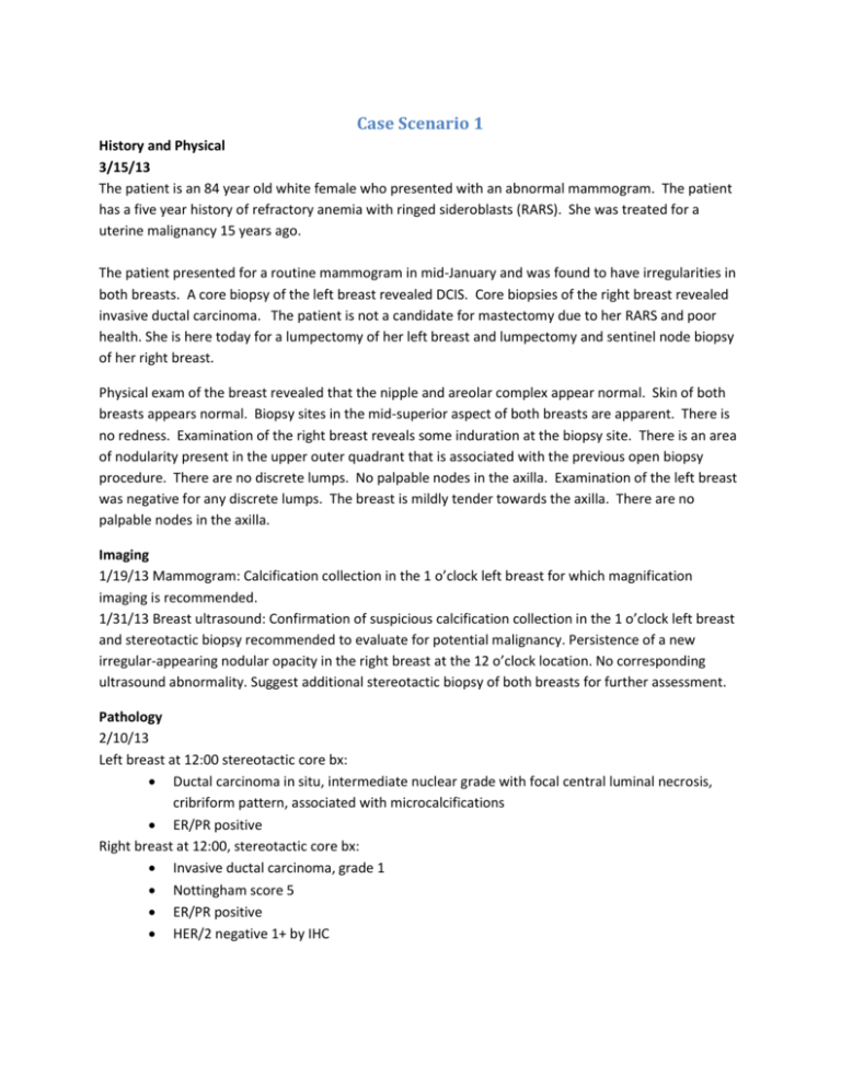
Case Scenario 1 History and Physical 3/15/13 The patient is an 84 year old white female who presented with an abnormal mammogram. The patient has a five year history of refractory anemia with ringed sideroblasts (RARS). She was treated for a uterine malignancy 15 years ago. The patient presented for a routine mammogram in mid-January and was found to have irregularities in both breasts. A core biopsy of the left breast revealed DCIS. Core biopsies of the right breast revealed invasive ductal carcinoma. The patient is not a candidate for mastectomy due to her RARS and poor health. She is here today for a lumpectomy of her left breast and lumpectomy and sentinel node biopsy of her right breast. Physical exam of the breast revealed that the nipple and areolar complex appear normal. Skin of both breasts appears normal. Biopsy sites in the mid-superior aspect of both breasts are apparent. There is no redness. Examination of the right breast reveals some induration at the biopsy site. There is an area of nodularity present in the upper outer quadrant that is associated with the previous open biopsy procedure. There are no discrete lumps. No palpable nodes in the axilla. Examination of the left breast was negative for any discrete lumps. The breast is mildly tender towards the axilla. There are no palpable nodes in the axilla. Imaging 1/19/13 Mammogram: Calcification collection in the 1 o’clock left breast for which magnification imaging is recommended. 1/31/13 Breast ultrasound: Confirmation of suspicious calcification collection in the 1 o’clock left breast and stereotactic biopsy recommended to evaluate for potential malignancy. Persistence of a new irregular-appearing nodular opacity in the right breast at the 12 o’clock location. No corresponding ultrasound abnormality. Suggest additional stereotactic biopsy of both breasts for further assessment. Pathology 2/10/13 Left breast at 12:00 stereotactic core bx: Ductal carcinoma in situ, intermediate nuclear grade with focal central luminal necrosis, cribriform pattern, associated with microcalcifications ER/PR positive Right breast at 12:00, stereotactic core bx: Invasive ductal carcinoma, grade 1 Nottingham score 5 ER/PR positive HER/2 negative 1+ by IHC Operative Report-3/15/13 Right lumpectomy with needle localization and sentinel lymph node biopsy and left lumpectomy with needle localization Pathology-3/15/13 Right lumpectomy with needle localization and sentinel lymph node biopsy. Left lumpectomy with needle localization. o Right sentinel lymph node (1): Metastatic ductal carcinoma. The carcinoma demonstrates focal extension into adipose tissue and measures approximately 0.8 cm. o Right lumpectomy: Invasive ductal carcinoma measuring 0.8 cm DCIS is focally present. Nottingham grade 1, Nottingham score 5 Lymphvascular invasion not identified Margins negative pT1bN1 (sn+) o Left lumpectomy: Focal ductal carcinoma in situ, cribriform type with focal central necrosis. DCIS approaches to within 0.5 cm of the 9:00 margin. Remaining margins are widely negative. Radiation Oncology The patient presents with a pT1bN1 (sn+) invasive ductal carcinoma of the right breast and a DCIS of the left breast. She is a not a candidate for mastectomy or chemotherapy due to underlying medical conditions and poor overall health. She started on Arimidex 5/25/13. Radiation Summary: Right Breast and axillary lymph nodes: mixture of 6 mv and 15 mv photons, 5040 cGy in 28 fractions from in 6/20/13 to 8/1/13, electron boost to tumor bed, 1080 cGy in 6 fractions from 8/3/13 to 8/10/13 Left breast: mixture of 6 mv and 15 mv photons, 5040 cGy in 28 fractions from 7/5/13 to 8/16/13, electron boost, 1080 cGy in 6 fractions from 8/17/13 to 8/24/13 How many primaries are present in this case scenario? 4-RARS, Uterine, Breast x 2 What is the diagnosis date? 02/10/2013 What is the sequence? 03 How would we code the histology of the primary you are currently abstracting? 8500/3 Stage/ Prognostic Factors CS Tumor Size CS Extension CS Tumor Size/Ext Eval 008 100 3 CS SSF 9 CS SSF 10 CS SSF 11 020 998 or 999 998 or 999 CS Lymph Nodes CS Lymph Nodes Eval Regional Nodes Positive Regional Nodes Examined CS Mets at Dx CS Mets Eval CS SSF 1 CS SSF 2 CS SSF 3 CS SSF 4 CS SSF 5 CS SSF 6 CS SSF 7 CS SSF 8 250 3 01 01 00 0 010 010 001 987 987 020 050 010 CS SSF 12 CS SSF 13 CS SSF 14 CS SSF 15 CS SSF 16 CS SSF 17 CS SSF 18 CS SSF 19 CS SSF 20 CS SSF 21 CS SSF 22 CS SSF 23 CS SSF 24 CS SSF 25 998 or 999 998 or 999 998 or 999 020 110 988 988 988 988 987 998 or 999 998 or 999 988 988 Treatment Diagnostic Staging Procedure Surgery Codes Surgical Procedure of Primary Site Scope of Regional Lymph Node Surgery Surgical Procedure/ Other Site Systemic Therapy Codes Chemotherapy Hormone Therapy Immunotherapy Hematologic Transplant/Endocrine Procedure 02 22 2 0 82 01 00 00 Radiation Codes Radiation Treatment Volume Regional Treatment Modality 19 27 Regional Dose Boost Treatment Modality Boost Dose Number of Treatments to Volume Reason No Radiation 05040 28 01080 034 0 How many primaries are present in this case scenario? 4-RARS, Uterine, Breast x 2 What is the diagnosis date? 02/10/2013 What is the sequence? 04 How would we code the histology of the primary you are currently abstracting? 8201/2 Stage/ Prognostic Factors CS Tumor Size CS Extension CS Tumor Size/Ext Eval 999 000 3 CS SSF 9 CS SSF 10 CS SSF 11 998 or 999 998 or 999 998 or 999 CS Lymph Nodes CS Lymph Nodes Eval Regional Nodes Positive Regional Nodes Examined CS Mets at Dx CS Mets Eval CS SSF 1 CS SSF 2 CS SSF 3 CS SSF 4 CS SSF 5 CS SSF 6 CS SSF 7 CS SSF 8 000 0 98 00 000 0 010 010 098 000 000 010 999 998 or 999 CS SSF 12 CS SSF 13 CS SSF 14 CS SSF 15 CS SSF 16 CS SSF 17 CS SSF 18 CS SSF 19 CS SSF 20 CS SSF 21 CS SSF 22 CS SSF 23 CS SSF 24 CS SSF 25 998 or 999 998 or 999 998 or 999 998 or 999 999 988 988 988 988 987 998 or 999 998 or 999 988 988 Treatment Diagnostic Staging Procedure Surgery Codes Surgical Procedure of Primary Site Scope of Regional Lymph Node Surgery Surgical Procedure/ Other Site Systemic Therapy Codes Chemotherapy Hormone Therapy Immunotherapy Hematologic Transplant/Endocrine Procedure 02 22 0 0 82 01 00 00 Radiation Codes Radiation Treatment Volume Regional Treatment Modality 18 27 Regional Dose Boost Treatment Modality Boost Dose Number of Treatments to Volume Reason No Radiation 05040 28 01080 034 0 Case Scenario 2 A 59 year old white female presents for partial mastectomy and sentinel lymph node biopsy. Approximately six months ago, she was found to have a complicated cyst in her left breast. She returned on 3/1/12 for a follow-up mammogram and was found to have new area of architectural distortion with a suggestion calcification. She returned on 3/4/12 for a targeted ultrasound and biopsy. The ultrasound showed an ill-defined hypoechoic shadowing nodule in the left breast at the 3:00 position/zone 2/posterior depth measuring 9 x 7 mm. The nodule has a maximum diameter estimated at 2.0 cm x 2.5 cm. An evaluation of the axilla demonstrated fatty-replaced lymph nodes which were not enlarged. A biopsy of the nodule revealed an invasive lobular carcinoma, ER/PR positive, HER/2 negative 1+ by IHC. Additional testing showed that the BRCA1 and BRCA2 were negative, Oncotype Dx score = 15. Physical exam: The right breast is negative for discrete palpable mass. There is no skin dimpling, retraction, or peau' d orange appearance. There is no nipple discharge or inversion. The left breast shows resolving ecchymosis at the 4 o'clock position related to the biopsy. Gentle palpation of these areas did not reveal any identified lump; however, a full-exam was difficult due to residual tenderness and discomfort. The remaining breast tissue shows no discrete palpable mass. No skin dimpling, retraction, or peau' d orange. No nipple discharge or inversion. Evaluation of the axilla demonstrates fatty-replaced lymph nodes which are not enlarged. No areas of irregular cortical thickening are identified. Pathology 3/4/12 Left breast needle core biopsy at 3:00: Invasive lobular carcinoma, ER/PR positive, HER/2 negative 1+ by IHC Operative Report 3/29/12 Left breast needle-directed partial mastectomy and left sentinel lymph node biopsy Pathology Left SLN Bx: A single LN, negative for carcinoma, IHC stains for keratin confirm the above impression. Left Breast Partial Mastectomy: Invasive lobular carcinoma, tumor forms multiple (4) masses within the specimen, the largest of which measures 1.4 cm. Nottingham grade 1, Nottingham score 5, lobular carcinoma in situ is present within the tumor and multi-focally within the specimen submitted. An area of ductal carcinoma in situ is also present. Lymph-vascular invasion is not appreciated. Final margins are within 0.1 cm. 4/27/12 Bilateral skin sparing mastectomy with bilateral tissue expander placement of the allomax. (Patient had implants placed 8/9) Adjuvant Treatment: Radiation not recommended. Patient started on letrozole 5/25. How many primaries are present in this case scenario? 1- M10 What is the diagnosis date? 03/04/2012 How would we code the histology of the primary you are currently abstracting? 8520/3 – H27 What is the sequence? 00 Stage/ Prognostic Factors CS Tumor Size CS Extension CS Tumor Size/Ext Eval 014 100 3 CS SSF 9 CS SSF 10 CS SSF 11 020 998 or 999 998 or 999 CS Lymph Nodes CS Lymph Nodes Eval Regional Nodes Positive Regional Nodes Examined CS Mets at Dx CS Mets Eval CS SSF 1 CS SSF 2 CS SSF 3 CS SSF 4 CS SSF 5 CS SSF 6 CS SSF 7 CS SSF 8 000 3 00 01 00 0 010 010 000 001 000 050 050 010 CS SSF 12 CS SSF 13 CS SSF 14 CS SSF 15 CS SSF 16 CS SSF 17 CS SSF 18 CS SSF 19 CS SSF 20 CS SSF 21 CS SSF 22 CS SSF 23 CS SSF 24 CS SSF 25 998 or 999 998 or 999 998 or 999 020 110 988 988 988 988 987 010 015 988 988 Treatment Diagnostic Staging Procedure Surgery Codes Surgical Procedure of Primary Site Scope of Regional Lymph Node Surgery Surgical Procedure/ Other Site Systemic Therapy Codes Chemotherapy Hormone Therapy Immunotherapy Hematologic Transplant/Endocrine Procedure 02 30 2 0 00 01 00 00 Radiation Codes Radiation Treatment Volume Regional Treatment Modality 00 00 Regional Dose Boost Treatment Modality Boost Dose Number of Treatments to Volume Reason No Radiation 00000 00 00000 000 1 Case Scenario 3 A 64 year old white female presents with an abnormal screening mammogram. She has a history of left breast cancer status post left partial mastectomy, axillary lymph node dissection and adjuvant radiation 20 years ago. Imaging 3/17/12 Mammogram: Postsurgical changes of the left breast. A potential area of developing density in the upper central portion of the left mid-breast was identified. Recommend diagnostic imaging for further assessment. 3/21/12 Left Breast Ultrasound: In the left breast superiorly at the 12 to 1:00 position/zone 3/mid to posterior depth postsurgical scarring is noted. In the left breast 12:00 position/zone 2/mid-depth an irregular hypoechoic vascular mass with irregular and micro-lobulated borders is identified, measuring 13 x 9 x 8 mm. This is considered highly suspicious for malignancy. Evaluation of the axilla demonstrates no suspicious appearing lymph nodes. 1. Highly suspicious mass in the left breast at 12:00. Biopsy is recommended for tissue diagnosis. 2. Left breast 1 to 2:00 far superior stable appearing post treatment changes. Assessment: BIRADS 5 - highly suggestive of malignancy appropriate action should be taken. 4/8/12 MRI Breasts: Focal area of abnormal irregular enhancement surrounding the biopsy site in the upperouter left breast at the junction of the middle and posterior one-third. This corresponds to the biopsy-proven invasive lobular carcinoma. The area of abnormal enhancement has a maximum diameter approximately 4 x 3 cm. No other abnormal enhancement and left breast. No significant abnormality demonstrated in the right breast. 4/8/12 Bone scan: No scintigraphic findings to suggest skeletal metastases. Operative Procedure 3/29/12 Ultrasound guided needle core biopsy Pathology 3/29/12 Left breast at 12:00 needle bx: Invasive lobular carcinoma, solid variant. Grade 2, Nottingham score 6, perineural invasion is identified. Features suspicious for lymph vascular invasion. ER/PR positive, HER/2 negative 1+ by IHC Operative Procedure 4/15/12 Left Total Mastectomy Pathology Final Diagnosis: A) BREAST, LEFT ANTERIOR MARGIN AT 11:00, EXCISION: o NEGATIVE FOR MALIGNANCY. B) BREAST, LEFT, TOTAL MASTECTOMY: o HISTOLOGIC TUMOR TYPE: INVASIVE LOBULAR CARCINOMA, SOLID VARIANT. o SIZE OF INVASIVE CARCINOMA: 1.3 X 1.2 X 1.0 CM. o COMPOSITE HISTOLOGIC GRADE: NOTTINGHAM GRADE II (OF III). Tubule formation score: 3 Nuclear pleomorphism score: 2 Mitotic count score: 1 Total Nottingham score: 6 o ANCILLARY STUDIES (PERFORMED ON CORE BIOPSY 3/29/2012): Estrogen Receptor: POSITIVE Progesterone Receptor: POSITIVE. HER2 Result: NEGATIVE. Oncotype Dx score = 4 o DUCTAL CARCINOMA IN-SITU: NOT IDENTIFIED. o LYMPH-VASCULAR INVASION: NOT IDENTIFIED. o MARGINS: THE SURGICAL MARGINS ARE NEGATIVE BY GREATER THAN 1.0 CM. o LYMPH NODES: NO LYMPH NODES ARE IDENTIFIED. o ADDITIONAL FINDINGS: TWO ADDITIONAL MICROSCOPIC FOCI OF INVASIVE LOBULAR CARCINOMA, NOTTINGHAM GRADE I (OF III), ARE IDENTIFIED SEPARATE FROM THE MAIN TUMOR MASS MEASURING 0.3 CM. AND 0.1 CM. o A MASS OF DENSE COLLAGENOUS TISSUE COMPATIBLE WITH SCAR FROM PRIOR SURGICAL EXCISION IS IDENTIFIED. o PATHOLOGIC TNM STAGE: AJCC pT1c NX C) SKIN, LEFT BREAST, EXCISION: NEGATIVE FOR MALIGNANCY. Adjuvant Treatment Summary Patient was started on a regimen of Arimidex beginning 4/28/12. How many primaries are present in this case scenario? 2 –M5 What is the diagnosis date? 03/21/2012 How would we code the histology of the primary you are currently abstracting? 8520/3 – H14 What is the sequence? 02 Stage/ Prognostic Factors CS Tumor Size CS Extension CS Tumor Size/Ext Eval 013 100 3 CS SSF 9 CS SSF 10 CS SSF 11 020 998 or 999 998 or 999 CS Lymph Nodes CS Lymph Nodes Eval Regional Nodes Positive Regional Nodes Examined CS Mets at Dx CS Mets Eval CS SSF 1 CS SSF 2 CS SSF 3 CS SSF 4 CS SSF 5 CS SSF 6 CS SSF 7 CS SSF 8 000 0 98 00 00 0 010 010 098 000 000 000 060 010 CS SSF 12 CS SSF 13 CS SSF 14 CS SSF 15 CS SSF 16 CS SSF 17 CS SSF 18 CS SSF 19 CS SSF 20 CS SSF 21 CS SSF 22 CS SSF 23 CS SSF 24 CS SSF 25 998 or 999 998 or 999 998 or 999 020 110 988 988 988 988 987 010 004 988 988 Treatment Diagnostic Staging Procedure Surgery Codes Surgical Procedure of Primary Site Scope of Regional Lymph Node Surgery Surgical Procedure/ Other Site Systemic Therapy Codes Chemotherapy Hormone Therapy Immunotherapy Hematologic Transplant/Endocrine Procedure 02 41 0 0 00 01 00 00 Radiation Codes Radiation Treatment Volume Regional Treatment Modality 00 00 Regional Dose Boost Treatment Modality Boost Dose Number of Treatments to Volume Reason No Radiation 00000 00 00000 000 1
