Differences in Predation Responses of Endangerd, Pseudemys
advertisement
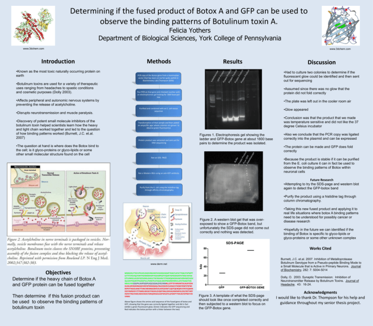
Determining if the fused product of Botox A and GFP can be used to observe the binding patterns of Botulinum toxin A. Felicia Yothers Department of Biological Sciences, York College of Pennsylvania www.3dchem.com www.3dchem.com Introduction •Known as the most toxic naturally occurring protein on earth •Botulinum toxins are used for a variety of therapeutic uses ranging from headaches to spastic conditions and cosmetic purposes (Dolly 2003). Methods Results •Had to culture two colonies to determine if the fluorescent glow could be identified and then sent out for sequencing PCR copy of the Botox-gene from a mammalian vector that has been on ice for quite awhile in Biochemistry Lab (Thompson 2009) •Assumed since there was no glow that the protein did not fold correctly Run PCR on that gene and checked reaction with an electrophoresis gel looking for 1800 base pair line •Affects peripheral and autonomic nervous systems by preventing the release of acetylcholine. •Disrupts neurotransmission and muscle paralysis. •Discovery of potent small molecule inhibitors of the botulinum toxin helped scientists learn how the heavy and light chain worked together and led to the question of how binding patterns worked (Burnett, J.C. et.al. 2007) •The question at hand is where does the Botox bind to the cell; is it glyco-proteins or glyco-lipids or some other small molecular structure found on the cell Discussion •The plate was left out in the cooler room air •Glow appeared Purified and combined with an E. coli vector sequence •Conclusion was that the product that we made was temperature sensitive and did not like the 37 degree Celsius incubator Transformation of that sample and then plated on ampicillin agar where colonies are grown to observe green fluorescence Protein product was extracted and sent out for DNA sequencing Figures 1. Electrophoresis gel showing the ladder and GFP-Botox gene at about 1800 base pairs to determine the product was isolated. Ran an SDS- PAGE •Also we conclude that the PCR copy was ligated correctly into the plasmid and can be expressed •The protein can be made and GFP does fold correctly •Because the product is stable if it can be purified from the E. coli culture it can in fact be used to observe the binding patterns of Botox within neuronal cells Ran a Western Blot using an anti-GFP antibody. Future Research •Attempting to try the SDS-page and western blot again to detect the GFP-botox band Purify from the E. coli using the histidine-tag through affinity chromatography •Purify the product using a histidine tag through column chromatography. Figure 2. A western blot gel that was overexposed to show a GFP-Botox band, but unfortunately the SDS-page did not come out correctly and nothing was detected. •Taking this new fused product and applying it to real life situations where botox A binding patterns need to be understood for possibly cancer or disease research •Hopefully in the future we can identified if the binding of Botox is specific to glyco-lipids or glyco-proteins or some other unknown complex Works Cited Burnett, J.C. et.al. 2007. Inhibition of Metalloprotease Botulinum Serotype from a Pseudo-peptide Binding Mode to a Small Molecule that is Active in Primary Neurons. Journal of Biochemistry. 282: 7: 5004-5014 www.derm.net Objectives Determine if the heavy chain of Botox A and GFP protein can be fused together Then determine if this fusion product can be used to observe the binding patterns of botulinum toxin MSKGEELFTGVVPILVELDGDVNGHKFSVSGEGEGDATYGKLTLKFICTTGKLPVPWPT LVTTFSYGVQCFSRYPDHMKRHDFFKSAMPEGYVQERTISFKDDGNYKTRAEVKFEG DTLVNRIELKGIDFKEDGNILGHKLEYNYNSHNVYITADKQKNGIKANFKIRHNIEDGSV QLADHYQQNTPIGDGPVLLPDNHYLSTQSALSKDPNEKRDHMVLLEFVTAAGITHG MDELYKSGSGPVLAVPSSDPLVQCGGIALGYELYKMRLLSTFTEYIKNIINTSILNLRYESN HLIDLSRYASKINIGSKVNFDPIDKNQIQLFNLESSKIEVILKNAIVYNSMYENFSTSFWI RIPKYFNSISLNNEYTIINCMENNSGWKVSLNYGEIIWTLQDTQEIKQRVVFKYSQMIN ISDYINRWIFVTITNNRLNNSKIYINGRLIDQKPISNLGNIHASNNIMFKLDGCRDTHRY IWIKYFNLFDKELNEKEIKDLYDNQSNSGILKDFWGDYLQYDKPYYMLNLYDPNKYVD VNNV Above figure shows the amino acid sequence of the fused gene of botox and GFP; showing that the gene was correctly ligated together and did in fact exhibit a green fluorescent glow. (Green indicates the GFP sequencing and Red indicates the botox portion with a linker between the two) Dolly, O. 2003. Synaptic Transmission: Inhibition of Neurotransmitter Release by Botulinum Toxins. Journal of Headache. 43: 16-24. Figure 3. A template of what the SDS-page should look like once completed correctly and then subjected to a western blot to focus on the GFP-Botox gene. Acknowledgments I would like to thank Dr. Thompson for his help and guidance throughout my senior thesis project.
