Mice and Their Spinal Cords
advertisement
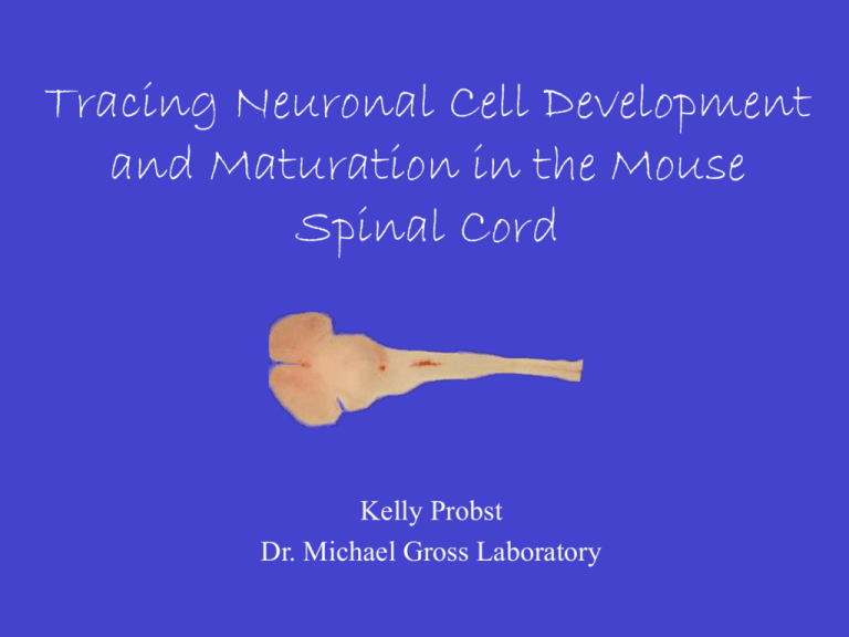
Tracing Neuronal Cell Development and Maturation in the Mouse Spinal Cord Kelly Probst Dr. Michael Gross Laboratory Sensory Information Somatosensory Visual Auditory Olfactory Gustatory Vestibular Somatosensory Information Exteroceptive Touch, Temperature, Pain Proprioceptive Body Position Interoceptive Status of Viscera Sensory Information Flow Visceral Afferents Brain Spinal Cord Body Spinal Cord Innervation by Sensory Neurons Dorsal Root Ganglion (DRG)) A A Spinal Cord Spinal Relay Interneurons Thalamus Hindbrain Synaptic Connection Sensory Neuron Relay Interneuron Cerebellum Neurons Born (E10.5 to E13.5) Ventricular Zone Mantle Zone PM G0 G1 M S G2 Early Embryonic Interneuron Populations Foxd3 = I2 Isl1-2 = I3 VZ Lbx1 = I4 MZ Project Objective • Match adult ascending tracts with embryonic neural tube populations ? • Identify when in development these relay axonal projections reach the brain Backfill Analysis Flour-Dextran Contralateral Ipsilateral Embryo Dissection Hindbrain C1 (Atlas) Cervical Vertebrae Fill TMR-dextran Fill Filled Neuron Incubation Spinal Cord/Brain Dissection Fill Site Hindbrain-Spinal Cord Junction Brachial Enlargement Cervical Region Abdominal Region Lumbar Region and Enlargement E18.5 Backfilled Spinal Cord Dorsal Section 27 (27 sections X 100 m = 2.7 mm) Ventral 1 cm E17.5 Backfilled Spinal Cord Dorsal Section 10 (10 sections X 100 m = 1.0 mm) Ventral 1 cm E16.5 Backfilled Lbx1 +/- Spinal Cord Dorsal Section 28 (28 sections X 40 m = 1.1 mm) Ventral 1 cm Conclusions • By embryonic day 16.5, the axons of some relay interneurons have reached the hindbrain • A subset of these relay interneurons express Lbx1 and therefore are derived from the I4, I5, or I6 embryonic populations Future Goals • Backfill younger spinal cords • Backfill from specific brain regions • Co-label backfills with more marker combinations Acknowledgements Dr. Michael Gross Dr. Kevin Ahern Howard Hughes Medical Institute


