Animal Evolution and Body Plans Name: Models of Multicellular
advertisement
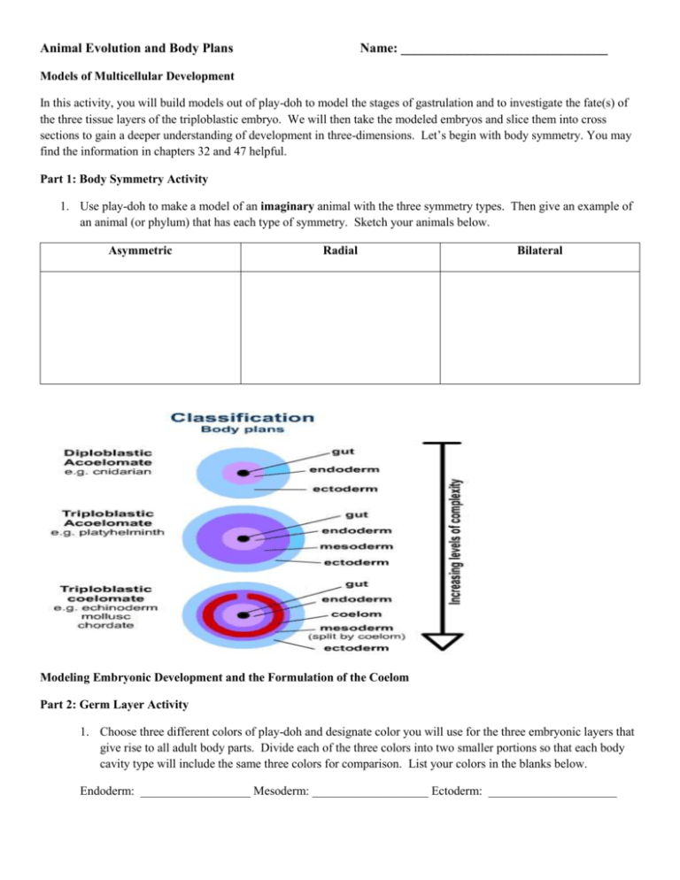
Animal Evolution and Body Plans Name: _______________________________ Models of Multicellular Development In this activity, you will build models out of play-doh to model the stages of gastrulation and to investigate the fate(s) of the three tissue layers of the triploblastic embryo. We will then take the modeled embryos and slice them into cross sections to gain a deeper understanding of development in three-dimensions. Let’s begin with body symmetry. You may find the information in chapters 32 and 47 helpful. Part 1: Body Symmetry Activity 1. Use play-doh to make a model of an imaginary animal with the three symmetry types. Then give an example of an animal (or phylum) that has each type of symmetry. Sketch your animals below. Asymmetric Radial Bilateral Modeling Embryonic Development and the Formulation of the Coelom Part 2: Germ Layer Activity 1. Choose three different colors of play-doh and designate color you will use for the three embryonic layers that give rise to all adult body parts. Divide each of the three colors into two smaller portions so that each body cavity type will include the same three colors for comparison. List your colors in the blanks below. Endoderm: __________________ Mesoderm: ___________________ Ectoderm: _____________________ 2. The above diagrams of the three body cavity types show an example of an animal (or phylum) that represents each type of body plan. Part 3: Modeling Gastrulation in a Diploblastic Organism 1. Make a pancake with a small ball of each color of play-doh. Layer two thin pancakes of play-doh on top of one another (ectoderm on the outside, endoderm on the inside), and fold them over into a hollow log shape. 2. Model the process of gastrulation by pressing one half of the ball into the other half so that it looks like a half moon. Use the plastic knife to cut the gastrula in half to see how it now has two germ layers; just a ball of dividing cells in a diploblastic organism has two cell layers. Practice Questions for Diploblastic Organisms 1. The early mitotic divisions of the zygote to form a ball of cells are called cleavages. What is unique about cleavage? 2. Sketch an illustration of the blastula stage of the early embryo. In your illustration, label and describe each of the following: blastula, blastocoel. 3. Sketch an illustration of the process of gastrulation. In your illustration, label and describe each of the following: gastrula, archenteron, and blastopore. 4. What is the name of the outer layer of cells? __________________________________ 5. What will the outer layer of cells eventually become? ___________________________________ 6. What is the name of the inner layer of cells? ____________________________________ 7. What will the inner layer of cells become? _____________________________________ 8. The simplest animal phyla only have two germ cell layers. Which phyla are they? ___________________ _____________________________________________________________________________________ 9. What is the space between the two layers called? _______________________________ 10. What is the opening to the outside called? ________________________________ Part 4: Modeling Gastrulation in a Triploblastic Organism 1. Make a pancake with a small ball of each color of play-doh. You will need three colors this time to include the addition of the mesoderm. Layer three thin pancakes of play-doh on top of one another (ectoderm on the outside, endoderm on the inside, mesoderm in the middle) and fold them over into a hollow ball. 2. Model the process of gastrulation by pressing one half of the ball into the other half so that it looks like a half moon. Use the plastic knife to the cut the gastrula in half to see how it now has three germ layers; just as a ball of dividing cells in triploblastic organism has three layers. Practice Questions for Triploblastic Organisms 1. What is the third layer of tissue called? ___________________________________ 2. From which cells in the gastrula does the mesoderm arise? ____________________________________ 3. What tissues will the mesoderm eventually give rise to? ______________________________________ 4. What will the blastopore eventually become? _________________________________________ 5. Which animal phyla are protostomes and which are deuterostomes? _________________________________ ________________________________________________________________________________________ 6. How else do protostomes and deuterostomes differ? ______________________________________________ ________________________________________________________________________________________ If we were to continue with the development of your embryo, neurulation would come next. You would start to see development of the notochord and neural plate. 7. What will the notochord eventually become? _______________________________________________ 8. Where will the spinal cord and brain be positioned in this model? ___________________________________ Part 4-Modeling Stem Cell Development 1. Zygote: Use a single color to make both the egg and a much smaller sperm. Mix them together to for a zygote. The tail of the sperm drops off and does not enter the egg. Place it on the petri dish to simulate in-vitro fertilization. The zygote is totipotent-this single cell will give rise to every cell type in the body and placenta. 2. Divide the single cell zygote in half, making two spheres. Divide each of those two cells in half, then each of those in half again, until there are 16 cells. 3. Morula: Push the 16 cells together to form a sphere. This represents the morula stage. Set this aside. 4. From the one-celled zygote to the 16-celled morula, the cells are considered totipotent. This means that any of the cells can differentiate into any tissue in the body. Identical cells arise from this stage of development. 5. Fill in the totipotent column on the table in part 5. 6. Blastula: the cells will continue to divide and now we will fast forward through the blastula stage, which occurs from the 3rd through 14th day of fertilization. At this point, some cells have differentiated into cells which will become the placenta. The pre-placental cells will form a hollow ball surrounding the embryonic cells. 7. Pick a new color and make a sphere about the size of a dollar coin. The idea is that this is a hollow ball; however, you can make a bowl shape. This represents the cells that become the placenta. Use the end of a pen to make indentations in the bowl that represent cells. 8. Use the clay from your original zygote model and make many small spheres (like a pea or smaller) to represent the cells growing inside the hollow ball (bowl). These represent the inner cell mass, or embryonic stem cells. Place a pile of these balls inside the bowl. Really these pre-placental cells would really form a hollow ball (trophoblast) completely surrounding the embryo. They originated from the morula, even though they are a different color. 9. The cells of the blastula are pluripotent. The cells have already gone through one “fate decision”; the hollow ball can only become the placenta and the inner cell mass is the source of embryonic stem cells. If the inner cell mass is removed, the blastula is destroyed and cannot continue to develop. 10. At this point, an embryonic stem cell line can be formed. Cells from the inner cell mass of a four to five day old blastula are transferred into a plastic lab culture dish and grown in a medium to support growth. When kept in this way, the inner cell mass can continue to divide and proliferate for long periods of time without differentiating or losing pluripotency. A stem cell line, directed to differentiate into specific cell types, offers the possibility of treating diseases including Parkinson’s and Alzheimer’s, spinal cord injuries, stroke, diabetes, rheumatoid arthritis, burns, heart disease, etc. 11. Set the blastula in the petri dish (paper plate). In real life, the embryo would go into the freezer if not implanted immediately during in vitro fertilization for pregnancy. 12. Fill in the pluripotent column on part 5. 13. Gastrula: Now we fast forward in time. The embryonic cells have continued to divide. After 14 days of division, the cells will begin to differentiate. 14. To build a gastrula, make a new early placenta bowl in the same color as the previous one. Take a small bit more of the original embryo color and form a ball the size of small pea. Flatten the a piece of a new color to wrap around the ball. Then add another layer of a new color around the outside. Now open a paper clip and use it to cut through the center of the gastrula. The three layer different tissue will be clearly visible on the inside. The three layer ball can be somewhat flattened before cutting with the paper clip. This more accurately represents the shape of the embryonic disc, from which the three layers of the earlier activity form. 15. The gastrula is multipotent. The early placenta cells can still only become placenta. The inner cell mass has undergone another “fate decision” and has differentiated into three layers, the endoderm, mesoderm, and ectoderm. Each of these layers becomes certain parts in your body. 16. Fill in the mulitpotent column on part 5. Part 5-Modeling Stem Cell Development Table Totipotent Stem Cells Pluripotent Stem Cells Multipotent Stem Cells Pluripotent Multipotent Zygote Blastula Gastrula Morula Embryonic stem cell Adult stem cell Diagrams of PlayDoh Creations Approximate Days Cell Division Occurs Approximate Number of Cells Definitions Totipotent of Important Terms Embryonic stem cell line Other Notes
