732
Complementary roles of basal ganglia and cerebellum in
learning and motor control
Kenji Doya
The classical notion that the basal ganglia and the cerebellum
are dedicated to motor control has been challenged by the
accumulation of evidence revealing their involvement in nonmotor, cognitive functions. From a computational viewpoint, it
has been suggested that the cerebellum, the basal ganglia,
and the cerebral cortex are specialized for different types of
learning: namely, supervised learning, reinforcement learning
and unsupervised learning, respectively. This idea of learningoriented specialization is helpful in understanding the
complementary roles of the basal ganglia and the cerebellum
in motor control and cognitive functions.
Addresses
Information Sciences Division, ATR International and CREST, Japan
Science and Technology Corporation, 2-2-2 Hikaridai, Seika, Soraku,
Kyoto 619-0288, Japan; e-mail: doya@isd.atr.co.jp
Current Opinion in Neurobiology 2000, 10:732–739
0959-4388/00/$ — see front matter
© 2000 Elsevier Science Ltd. All rights reserved.
Abbreviations
GP
globus pallidus
LTD
long-term depression
RL
reinforcement learning
SMA
supplementary motor area
SNc
substantia nigra pars compacta
SNr
substantia nigra pars reticulata
TD
temporal difference
VTA
ventral tegmental area
Introduction
The basal ganglia and the cerebellum have long been
known to be involved in motor control because of the
marked motor deficits associated with their damage.
However, the exact aspects of motor control that they are
involved in under normal conditions has not been clear.
Traditionally, the cerebellum was supposed to be involved
in real-time fine tuning of movement [1,2], whereas the
basal ganglia were thought to be involved in the selection
and inhibition of action commands [3]. However, these
distinctions were by no means clear-cut [4]. Furthermore,
an ever-increasing number of brain-imaging studies show
that the basal ganglia and the cerebellum are involved in
non-motor tasks, such as mental imagery [5,6], sensory processing [7–9], planning [10,11•,12], attention [13], and
language [14–17].
Both the basal ganglia and the cerebellum have recurrent
connections with the cerebral cortex [1,3]. Anatomical
studies using transneuronally transported viruses
[18–20,21••] have clearly shown that the projections from
the basal ganglia and the cerebellum through the thalamus
to the cortex constitute multiple ‘parallel’ channels. The
diversity of the target cortical areas — not only the motor
and premotor cortices but also the prefrontal [18], temporal [22], and parietal cortices [19] — is in agreement with
their involvement in diverse functions. However, the neural activity tuning and the lesion effects of a subsection of
the basal ganglia or the cerebellum tend to be similar to
those of the cortical area to which it projects [21••]. This
makes it difficult to distinguish the specific roles of the
basal ganglia and the cerebellum simply from recording or
lesion results.
Is it then impossible to characterize the specific information processing in the basal ganglia and the cerebellum?
Despite their diverse, overlapping target cortical areas, the
basal ganglia and the cerebellum have unique local circuit
architectures and synaptic mechanisms. This strongly suggests that each structure is specialized for a particular type
of information processing [23]. In this review, a novel view
is introduced in which the basal ganglia and the cerebellum are specialized for different types of learning
algorithms: reward-based learning in the basal ganglia and
error-based learning in the cerebellum [24••]. Recent
experimental as well as modeling studies on the basal ganglia and the cerebellum are reviewed from this viewpoint
of learning-oriented specialization.
Learning-oriented specialization
Theoretical models of learning in different parts of the
brain suggest that the cerebellum, the basal ganglia, and
the cerebral cortex are specialized for different types of
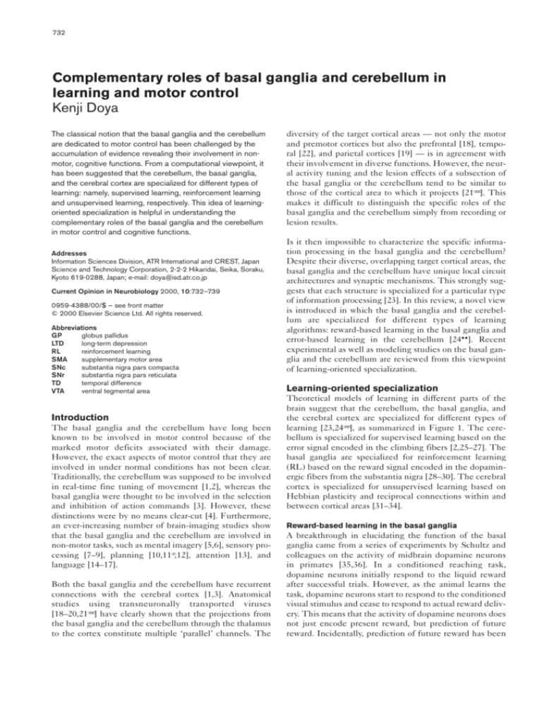
learning [23,24••], as summarized in Figure 1. The cerebellum is specialized for supervised learning based on the
error signal encoded in the climbing fibers [2,25–27]. The
basal ganglia are specialized for reinforcement learning
(RL) based on the reward signal encoded in the dopaminergic fibers from the substantia nigra [28–30]. The cerebral
cortex is specialized for unsupervised learning based on
Hebbian plasticity and reciprocal connections within and
between cortical areas [31–34].
Reward-based learning in the basal ganglia
A breakthrough in elucidating the function of the basal
ganglia came from a series of experiments by Schultz and
colleagues on the activity of midbrain dopamine neurons
in primates [35,36]. In a conditioned reaching task,
dopamine neurons initially respond to the liquid reward
after successful trials. However, as the animal learns the
task, dopamine neurons start to respond to the conditioned
visual stimulus and cease to respond to actual reward delivery. This means that the activity of dopamine neurons does
not just encode present reward, but prediction of future
reward. Incidentally, prediction of future reward has been
Complementary roles of basal ganglia and cerebellum in learning and motor control Doya
733
Figure 1
Specialization of the cerebellum, the basal
ganglia, and the cerebral cortex for different
types of learning [24••]. The cerebellum is
specialized for supervised learning, which is
guided by the error signal encoded in the
climbing fiber input from the inferior olive. The
basal ganglia are specialized for
reinforcement learning, which is guided by the
reward signal encoded in the dopaminergic
input from the substantia nigra. The cerebral
cortex is specialized for unsupervised
learning, which is guided by the statistical
properties of the input signal itself, but may
also be regulated by the ascending
neuromodulatory inputs [80].
Unsupervised learning
Output
Input
Cerebral cortex
Reinforcement learning
Reward
Basal
Thalamus
ganglia
Substantia
nigra
Inferior
olive
Output
Input
Cerebellum
Target
Supervised learning
Error
+
–
Input
Output
Current Opinion in Neurobiology
the main issue in the computational theory of RL [37,38].
In the RL theory, a policy (sensory–motor mapping) is
sought so that the expected sum of future reward
predicting the future reward from the present sensory state
whereas the matrix is responsible for predicting future
rewards associated with different candidate actions.
V(t) = E[ r(t + 1) + r(t + 2) + ...]
Through a competition of the matrix outputs, the motor
action with the highest expected future reward is selected
in SNr/GP and sent to the motor nuclei in the brain stem
as well as thalamo-cortical circuits. The TD error is computed in SNc, based on the limbic input coding the
present reward and the striatal input coding the future
reward. The nigrostriatal dopaminergic projection, which
terminates at the cortico-striatal synaptic spines, modulates the synaptic plasticity and serves as the error signal in
the striosome prediction network and the reinforcement
signal for the matrix action network. Such models have
successfully replicated the learning of conditioning tasks as
well as sequence learning tasks [39,40,41•].
is maximized. Here, r(t) denotes the reward acquired at
time t and V(t) represents the ‘value’ of the present state.
RL generally involves two issues: prediction of cumulative
future reward and improvement of state–action mapping to
maximize the cumulative future reward. A so-called ‘temporal difference’ (TD) signal δ(t)
δ(t) = r(t) + V(t) – V(t – 1),
takes dual roles as the error signal for reward prediction and
the reinforcement signal for sensory–motor mapping [37].
The reward-predicting response of dopamine neurons was
a big surprise because it was exactly how the TD signal
would respond in reward-prediction learning — initially
responding to the reward itself [δ(t) = r(t)], and later to the
change in the predicted reward [δ(t) = V(t) – V(t – 1)] even
without a reward. Dopamine was also well known to have
the role of behavioral reinforcer. Accordingly, a number of
models of the basal ganglia as a RL system have been proposed in which the nigrostriatal dopamine neurons
represent the TD signal [28–30,39,40,41•].
Figure 2 summarizes the outline of an RL-based model. The
input site of the basal ganglia, the striatum, comprises two
compartments: the striosome, which projects to the dopamine
neurons in the substantia nigra pars compacta (SNc), and the
matrix, which projects to the output site of the basal ganglia,
the substantia nigra pars reticulata (SNr) and the globus pallidus (GP). In the RL model, the striosome is responsible for
However, there still remain several issues to be clarified
in the RL hypothesis of the basal ganglia. First, it is not
clear how the ‘temporal difference’ of expected future
reward is calculated in the circuit leading to the substantia nigra, although a few possible mechanisms have
been suggested [28,29,40,42]. A recent review by Joel
and Weiner [43••] provides a comprehensive picture of
the connections to and from the dopaminergic neurons
in SNc and the ventral tegmental area (VTA). According
to their view, there are two major pathways from the
striatum to the SNc dopamine neurons: direct inhibition
by striosome neurons through slow GABAB-type synapses, and indirect disinhibition by matrix neurons through
inhibition of GABAA-type synaptic inputs from SNr
neurons [44]. Thus it is possible that the fast disinhibition by the matrix and the slow inhibition by the
striosome provide the temporal difference component
V(t) – V(t – 1).
734
Motor systems
Figure 2
Sensory
input
Action
output
Cerebral cortex
State representation
Striatum
Evaluation
Thalamus
TD signal
Dopamine neurons
Reward
SNr, GP
A schematic diagram of the cortico-basal
ganglia loop and the possible roles of its
components in a reinforcement learning
model. The neurons in the striatum predict the
future reward for the current state and the
candidate actions. The error in the prediction
of future reward, the TD error, is encoded in
the activity of dopamine neurons and is used
for learning at the cortico-striatal synapses.
One of the candidate actions is selected in
SNr and GP as a result of competition of
predicted future rewards. The direct and
indirect pathways within the globus pallidus
are omitted for simplicity. The filled and open
circles denote inhibitory and excitatory
synapses, respectively.
Action selection
Current Opinion in Neurobiology
Another open issue in TD-based models is the plasticity of
cortico-striatal synapses. The above RL model predicts
that there should be different plastic mechanisms for the
striosome and the matrix, which are respectively involved
in the evaluation of current state and possible actions.
Although it has been shown that cortico-striatal synaptic
plasticity is strongly modulated by dopamine [45,46,47•], it
remains to be clarified whether there are different plastic
mechanisms in different compartments.
Reward-related activities have also been found in frontal
cortices, including dorsolateral prefrontal cortex [48],
orbitofrontal cortex [49], and cingulate cortex [50].
Dopaminergic neurons in the VTA project to those cortical
areas. What is the difference between reward processing in
the cerebral cortex and that in the basal ganglia? A systematic comparison of the reward-related activities in the
orbitofrontal cortex, the striatum, and the dopamine neurons revealed the following characteristics [51••]: the
cortical neurons retain more information about sensory
input; the striatal neurons show a richer variety of activation in relation to task progress; and, the dopamine
neurons respond mainly to unpredicted reward or sensory
stimuli. This suggests that the cortex is responsible for
analyzing sensory input, the striatum is involved in the
production of actions, and the dopamine neurons are particularly responsible for learning new behaviors.
Error-based learning in the cerebellum
The idea that the cerebellum is a supervised learning system
dates back to the hypotheses of Marr [25] and Albus [26]. It
was shown by Ito in vestibulo-ocular reflex (VOR) adaptation
experiments [2,52] that long-term depression (LTD) of the
Purkinje cell synapses dependent on the climbing fiber
input is the neural substrate of such error-driven learning
(Figure 3). Although it is still controversial whether the LTD
in Purkinje cells is the only locus of plasticity and memory
storage for the VOR [53], recent results are in accordance
with the cerebellar error-based learning hypothesis.
During ocular following response movements, Kobayashi
and colleagues [54] showed that the response tuning of
complex spikes is the mirror image of that of simple spikes
[55]. This is in agreement with the hypothesis that the
simple spike responses of Purkinje cells are shaped by
LTD of parallel fiber synapses with the error signal provided by the climbing fibers. These authors also showed
that the modulation of simple spikes by complex spikes is
too weak to be useful for real-time motor control.
Kitazawa et al. [56] analyzed the information content of
complex spikes in arm-reaching movement in monkeys.
The results showed that complex spike firing carries
information about the target direction in the early phase
of the movement, whereas it carries information about
the end-point error near the end of the movement. The
coding of end-point error is consistent with the LTD
hypothesis. What is the role of the target-related activity
at the beginning of a movement? The low probability of
firing of complex spikes near the beginning of a movement (less than one spike per trial) suggests that the
signal may not be useful for on-line movement control.
One possibility is that the spikes are potential error signals that could be used for further improvement of the
performance should there be any preceding sensory cues
that enable movement preparation.
Collaboration of learning modules
In the above framework of learning-orientated specialization, each organization is not specialized in what to do, but
in how to learn it. Specific behaviors or functions can be
realized by a combination of multiple learning modules
Complementary roles of basal ganglia and cerebellum in learning and motor control Doya
735
Figure 3
A schematic diagram of the cortico-cerebellar
loop. In a supervised learning model of the
cerebellum, the climbing fibers from the
inferior olive provide the error signal for the
Purkinje cells. Coincident inputs from the
inferior olive and the granule cells result in
LTD of the granule-to-Purkinje synapses. The
filled and open circles denote inhibitory and
excitatory synapses, respectively.
Cerebral cortex
Granule cells
Purkinje cells
Thalamus
Cerebellar
nucleus
Error signal
Cerebral cortex
red nucleus
Inferior olive
Current Opinion in Neurobiology
distributed among the basal ganglia, the cerebellum, and
the cerebral cortex [23,24••].
cerebellum: only the synaptic weights for the inputs that
are associated with the retinal error signal are modified [61].
The use of internal models of the body and the environment can improve the performance of motor control
[57,58•]. Such internal models could be acquired by supervised learning with the motor command as the input and
the sensory outcome as the teacher signal. Furthermore,
for supervised or reinforcement leaning, it is often helpful
to use unsupervised learning algorithms to extract the
essential information in the raw sensory input. Such methods of combination of different learning modules could be
helpful in exploring possible collaborations of the cerebellum, the basal ganglia, and the cerebral cortex [24••].
Table 1 summarizes possible roles of the learning modules
in these three areas.
In many brain-imaging experiments, different parts of the
cerebellum, the basal ganglia and the cerebral cortex are
activated simultaneously. The above hypotheses concerning the specialization of these areas for the different
frameworks of learning can provide us with a helpful hint
as to the different roles of simultaneously activated brain
areas. Below, we review recent studies from the viewpoint
of this learning-oriented specialization.
Neurons in the caudate nucleus are known to be activated
during memory-guided saccades toward a certain preferred
direction [62,63•]. Recently, Kawagoe and colleagues performed delayed-saccade experiments in which reward was
given in only one of four possible saccade directions [64].
Surprisingly, the direction tuning of caudate neurons was
strongly modulated by the reward condition. In some neurons, direction tuning was enhanced when the preferred
direction coincided with the rewarded direction. In others,
the preferred direction changed with the reward direction
[64]. These data suggest that the striatal neurons do not
simply represent motor action itself, but the reward associated with the state and actions. In reference to the above
reinforcement learning model, it is tempting to speculate
that the reward-following neurons are in the striosome and
the reward-modulated neurons are in the matrix [65] and
that the change in their behaviors is based on the
dopamine-dependent plasticity of cortico-striatal synapses
[63•]. However, it remains to be tested if such a change in
the striatal response tuning is solely due to the plasticity
within the striatum or also due to the reward-dependent
responses of the cortical neurons [48–50,51••].
Eye movements
Arm reaching
Recent experiments on saccadic eye movements have
shown separate gain adaptation for different types of saccades, such as visually guided and memory-guided [59,60].
Such input-dependent adaptation of eye movement is naturally expected from supervised learning models of the
It has been shown in arm-reaching studies in monkeys
that the cerebellum is involved in externally driven (e.g.
visually guided) movement, whereas the basal ganglia are
involved in internally generated (e.g. memory-guided)
movement [66]. Recent recording and inactivation studies
736
Motor systems
Table 1
Possible roles of different learning modules.
Cerebellum: supervised learning
Internal models of the body and the environment.
Replication of arbitrary input–output mapping that was learned
elsewhere in the brain.
Basal ganglia: reinforcement learning
Evaluation of current situation by prediction of reward.
Selection of appropriate action by evaluation of candidate actions.
Cerebral cortex: unsupervised learning
Concise representation of sensory state, context, and action.
Finding appropriate modular architecture for a given task.
of motor thalamus [67•,68•] further confirmed this contrasting involvement. In the part of the thalamus that
receives input from the cerebellum and projects to the
ventral premotor cortex, the majority of neurons are selectively activated during visually triggered movements. On
the other hand, in the part of the thalamus that receives
input from the basal ganglia and projects to the prefrontal
cortex, the majority of neurons are selective for internally
generated movements.
What is the reason for such differential involvement?
During visually guided movements, the most critical computation is the coordinate transformation of visual input to
corresponding motor output. Such mapping could be
learned in a form of supervised learning in the cerebellum.
During memory-guided or internally generated movements, what is most critical is the selection of an
appropriate action and the suppression of unnecessary
actions, both of which require prediction of reward value.
Sequence learning
It has been shown by trial-and-error learning of sequential movement that cortico-basal ganglia loops are
differentially involved in early and late stages of learning.
Brain areas in the prefrontal loop (prefrontal cortex,
preSMA [supplementary motor area], caudate head) are
involved in the learning of new sequences, whereas those
in the motor loop (SMA, putamen body) are involved in
the execution of well-learned movements [69,70]. Why
should the information about the sequence learned initially in the prefrontal loop be copied to the motor loop?
We hypothesized that the two cortico-basal ganglia loops
learn a sequence using different representations: visuospatial coordinates in the prefrontal loop and motor
coordinates in the motor loop [41•,71••]. A RL model of
sequence learning based on this hypothesis could replicate many experimental findings — for example, the time
course of learning, the performance for modified
sequences, and the results of lesion experiments [41•]. In
a recent psychophysical experiment motivated by this
hypothesis, it was confirmed in a sequential key-press
task that human subjects depend increasingly on bodyspecific representation than on visual representation with
the progress of learning [72•].
Timing and rhythm
In a series of imaging studies by Sakai and colleagues [73,74•],
it was shown that the memory of simple rhythms involves the
anterior cerebellum, whereas the memory of complex rhythms
and the adjustment of movement timing in response to irregular external triggers involve the posterior cerebellum. A
possible reason for such differential involvement is the use of
different representations [71••]. The anterior cerebellum can
provide internal models of body dynamics, which can be helpful in the prediction of regular timing as well as in the control
of detailed movement parameters. The posterior cerebellum
may provide internal models for prediction of sensory events,
which may be useful in timing perception and adjustment.
Cognitive processing
Involvement of the basal ganglia and the cerebellum in
cognitive functions was once a controversial issue [14,75].
However, there are now abundant brain-imaging data
showing their involvement in mental imagery [5,6], sensory discrimination [7–9], planning [10,11•,12], attention
[13], and language [14–17]. Careful studies of patients with
cerebellar and basal ganglia damage have also revealed that
their impairments are not limited to motor control but also
extend to cognitive functions [21••,76,77]. Lesion studies
in rodents also suggest the involvement of the basal ganglia in rule-based learning [78] and spatial navigation [79].
In a recent positron emission tomography (PET) study
using the Tower of London task, Dagher and colleagues
found that the activity of the caudate nucleus, as well as the
premotor and prefrontal cortices, is correlated with the task
complexity [11•]. This suggests that the cortico-basal ganglia loops may be involved in multi-step planning of actions.
Conclusion
A new hypothesis concerning the specialization of brain
structures for different learning paradigms provides helpful clues as to the differential roles of the basal ganglia and
the cerebellum [24••]. Whereas the use of different learning algorithms is associated with differential involvement
of the cerebellum, the basal ganglia and the cerebral cortex, the use of different representations is associated with
differential involvement of the channels in cortico-basal
ganglia loops and cortico-cerebellar loops.
The frontal cortex has been regarded as the site of high-level
information processing because of its activity related to working memory, action planning, and decision making. However,
what has been found in the cerebral cortex could be just the tip
of an iceberg. The activities of the cortical neurons could be the
result of the recurrent dynamics of the cortico-basal ganglia and
cortico-cerebellar loops. An important role of the cerebral cortex is to provide common representations upon which both the
basal ganglia and the cerebellum can work together.
Acknowledgements
The author is grateful to Mitsuo Kawato and Hiroyuki Nakahara for their
helpful comments on the manuscript.
Complementary roles of basal ganglia and cerebellum in learning and motor control Doya
References and recommended reading
Papers of particular interest, published within the annual period of review,
have been highlighted as:
• of special interest
•• of outstanding interest
1.
Allen GI, Tsukahara N: Cerebro cerebellar communication systems.
Physiol Rev 1974, 54:957-1006.
2.
Ito M: The Cerebellum and Neural Control. New York: Raven Press; 1984.
3.
Alexander GE, Crutcher MD: Functional architecture of basal
ganglia circuits: neural substrates of parallel processing. Trends
Neurosci 1990, 13:266-271.
4.
Thach WT, Mink JW, Goodkin HP, Keating JG: Combining versus
gating motor programs: differential roles for cerebellum and basal
ganglia? In Roles of Cerebellum and Basal Ganglia in Voluntary
Movement. Edited by Mano M, Hamada I, DeLong MR. Amsterdam:
Elsevier; 1993:235-245.
5.
Parsons LM, Fox PT, Downs JH, Glass T, Hirsch TB, Martin CC,
Jerabek PA, Lancaster JL: Use of implicit motor imagery for visual
shape discrimination as revealed by PET. Nature 1995, 375:54-58.
6.
Lotze M, Montoya P, Erb M, Hulsmann E, Flor H, Klose U,
Birbaumer N, Grodd W: Activation of cortical and cerebellar motor
areas during executed and imagined hand movements: an fMRI
study. J Cogn Neurosci 1999, 11:491-501.
7.
Gao JH, Parsons LM, Bower JM, Xiong J, Li J, Fox PT: Cerebellum
implicated in sensory acquisition and discrimination rather than
motor control. Science 1996, 272:545-547.
8.
Poldrack RA, Prabhakaran V, Seger CA, Gabrieli JD: Striatal
activation during acquisition of a cognitive skill. Neuropsychology
1999, 13:564-574.
9.
Parsons LM, Denton D, Egan G, McKinley M, Shade R, Lancaster J,
Fox PT: Neuroimaging evidence implicating cerebellum in support
of sensory/cognitive processes associated with thirst. Proc Natl
Acad Sci USA 2000, 97:2332-2336.
10. Kim S-G, Ugurbil K, Strick PL: Activation of a cerebellar output
nucleus during cognitive processing. Science 1994, 265:949-951.
11. Dagher A, Owen AM, Boecker H, Brooks DJ: Mapping the network
•
for planning: a correlational PET activation study with the Tower
of London task. Brain 1999, 122:1973-1987.
In this positron emission tomography (PET) study, the authors used not only
the subtraction between the test and control conditions, but also the correlation with the complexity of the task (in this case, the number of steps needed to solve a puzzle), to identify the areas that are involved in multi-step
action planning. These areas include lateral premotor, rostral anterior cingulate, and dorsolateral prefrontal cortices, as well as dorsal caudate nucleus.
12. Weder BJ, Leenders KL, Vontobel P, Nienhusmeier M, Keel A,
Zaunbauer W, Vonesch T, Ludin HP: Impaired somatosensory
discrimination of shape in Parkinson’s disease: association with
caudate nucleus dopaminergic function. Hum Brain Mapp 1999,
8:1-12.
13. Allen G, Buxton RB, Wong EC, Courchesne E: Attentional activation
of the cerebellum independent of motor involvement. Science
1997, 275:1940-1943.
14. Leiner JC, Leiner AL, Dow RS: Cognitive and language functions of
the cerebellum. Trends Neurosci 1993, 16:444-447.
15. Kim JJ, Andreasen NC, O’Leary DS, Wiser AK, Ponto LL, Watkins GL,
Hichwa RD: Direct comparison of the neural substrates of recognition
memory for words and faces. Brain 1999, 122:1069-1083.
16. Price CJ, Green DW, von Studnitz R: A functional imaging study of
translation and language switching. Brain 1999, 122:2221-2235.
17.
Dong Y, Fukuyama H, Honda M, Okada T, Hanakawa T, Nakamura K,
Nagahama Y, Nagamine T, Konishi J, Shibasaki H: Essential role of
the right superior parietal cortex in Japanese kana mirror reading:
an fMRI study. Brain 2000, 123:790-799.
737
20. Hoover JE, Strick PL: The organization of cerebellar and basal
ganglia outputs to primary motor cortex as revealed by retrograde
transneuronal transport of herpes simplex virus type 1. J Neurosci
1999, 19:1446-1463.
21. Middleton FA, Strick PL: Basal ganglia and cerebellar loops: motor
•• and cognitive circuits. Brain Res Brain Res Rev 2000, 31:236-250.
A thorough review of the anatomy of the outputs of the basal ganglia and
cerebellum to the cerebral cortex. Multiple output channels that target different cortical areas are revealed by the group’s studies using transneuronally transported herpes virus. The paper also includes a comprehensive
review of the physiology and the pathology of the basal ganglia and the
cerebellum in motor control and cognitive functions.
22. Middleton FA, Strick PL: The temporal lobe is a target of output from
the basal ganglia. Proc Natl Acad Sci USA 1996, 93:8683-8687.
23. Houk JC, Wise SP: Distributed modular architectures linking basal
ganglia, cerebellum, and cerebral cortex: their role in planning and
controlling action. Cereb Cortex 1995, 2:95-110.
24. Doya K: What are the computations of the cerebellum, the basal
•• ganglia, and the cerebral cortex. Neural Networks 1999, 12:961-974.
This paper presents a novel view that the cerebellum, the basal ganglia, and
the cerebral cortex are specialized for different types of learning: namely,
supervised, reinforcement and unsupervised learning, respectively. The paper
reviews basic algorithms for three learning paradigms and presents the way
in which they could be implemented in the neural circuits. Possible control
architectures that combine different learning modules are also considered.
25. Marr D: A theory of cerebellar cortex. J Physiol 1969, 202:437-470.
26. Albus JS: A theory of cerebellar function. Math Biosci 1971,
10:25-61.
27.
Kawato M, Gomi H: A computational model of four regions of the
cerebellum based on feedback-error learning. Biol Cybern 1992,
68:95-103.
28. Houk JC, Adams JL, Barto AG: A model of how the basal ganglia
generate and use neural signals that predict reinforcement.
In Models of Information Processing in the Basal Ganglia. Edited by
Houk JC, Davis JL, Beiser DG. Cambridge, MA: MIT Press;
1995:249-270.
29. Montague PR, Dayan P, Sejnowski TJ: A framework for
mesencephalic dopamine systems based on predictive Hebbian
learning. J Neurosci 1996, 16:1936-1947.
30. Schultz W, Dayan P, Montague PR: A neural substrate of prediction
and reward. Nature 1997, 275:1593-1599.
31. Malsburg CVD: Self-organization of orientation sensitive cells in
the striate cortex. Kybernetik 1973, 14:85-100.
32. Amari S, Takeuchi A: Mathematical theory on formation of category
detecting nerve cells. Biol Cybern 1978, 29:127-136.
33. Sanger TD: Optimal unsupervised learning in a single-layer linear
feedforward neural network. Neural Networks 1989, 2:459-473.
34. Olshausen BA, Field DJ: Emergence of simple-cell receptive field
properties by learning sparse code for natural images. Nature
1996, 381:607-609.
35. Schultz W, Apicella P, Ljungberg T: Responses of monkey
dopamine neurons to reward and conditioned stimuli during
successive steps of learning a delayed response task. J Neurosci
1993, 13:900-913.
36. Schultz W: Predictive reward signal of dopamine neurons.
J Neurophysiol 1998, 80:1-27.
37.
Barto AG: Adaptive critics and the basal ganglia. In Models of
Information Processing in the Basal Ganglia. Edited by Houk JC,
Davis JL, Beiser DG. Cambridge, MA: MIT Press; 1995:215-232.
38. Sutton RS, Barto AG: Reinforcement Learning. Cambridge, MA: MIT
Press; 1998.
18. Middleton FA, Strick PL: Anatomical evidence for cerebellar and
basal ganglia involvement in higher cognitive function. Science
1994, 266:458-461.
39. Suri RE, Schultz W: Learning of sequential movements by neural
network model with dopamine-like reinforcement signal. Exp
Brain Res 1998, 121:350-354.
19. Middleton FA, Strick PL: Cerebellar output: motor and cognitive
channels. Trends Cogn Sci 1998, 2:348-354.
40. Berns GS, Sejnowski TJ: A computational model of how the basal
ganglia produce sequences. J Cogn Neurosci 1998, 10:108-121.
738
Motor systems
41. Nakahara H, Doya K, Hikosaka O: Parallel cortico-basal ganglia
•
mechanisms for acquisition and execution of visuo-motor
sequences: a computational approach. J Cogn Neurosci 2001,
in press.
This paper explores how the prefrontal and motor cortico-basal ganglia loops
can work together in order to realize quick acquisition and robust execution
of sequential movement. The authors used a RL model of the cortico-basal
ganglia loops to simulate the experiments of the ‘2 × 5 task’ in monkeys
[71••]. The model successfully replicated the time-course of short-term and
long-term learning as well as the differential effects of lesions to the basal
ganglia loops.
42. Brown J, Bullock D, Grossberg S: How the basal ganglia use
parallel excitatory and inhibitory learning pathways to selectively
respond to unexpected rewarding cues. J Neurosci 1999,
19:10502-10511.
43. Joel D, Weiner I: The connections of the dopaminergic system with
•• striatum in rats and primates: an analysis with respect to the
functional and compartmental organization of the striatum.
Neuroscience 2000, 96:451-474.
This paper reviews the connections of the dopaminergic neurons in SNc and
VTA to and from the striatum. In the authors’ view, the motor striatum can
affect the dopaminergic neurons in two opposite ways: striosome neurons
directly inhibit SNc neurons through GABAB-type receptors, while matrix neurons indirectly disinhibit SNc neurons by removing GABAA-type inhibitory
inputs from SNr neurons. They also postulate an open-interconnected loop
organization of the basal ganglia circuit, rather than closed, segregated loops.
44. Tepper J, Martin LP, Anderson DR: GABAA receptor-mediated
inhibition of rat substantia nigra dopaminergic neurons by pars
reticulata projection neurons. J Neurosci 1995, 15:3092-3103.
45. Wickens JR, Begg AJ, Arbuthnott GW: Dopamine reverses the
depression of rat corticostriatal synapses which normally follows
high-frequency stimulation of cortex in vitro. Neuroscience 1996,
70:1-5.
46. Calabresi P, Pisani A, Mercuri NB, Bernardi G: The corticostriatal
projection: from synaptic plasticity to dysfunctions of the basal
ganglia. Trends Neurosci 1996, 19:19-24.
47.
•
Reynolds JNJ, Wickens JR: Substantia nigra dopamine regulates
synaptic plasticity and membrane potential fluctuations in the rat
neostriatum, in vivo. Neuroscience 2000, 99:199-203.
It is shown in in vivo intracellular recording of rat striatal neurons that highfrequency stimulation of cortical input without nigral dopamine input results
in LTD of the cortico-striatal synapse. This LTD is suppressed or reversed if
the SNc is concurrently stimulated. The result reconfirms the authors’ and
others’ previous results in in vitro experiments.
48. Watanabe M: Reward expectancy in primate prefrontal neurons.
Nature 1996, 382:629-632.
49. Hikosaka K, Watanabe M: Delay activity of orbital and lateral
prefrontal neurons of the monkey varying with different rewards.
Cereb Cortex 2000, 10:263-271.
50. Shima K, Tanji J: Role for cingulate motor area cells in voluntary
movement selection based on reward. Science 1998,
282:1335-1338.
56. Kitazawa S, Kimura T, Yin P-B: Cerebellar complex spikes encode
both destinations and errors in arm movements. Nature 1998,
392:494-497.
57.
Wolpert DM, Miall RC, Kawato M: Internal models in the
cerebellum. Trends Cogn Sci 1998, 2:338-347.
58. Kawato M: Internal models for motor control and trajectory
•
planning. Curr Opin Neurobiol 1999, 9:718-727.
This paper provides a comprehensive review of how the internal models of
the body and the environment can be acquired in the cerebellum and how
they can be used for feedforward control, trajectory planning, and selection
of appropriate control modules.
59. Deubel H: Separate adaptive mechanisms for the control of
reactive and volitional saccadic eye movements. Vision Res 1995,
35:3529-3540.
60. Fujita, M, Amagai A, Minakawa F: Selective and delayed
adaptations of human saccades. In Proceedings of the International
Joint Conference on Neural Networks. Como, Italy; July 24–27, 2000.
5:460-463.
61. Gancarz G, Grossberg S: A neural model of saccadic eye
movement control explains task-specific adaptation. Vision Res
1999, 39:3123-3143.
62. Hikosaka O, Wurtz RH: Visual and oculomotor functions of
monkey Macaca mulatta substantia nigra pars reticulata. J Physiol
1983, 49:1230-1301.
63. Hikosaka O, Takikawa Y, Kawagoe R: Role of the basal ganglia in
•
the control of purposive saccadic eye movements. Physiol Rev
2000, 80:953-978.
This paper reviews the anatomy and physiology of the network linking the
cortical areas, the basal ganglia, and the superior colliculus. In addition to
the classical notion of inhibition and disinhibition of movement, the role of the
basal ganglia in associating the sensory input from the cortex and the reward
input from the dopamine neurons are stressed.
64. Kawagoe R, Takikiwa Y, Hikosaka O: Expectation of reward
modulates cognitive signals in the basal ganglia. Nat Neurosci
1998, 1:411-416.
65. Trapenberg T, Nakahara H, Hikosaka O: Modeling reward dependent
activity pattern of caudate neurons. In Proceedings of the
International Conference on Artificial Neural Networks. Skövde,
Sweden; Sept 21–24, 1998:973-978.
66. Mushiake H, Strick PL: Pallidal neuron activity during sequential
arm movements. J Neurophysiol 1995, 74:2754-2758.
67.
•
van Donkelaar P, Stein JF, Passingham RE, Miall RC: Neuronal
activity in the primate motor thalamus during visually triggered
and internally generated limb movements. J Neurophysiol 1999,
82:934-945.
This and the subsequent paper [68•] compare the activities and the lesion
effects of different parts of the thalamus during limb movements. While area
X, which receives major input from the cerebellum, is specifically involved in
visually guided movements, VApc (parvocellular portion of the ventral anterior nucleus), which receives major input from the basal ganglia, is involved in
internally generated movements.
51. Schultz W, Tremblay L, Hollerman JR: Reward processing in primate
•• orbitofrontal cortex and basal ganglia. Cereb Cortex 2000,
10:272-284.
This paper summarizes the results of the authors’ own studies on the activities of dopaminergic, striatal, and orbitofrontal neurons in goal-directed motor
control and learning. All these neurons show reward-predictive activities, but
the cortical neurons tend to retain specific sensory information, the striatal
neurons show variety of temporal response profiles during a trial, and the
dopaminergic neurons are activated only in response to unexpected rewards.
69. Miyachi S, Hikosaka O, Miyashita K, Karadi Z, Rand MK: Differential
roles of monkey striatum in learning of sequential hand
movement. Exp Brain Res 1997, 151:1-5.
52. Ito M, Sakurai M, Tongroach P: Climbing fibre induced depression
of both mossy fibre responsiveness and glutamate sensitivity of
cerebellar Purkinje cells. J Physiol 1982, 324:113-134.
70. Jueptner M, Frith CD, Brooks DJ, Frackowiak RSJ, Passingham RE:
Anatomy of motor learning. II. Subcortical structures and learning
by trial and error. J Neurophysiol 1997, 77:1325-1337.
53. Raymond JL, Lisberger SG, Mauk MD: The cerebellum: a neuronal
learning machine? Science 1996, 272:1126-1131.
71. Hikosaka O, Nakahara H, Rand MK, Sakai K, Lu X, Nakamura K,
•• Miyachi S, Doya K: Parallel neural networks for learning sequential
procedures. Trends Neurosci 1999, 22:464-471.
This paper summarizes this group’s series of experiments using the ‘2 × 5
task’ paradigm, in which sequential key-press movements are learned by trial
and error. They propose a parallel learning mechanism by multiple corticobasal ganglia loops. While the network linking the prefrontal cortex and the
anterior part of the basal ganglia is specifically involved in acquisition of a
new sequence with the use of visuospatial representation, the network linking the supplementary cortex and the posterior part of the basal ganglia is
mainly involved in quick, robust execution of well-learned sequences with the
use of somatosensory representations.
54. Kobayashi Y, Kawano K, Takemura A, Inoue Y, Kitama T, Gomi H,
Kawato M: Temporal firing patterns of Purkinje cells in the
cerebellar ventral paraflocculus during ocular following
responses in monkeys. II. Complex spikes. J Neurophysiol 1998,
80:832-848.
55. Gomi H, Shidara M, Takemura A, Inoue Y, Kawano K, Kawato M:
Temporal firing patterns of Purkinje cells in the cerebellar ventral
paraflocculus during ocular following responses in monkeys. I.
Simple spikes. J Neurophysiol 1998, 80:818-831.
68. van Donkelaar P, Stein JF, Passingham RE, Miall RC: Temporary
•
inactivation in the primate motor thalamus during visually
triggered and internally generated limb movements.
J Neurophysiol 2000, 83:2780-2790.
See annotation to [67•].
Complementary roles of basal ganglia and cerebellum in learning and motor control Doya
72. Bapi RS, Doya K, Harner AM: Evidence for effector independent
•
and dependent representations and their differential time course
of acquisition during motor sequence learning. Exp Brain Res
2000, 132:149-162.
In order to test the above hypothesis [71••], the authors tested the generalization performance of sequential finger movement under two conditions: the
same spatial targets pressed with different finger movement (visual condition) and altered spatial targets pressed with the same finger movement
(motor condition). It was shown that after extended training, the response
time was significantly shorter in the motor condition than in the visual condition, which is in accordance with the above hypothesis that the motor representation is established and used in the late stage of sequence learning.
739
Furthermore, lateral premotor cortex was most active when both response
selection and timing adjustment were required, suggesting its role in integrating the outputs of preSMA and the posterior cerebellum.
75. Ito M: Movement and thought: identical control mechanisms by
the cerebellum. Trends Neurosci 1993, 16:448-450.
76. Knowlton BJ, Mangels JA, Squire LR: A neostriatal habit learning
system in humans. Science 1996, 273:1399-1402.
77.
Middleton FA, Strick PL: Basal ganglia output and cognition:
evidence from anatomical, behavioral, and clinical studies. Brain
Cogn 2000, 42:183-200.
73. Sakai K, Hikosaka O, Miyauchi S, Takino R, Tamada T, Iwata NK,
Nielsen M: Neural representation of a rhythm depends on its
interval ratio. J Neurosci 1999, 19:10074-10081.
78. Van Golf Racht-Delatour B, El Massioui N: Rule-based learning
impairment in rats with lesions to the dorsal striatum. Neurobiol
Learn Mem 1999, 72:47-61.
74. Sakai K, Hikosaka O, Takino R, Miyauchi S, Nielsen M, Tamada T:
•
What and when: parallel and convergent processing in motor
control. J Neurosci 2000, 20:2691-2700.
The brain activity during a choice reaction-time task in four conditions (with
and without response selection and timing adjustment) was measured using
fMRI. While the preSMA was selectively active in response selection, the
posterior cerebellum was selectively active in timing adjustment.
79. Devan BD, White NM: Parallel information processing in the dorsal
striatum: relation to hippocampal function. J Neurosci 1999,
19:2789-2798.
80. Doya K: Metalearning, neuromodulation, and emotion. In Affective
Minds. Edited by Hatano G, Okada N, Tanabe H. Amsterdam:
Elsevier; 2000:101-104.

