Epithelial Tissue Histology: Instructor's Guide & Worksheet
advertisement
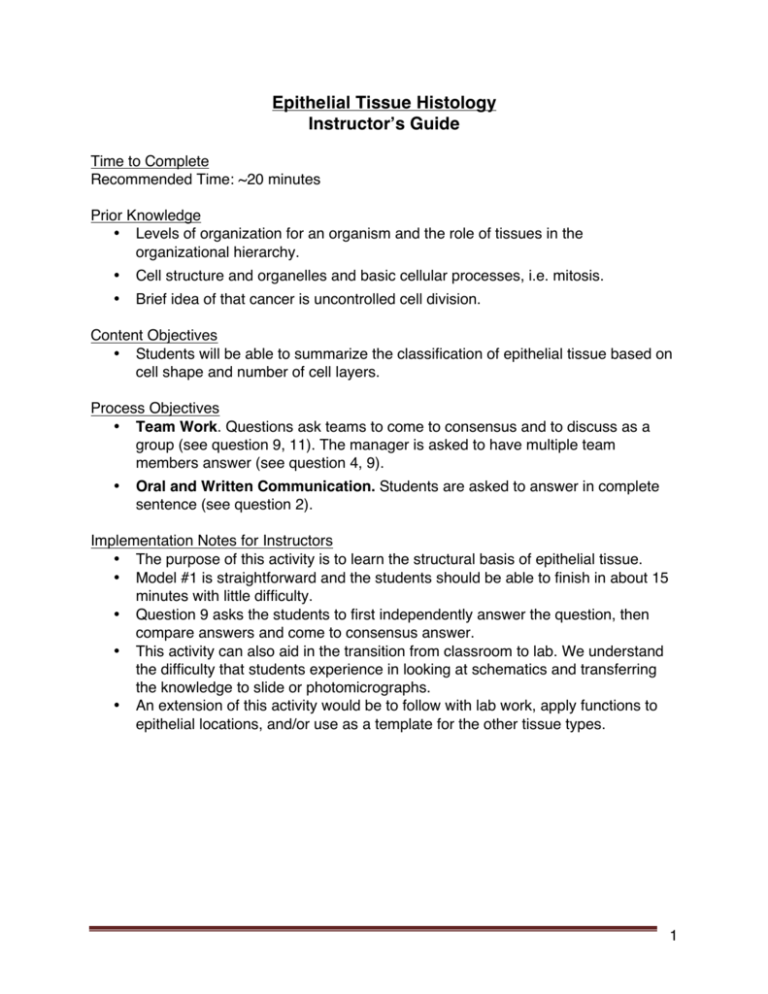
Epithelial Tissue Histology Instructor’s Guide Time to Complete Recommended Time: ~20 minutes Prior Knowledge • Levels of organization for an organism and the role of tissues in the organizational hierarchy. • Cell structure and organelles and basic cellular processes, i.e. mitosis. • Brief idea of that cancer is uncontrolled cell division. Content Objectives • Students will be able to summarize the classification of epithelial tissue based on cell shape and number of cell layers. Process Objectives • Team Work. Questions ask teams to come to consensus and to discuss as a group (see question 9, 11). The manager is asked to have multiple team members answer (see question 4, 9). • Oral and Written Communication. Students are asked to answer in complete sentence (see question 2). Implementation Notes for Instructors • The purpose of this activity is to learn the structural basis of epithelial tissue. • Model #1 is straightforward and the students should be able to finish in about 15 minutes with little difficulty. • Question 9 asks the students to first independently answer the question, then compare answers and come to consensus answer. • This activity can also aid in the transition from classroom to lab. We understand the difficulty that students experience in looking at schematics and transferring the knowledge to slide or photomicrographs. • An extension of this activity would be to follow with lab work, apply functions to epithelial locations, and/or use as a template for the other tissue types. 1 Epithelial Tissue Histology Model #1: Structure of Epithelial Tissue SHAPE Cuboidal Columnar Other Simple Squamous Simple Cuboidal Simple Columnar Pseudostratified Columnar Stratified Squamous Stratified Cuboidal Stratified Columnar Transitional Stratified LAYERS Simple Squamous QUESTIONS: 1. Describe the morphology (shape) of the following epithelial tissue cells. a) Cuboidal: cube / square b) Columnar: column / rectangle c) Squamous: flat 2 2. Write a grammatically correct sentence to describe the difference between simple and stratified epithelial tissue: Sentence should discuss that simple epithelium contains one layer (row) of cells, while stratified epithelium contains two or more layers of cells. 3. Reviewing your answers to questions #1 and #2, what two characteristics do you think are used to classify epithelial tissue? shape of cell and layers of cells 4. Based on your examination of Model 1, which epithelial tissues do not conform to the naming classification rules you described in question #3? (The manager should have two different team members state a tissue and describe its structure). Pseudostratified columnar and transitional epitheliums do not follow normal naming convention. Pseudostratified columnar contains rectangle (column) shaped cells arranged in only layer. Transitional epithelium contains multiple layers of cube shaped cells the apical surface having larger cells then the basal surface. 5. Compare pseudostratified columnar epithelium and simple columnar epithelium and answer the following questions: a) How are these two tissues the same? Both are simple tissues with column shaped cells. b) What differentiates pseudostratified columnar epithelium from simple columnar? In pseudostratified columnar, not all the cells appear to reach from top to bottom and nucleus is found at different levels giving it a layered appearance 6. The bottom (basal) layer of stratified epithelial tissue may have different cell shapes from the top (apical) layer. Which layer determines the shape classification of the tissue? Provide evidence to support your answer. Top layer. Both stratified squamous and stratified columnar contain cube shaped cells on the basal surface, but apical shape matches the name of the tissue. 7. Name the specific type of epithelial described by the following: a tissue that consists of multiple layers of cells, in which the top layer is composed of columnar cells. Stratified columnar epithelial. 3 8. Before answering as a group, each person should individually complete this question by themselves. Once everyone is done, complete Question #9 as a group. a. Label the apical surface on each epithelial tissue photomicrograph. b. Label the basal surface on each epithelial tissue photomicrograph. c. Draw a bracket to indicate the location of the epithelial tissue. d. Name the specific epithelial type under both Tissue a and Tissue b. APICAL BASAL a. b. Images courtesy of The University of Kansas Tissue = simple columnar epithelial Tissue = stratified squamous epithelial 9. Discuss and compare your individual answers to the above question with your group. (The manager should ensure that all group members share their answers). Develop a group consensus and write the best answer below: Group consensus to question eight above. 10. The majority of dust is composed of human skin cells. What does this indicate about the rate of mitosis for epithelial tissue? Answers should discuss the exposure of skin to the outside world and lead to an idea that skin cells have a relatively rapid rate of mitosis. 11. Epithelial cancers are the most common types of cancer. Answer the following questions together as a group: a) Briefly explain what cancer is. An abnormal growth of cells which tend to proliferate in an uncontrolled way and, in some cases, to metastasize (spread). b) What do you think makes skin so susceptible to uncontrolled cellular growth? Answers could discuss either or both the exposure to the external environment and possible carcinogens, or the rapid rate of mitosis that would allow the cancer to spread quickly. 4
