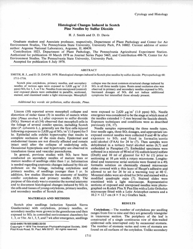
Cytology and Histology
Histological Changes Induced in Scotch
Pine Needles by Sulfur Dioxide
H. J. Smith and D. D. Davis
Graduate student and Associate professor, respectively, Department of Plant Pathology and Center for Air
Environment Studies, The Pennsylvania State University, University Park, PA 1680.2. Current address of senior
author: Argonne National Laboratory, Argonne, IL 60439.
Contribution 1023, Department of Plant Pathology, The Pennsylvania Agricultural Experiment Station.
Authorized for publication 10 March 1978 as Journal Series Paper 5465, and Contribution 498-78, Center for Air
Environment Studies. The Pennsylvania State University, University Park.
Accepted for publication 6 July 1978.
ABSTRACT
SMITH, H. J., and D. D. DAVIS. 1978. Histological changes induced in Scotch pine needles by sulfur dioxide. Phytopathology 68:
1711-1716.
Scotch pine cotyledons, primary needles, and secondary
collapse was the most common structural change induced by
needles of various ages were exposed to 2,620 ug/m' (1.0
SO 2 in all three needle types. Resin canal occlusion also was
ppm) SO2 for 1,3, or 5 hr. Needles from unexposed (control)
observed in primary and secondary needles exposed to SO2.
and exposed plants were embedded in paraffin, sectioned,
Increased dosages of SO 2 did not induce additional
stained, and examined under a light microscope. Mesophyll
symptoms but intensified those already present.
Additional key words: air pollution, sulfur dioxide, Pinus.
Linzon (10) reported severe mesophyll collapse and
distortion of stelar tissue (5) in needles of eastern white
pine (Pinus strobus L.) after exposure to sulfur dioxide
(SO 2). Stewart et al (14) observed that mesophyll cells in
the foliage of several conifer species, including Scotch
pine (P. sylvestris L.), generally are the first cells injured
following exposure to 2,620 /tg of S0 2 / m3 (1.0 ppm) for 5
hr. Epithelial cells exhibit hypertrophy that results in
complete occlusion of the resin ducts. Epidermal and
endodermal tissues are not directly affected and remain
intact until after the collapse of underlying cells.
Occasional hyperplasia and hypertrophy are observed in
transfusion tissue and vascular parenchyma.
In general, previous studies with SO 2 have been
conducted on secondary needles of mature trees or
mature needles of seedlings older than 1 yr. Information
is needed regarding anatomic changes induced by this air
pollutant on* the foliage, including cotyledons and
primary needles, of seedlings younger than 1 yr. In
addition, few studies illustrate the anatomy of healthy
conifer cotyledons and primary needles (8,9). The
purpose of this study was to examine the normal anatomy
and to document histological changes induced by SO 2 in
the cells and tissues of young cotyledons, primary needles,
and secondary needles of Scotch pine.
were exposed to 2,620 #g/m 3 (1.0 ppm) SO 2 . Needle
emergence was considered to be the stage at which most of
the needles extended 1-2 mm beyond the fascicle sheath.
Exposure techniques and conditions were as described
previously (16, 17).
Needle samples representing the three needle types,
four needle ages, three SO 2 dosages, and appropriate unexposed control needles were collected 0 and 48 hr after
exposure to SO 2 and fixed in formalin/acetic
acid/alcohol (FAA) for 24 hr (7). Then samples were
dehydrated in a tertiary butyl alcohol series (6,7) and
embedded in Paraplast (7). Embedded specimens were
softened in a mixture of 90 ml of 1% sodium lauryl sulfate
(Dreft) and 10 ml of glycerol for 6.5 hr (1) prior to
sectioning at 10 /m with a rotary microtome. Longitudinal and transverse serial sections were floated in a 4%
formalin solution on chemically cleaned microscope
slides covered with a thin layer of Haupt's adhesive and
allowed to set for 24 hr on a warming tray at 40 C.
Mounted slides were air-dried for 24 hr and stained with a
modified quadruple stain (7). Stained slides were
examined with a light microscope. Representative
sections of exposed and unexposed needles were photographed on Kodak Plus X Pan film with a Leitz Ortholux
microscope fitted with a Leitz Aristophot camera and a
10.2 X 12.7 cm (4 X 5 in.) Graflex back.
MATERIALS AND METHODS
RESULTS
Scotch pine seedlings (selection Spanish Sierra
Guadarrama) with cotyledons, primary needles, or
Cotyledons.-The number of cotyledons per seedling
secondary needles were grown in the greenhouse and then ranges from five to nine and the are enerall triangular
exposed to SO 2 in controlled environment chambers for in g
ry
gp y
g
1, 3, or 5 hr. At 1,3, 5, and 7 wk after emergence, seedlings constructed of a single continuous row of thin-walled
0032-949X/78/000309$3.00/00
Copyright © 1978 The American Phytopathological Society, 3340
Pilot Knob Road, St. Paul, MN 55121. All rights reserved,
1711
epidermal cells interrupted only by occasional stomata.
The number of stomata varies and rows of stomata are
found on all surfaces of the cotyledon. Unlike secondary
1712
PHYTOPATHOLOGY
[Vol. 68
ff
II
Fig. 1-2. Normal (unexposed to SO 2) Scotch pine cotyledon tissue in 1) transverse and 2) longitudinal sections showing the
arrangement of the epidermal cells. Legend: epidermis, ep; mesophyll, m; endodermis, en; transfusion tissue, tt; vascular tissue, vt;
and stoma, s. Magnifications: Fig. 1 X378 and Fig. 2 X337.
34
ep
ep
Fig. 3-4. Scotch pine cotyledon tissue exposed for 5 hr to 2,620 ,g SO 2/ i 3 . 3) Transverse section showing collapse of mesophyll
(m), distortion of epidermis (ep) and endodermis (en), and compression of the transfusion tissue (tt) (X314). 4) Longitudinal section
showing collapse of necrotic mesophyll tissue and distortion of the epidermis and endodermis (X362).
December 1978]
SMITH AND DAVIS: SCOTCH PINE/NEEDLE HISTOPATHOLOGY/SO 2
needles, cotyledons do not contain a hypodermal layer or
resin canals. The mesophyll is composed of looselypacked, spherical cells that lack the typical internal ridges
of primary and secondary needles (Fig. 1-2). A circular,
single row of thin-walled cells forms the endodermis. A
central single vascular bundle of primary xylem and
primary phloem tissue is surrounded by one or two layers
of transfusion parenchyma.
The most common anatomic change in cotyledons
exposed to SO 2 was collapse of mesophyll cells (Fig. 3--4).
Occasional hypertrophy of these cells was observed in the
abrupt 1-2 mm transition zone between necrotic and
apparently uninjured tissue. The epidermis did not
appear to be directly affected by SO 2 but eventually
became distorted after the underlying mesophyll cells
collapsed. Distortion of the stelar region was observed
only after the initiation of mesophyll collapse and
hypertrophy. This response was observed in the
endodermis, transfusion tissue, and phloem cells. Xylem
distortion occurred only after all other structures
collapsed.
Changes were not observed in any cells of cotyledons
exposed to SO 2 for 1 hr or in any samples exposed at the
time of seed coat shedding regardless of the dosage used.
The types of changes recorded were similar for all ages
from 1to 7 wk for both 3-hr and 5-hr exposures. The only
variation noted was with the total amount of tissue
injured,
Primary and secondary needles.-As the cotyledons,
1713
the periphery of the primary needle is composed of a
single continuous row of thin-walled epidermal cells and
is devoid of a hypodermis (Fig. 5 -6). Typical plicate
mesophyll cells (4,15) are present in a closely-packed
arrangement (Fig. 6). Resin canals are present and are
circumscribed by a well-defined single layer of
sclerenchyma cells. The endodermis is formed by a single
row of thin-walled cells arranged in a circle when viewed
in cross 'section (Fig. 5). As observed in the cotyledons,
primary needles contain only one vascular bundle that is
surrounded by several layers of transfusion tissue.
Secondary needles are semicircular in cross section
with a periphery constructed of a single row of thickwalled epidermal cells and a single row of sclerified,
fibrous hypodermal cells (Fig. 7-8). Mesophyll cells are
plicate and contain several deep internal ridges. The
endodermis of the secondary needles consists of one row
of thick-walled cells. Two vascular bundles are present
and surrounded by several layers of transfusion
parenchyma and tracheids. Each needle has six or seven
peripheral resin canals. These canals are surrounded by a
single row of sclerenchyma cells attached to the
hypodermis and are lined with a single row of thin-walled
epithelial cells.
Both primary and secondary needle types responded
similarly regardless of age of SO 2 dosage (Fig. 9-10,1112). As with the cotyledons, the most common change in
needle anatomy was collapse of the mesophyll tissue with
hypertrophy (Fig. 8, 12). Hypertrophy was limited to the
5€
CS
Fig. 5-6. Normal (unexposed to SO2) Scotch pine primary needle tissue. 5) Transverse section illustrating the arrangement of
epidermis (ep), plicate mesophyll (m), resin canals (rc), stoma (s), endodermis (en) with Casparian strips (cs), transfusion tissule (tt),
xylem (x), phloem (p), and sclerenchyma layer (sl) (X244). 6) Longitudinal section illustrating the arrangement of plicate mesophyll,
endodermis, and transfusion tissue (X272).
1714
PHYTOPATHOLOGY
7
[Vol. 68
rc
S}
Fig. 7-8. Normal (unexposed to SO 2) Scotch pine secondary needle tissue. 7) Transverse section showing needle shape and internal
structure including epidermis (ep), hypodermis (h), mesophyll (m), resin canal (rc), stomata (s), endodermis (en), transfusion tissue
(tt), and vascular bundles (vb) (X106). 8) Longitudinal section showing epidermis, plicate mesophyll (pm), endodermis, and vascular
tissue (vt) (X270).
ell-n
Fig. 9-10. Scotch pine primary needle tissue exposed for 3 hr to 2,620 ltg S0 2 /m 3 . 9) Transverse section showing collapsed
mesophyll (m) and the resulting distortion of the epidermis (ep), compression of endodermis (en), transfusion tissue (tt), and vascular
tissue (vt) (X266). 10) Longitudinal section illustrating mesophyll (m) collapse, distortion of epidermis, and compression of the
endodermis (X430). Section is from the transition zone between asymptomatic and necrotic (tip necrosis) tissue.
December 1978]
SMITH AND DAVIS: SCOTCH PINE/NEEDLE HISTOPATHOLOGY/SO
transition zone between healthy and injured tissue and
did not vary appreciably between primary and secondary
needles. However, the extent of collapse appeared to be
slightly greater in the secondary needles. Again, the
epidermis was not directly affected by SO 2 but became
distorted after the underlying cells collapsed. Epithelial
cells showed severe hypertrophy resulting in total
occlusion of the resin canals (Fig. 11). The layer of
sclerenchyma cells surrounding the canals was
compressed as a result of simultaneous mesophyll and
epithelial cell hypertrophy. This occlusion of the resin
ducts continued far into the portion of the needle that did
not show macroscopic symptoms. As with the epidermis,
the endodermis did not generally show direct injury as a
result of exposure to SO 2 . Compression and collapse of
the endodermis became evident as a result of the hypertrophy of adjacent mesophyll and transfusion tissue.
Changes in the vascular region were characterized by
hypertrophy and associated compression of transfusion
and phloem parenchyma. Xylem tissue was the last of the
vascular tissues to collapse (Fig. 7).
As with the cotyledons, no variation in the type of
structural changes was observed from one age to another
or between the 3-hr and 5-hr doses of SO 2 . The primary
needles did not exhibit changes when exposed to SO 2 at
the time of seed coat shedding (0 wk); secondary needles
exposed at the time of needle emergence from fascicle
2
1715
sheaths (0 wk) showed anatomic changes for both 3-hr
and 5-hr dosages. No significant changes in anatomy were
observed in cotyledons, primary needles, and secondary
needles collected immediately after exposure to SO 2.
Detectable changes were induced only after 3 or 5 hr of
exposure to SO 2 . Microscopic injury was observed only
on needles that exhibited macroscopic symptoms. The
most common macroscopic symptom on Scotch pine
cotyledons, primary needles, and secondary needles was a
tan necrosis, extending from the needle tip toward the
base. Necrotic areas were separated from the uninjured
green base by an abrupt line of demarcation.
DISCUSSION
Sutherland (15) and others (2-5,10,12-14) have
provided information on the anatomy of mature conifer
foliage, but few reports deal with the structure of
cotyledons and primary needles. Such a lack of basic
information can create problems, as illustrated by
comparing the study conducted by Kozlowski and
Clausen (8) with that of Krugman and Critchfield (9).
The anatomy of healthy cotyledons, primary needles,
and secondary needles of Scotch pine were quite similar
to those reported for red pine by Krugman ,and
Critchfield (9). Both studies reveal histological
differences among needle types. Both primary and
12
ep
:~tt
Fig. 11-12. Scotch pine secondary needle tissue exposed for 5 hr to 2,620 ,g SO2.11) Transverse section showing occlusion of resin
canals (rc), compression of the sclerenchyma layer (sl) surrounding the resin canal, compression of the endodermis (en) and
transfusion tissue (tt), and distortion of the phloem tissue (p) (X116). 12) Longitudinal section showing hypertrophy and collapse of
mesophyll (m), compression of endodermal cells, and distortion of epidermis, transfusion tissue, and vascular tissue (vt)(X383).
Section was taken in the transition zone between asymptomatic and necrotic (tip necrosis) tissue.
1716
PHYTOPATHOLOGY
secondary needles contain resin canals, but the
cotyledons do not. The secondary needles have a thickwalled epidermis accompanied by a fibrous sclerified
hypodermis, and the cotyledons and primary needles
have a thin-walled epidermis and lack a hypodermis.
Such differences are uniform, and the needle types do not
vary with age. These findings are similar for Scotch pine
(14) and red pine (9) and may be true for the genus Pinus
ngeneral.
In this study most changes on needles injured by SO 2
were similar to previously described alterations of
secondary needles of Scotch pine and other conifers
exposed to this pollutant (2,10,11,14). The extent of most
changes did not vary appreciably with needle types or
ages. The most frequent change was the severe collapse of
the mesophyll cells that may have been responsible for the
distortion of the epidermis and stelar region in the
endodermis. An interesting observation was the
extension of the resin canal occlusion far into the
apparently uninjured portion of symptomatic primary
and secondary needles; all other responses terminated at
the end of the 1-2 mm transition zone. A similar response
was reported in mature secondary Scotch pine needles
exposed to SO 2 (14).
Stewart et al (14) reported that SO 2 induces severe
hypertrophy of the tissues of Scotch pine needles in the
transfusion and vascular regions. In this study only minor
swelling was observed on the needles and the most
common change in those areas was compression or
collapse but not hypertrophy. In addition, Stewart et al
(14)
observed
esopylland1973.
bth mesophyll
n both
(14)obsrvedgraulaion
granulation in
and
transfusion tissue cells. No evidence of granulation was
observed in this study. Some of these differences,
however, may reflect the differences in needle age in the
two studies or a genetic difference in the selections
investigated.
LITERATURE CITED
1. ALCORN, S. M., and P. A. ARK. 1953. Softening paraffinembedded plant tissue. Stain Technol. 28:55-56.
[Vol. 68
2. EVANS, L. S., and P. R. MILLER. 1972. Comparative
needle anatomy and relative ozone sensitivity of four pine
species. Can. J. Bot. 50:1067-1077.
3. EVANS, L. S., and P. R. MILLER. 1972. Ozone damage to
ponderosa pine: A histological and histochemical
appraisal. Am. J. Bot. 59:297-304.
4. ESAU, K. 1960. Anatomy of seed plants. John Wiley &
Sons, New York. 376 p.
5. ESAU,
1965. Plant anatomy. John Wiley & Sons, New
Ybrk.K.767
p.
6. JENSEN, W. A. 1962. Botanical histochemistry. W. H.
Freeman and Co., San Francisco. 408 p.
7. JOHANSEN, D. A. 1940. Plant microtechnique. McGrawHill, New York. 523 p.
8. KOZLOWSKI, T. T., and J. J. CLAUSEN. 1966.
Anatomical responses of pine needles to herbicides.
Nature 209:486-487.
9. KRUGMAN, S. L., and W. B. CRITCHFIELD. 1968. Red
pine needle structure. Nature 217:685-686.
10. LINZON, S. N. 1967. Histological studies of symptoms in
semimature-tissue needle blight of eastern white pine.
Can. J. Bot. 45:133-143.
11. SCURFIELD, G. 1960. Air pollution and tree growth. For.
Abstr. 21:339-347, 517-528.
12. SOLBERG, A., and D. F. ADAMS. 1955. Some effects of
hydrogen fluoride on the internal structure of Pinus
ponderosa. Natl. Air Pollut. Symp. 4:164-176.
13. SOLBERG, A., and D. F. ADAMS. 1956. Histological
leaves to hydrogen fluoride and
of some
responses
sulfur dioxide.
Am.plant
J. Rot. 43:755-766.
and F.:M.5ARNER
H
e. m. T
14. STEAR,
n eedle ner.
of conifer
ato
1 93 PATh
anatomy of conifer needle necrosis.
Can. J.Pathological
Rot. 51:983-988.
15. SUTHERLAND, M. 1933. A microscopic study of the
structure of the leaves of the genus Pinus. N. Z. Inst.
Trans. Proc. 63:517-568.
16. SMITH, H. J. 1976. Influence of SO 2 on conifers with
emphasis on morphology and histology of Scotch pine.
M.S. Thesis, The Penn. State Univ., University Park. 74
P.
17. SMITH, H. J., and D. D. DAVIS. 1977. The influence of
needle age on sensitivity of Scotch pine to acute doses of
SO 2 . Plant Dis. Rep. 61:870-874.

