Purification and Identification of Paenibacillus sp., Isolated from
advertisement
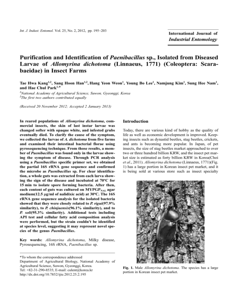
Int. J. Indust. Entomol. Vol. 25, No. 2, 2012, pp. 195~203 International Journal of Industrial Entomology Purification and Identification of Paenibacillus sp., Isolated from Diseased Larvae of Allomyrina dichotoma (Linnaeus, 1771) (Coleoptera: Scarabaeidae) in Insect Farms Tae Hwa Kang1,2, Sang Hoon Han1,2, Hang Yeon Weon1, Young Bo Lee1, Namjung Kim1, Sung Hee Nam1, and Hae Chul Park1,* 1 National Academy of Agricultural Science, Suwon, Gyeonggi, Korea The first two authors contributed equally 2 (Received 20 November 2012; Accepted 2 January 2013) In reared populations of Allomyrina dichotoma, commercial insects, the skin of last instar larvae was changed softer with opaque white, and infested grubs eventually died. To clarify the cause of the symptom, we collected the larvae of A. dichotoma from five farms and examined their intestinal bacterial florae using pyrosequencing technique. From those results, a member of Paenibacillus was found only in the larvae showing the symptom of disease. Through PCR analysis using a Paenibacillus specific primer set, we obtained the partial 16S rRNA gene sequence and confirmed the microbe as Paenibacillus sp. For clear identification, a whole guts was extracted from each larva showing the sign of the disease and incubated at 70oC for 15 min to isolate spore forming bacteria. After then, each content of guts was cultured on MYPGPNAL agar medium(12.5 µg/ml of nalidixic acid) at 30oC. The 16S rRNA gene sequence analysis for the isolated bacteria showed that they were closely related to P. rigui(97.9% similarity), to P. chinjuensis(96.1% similarity), and to P. soli(95.3% similarity). Additional tests including API test and cellular fatty acid composition analysis were performed, but the strain couldn't be identified at species level, suggesting it may represent novel species of the genus Paenibacillus. Introduction Today, there are various kind of hobby as the quality of life as well as economic development is improved. Keeping insects such as dynastid beetles, stag beetles, crickets, and ants is becoming more popular. In Japan, of pet insects, the size of stag beetles market approached to over two or three hundred billion KRW, and the insect pet market size is estimated as forty billion KRW in Korea(Choi et al., 2011). Allomyrina dichotoma (Linnaeus, 1771)(Fig. 1) has a large portion in Korean insect pet market, and it is being sold at various store such as insect specialty Key words: Allomyrina dichotoma, Milky disease, Pyrosequencing, 16S rRNA, Paenibacillus sp. *To whom the correspondence addressed Department of Agricultural Biology, National Academy of Agricultural Science, Suwon, Gyeonggi, Korea. Tel: +82-31-290-8535; E-mail: culent@korea.kr http://dx.doi.org/10.7852/ijie.2012.25.2.195 Fig. 1. Male Allomyrina dichotoma. The species has a large portion in Korean insect pet market. 196 Tae Hwa Kang et al. reached at between 2 × 10⁹ and 5 × 10⁹(Zhang et al.,1997). The object of this study is to purify and identify the caused bacteria of the disease appearing in Korean A. dichotoma larvae. For this, firstly, the intestinal bacterial florae of the healthy and the diseased larvae were surveyed. On the basis of the bacterial florae, we tried to search different points between the healthy and the diseased larva and, lastly, to purify and to identify the microbe appearing only in the diseased larvae. Materials and Methods Fig. 2. Symptoms of the bacterial disease in Allomyrina dichotoma. (1) healthy larva; (2) rough epidermis; (3) the body color changes to milky white, and the body becomes soften; (4) finally, the larva dies. stores and hypermarkets. In 2010, however, a pandemic disease spreads at several insect farms, and in 2012, a case of mass mortality of Japanese rhinoceros beetles showed through the whole country of Korea. The symptoms of the disease mainly emerge at the third instar-end larvae of the beetle, and the larva dies before or after making pupa room. The symptoms of the disease were as follows(Fig. 2): (1) rough epidermis; (2) the body color changes from shiny white to milky white, and the body becomes soften; (3) finally, the larva dies. In general, when the larva of the beetle was bred at an insect farm, about 60 individuals with fermented sawdust were put into 120L plastic box. In this case, if a diseased larva is found in the rearing box, almost all individuals die of a disease which is similar to a bacterial infection. Several bacterial diseases were reported in insects, such as American Foulbrood caused by Paenibacillus larvae in European honeybee(Apis mellifera Linnaeus, 1758), Sotto disease by Bacillus thuringiensis in silkworm(Bombix mori (Linnaeus, 1758)), milky disease by Paenibacillus popilliae and P. lentimorbus in Scarabaeid beetles, and amber disease by Serratia entomophila and S. proteamaculans in Scarabaed beetles(Dutky, 1940; Grkovic et al., 1995; Han et al., 2008; Ibrahim et al., 2010; Zhang et al., 1997). Most of the diseases were caused by Bacillus and Paenibacillus, and the only one case, amber disease by Serratia spp. Specially, milky disease appearing in Scarabaeid beetles is very similar to that of Korean A. dichotoma in the symptoms, which the body color of A. dichotoma larvae was changed to milky white and died when the number of spore of P. popilliae Genomic DNA extraction from the gut of A. dichotoma larvae For DNA extraction, A. dichotoma larva was washed as follows: the larva was soaked in 70% ethanol for 1 min and then in 0.8% HOCl solution for 1 min. And solution residues were removed by sinking in DW for 1 min. The larva was dissected in a sterilized petri dish. The gut extracted from the larva was mashed by a mortar. Genomic DNA was extracted from 200 mg of the mashed gut material with QiaAmp DNA mini kit(Qiagen, Germany) according to the manufacturer’s instruction. Pyrosequencing on the gut microbiota of A. dichotoma larvae The intestinal bacterial florae was surveyed in three healthy(two individuals from insect farm in Yuseong of Daejeon, and one from Hwingseong of Gangwondo) and three diseased larvae(each individual from Hwingseong of Gangwondo, Namyangju of Gyeonggido, and Yeongam of Jeollanamdo) by using a pyrosequencing technique. For pyrosequencing, bacterial 16S rRNA genes were amplified using the primers V1-9F(5-X-ACGAGTTTGATCMTGGCTCAG-3) and V3-541R(5-XAC-WTTACCGCGGCTGCT GG-3)(Chun et al., 2010) where X denotes a 7–11 nucleotide long barcode followed by a common linker AC. PCR amplification was performed using the following PCR protocol: initial denaturation(5 min at 94°C), 30 cycles at 94°C for 30 s, 55°C for 30 s, and 72°C for 60 s and the final extension at 72°C for 5 m. The PCR amplification curves were constructed by sampling the PCR product at 5 cycle intervals and quantifying it using a Qubit 2.0 fluorometer and Qubit dsDNA BR assay kit(Invitrogen, USA). The PCR products obtained at 20 cycles and 30 cycles were selected. The PCR products were gel-purified with the QIAquick Gel extraction kit(Qiagen, Germany) and subjected to pyrosequencing, which was performed by National Instru- Purification and identification of Paenibacillus sp. from A. dichotoma mentation Center for Environmental Management(Seoul, Korea) using a 454 GS FLX Titanium Sequencing System(Roche, Germany), according to the manufacturer’s instructions. Pyrosequencing data were analyzed using the Mothur software package(version 1.23.1)(Schloss et al., 2009). Pre-filtered flowgrams of the pyrosequencing reads were clustered using the PyroNoise algorithm(Quince et al., 2011), and chimeric sequences were removed using UCHIME(Edgar et al., 2011). UCHIME was performed in De novo mode. Polymerase chain reaction using Paenibacillus specific primer Paenibacillus sp. which may be one of causative pathogens of A. dichotoma larva was detected from the gut of the larvae from Yuseong of Daejeon. Thus, with Paenibacillus specific primer set, PAEN515F(Shida et al., 1997) and RNA1484R(Petterson et al., 1999), PCR detection was conducted for the larvae collected from insect farms of six regions, Hwingseong of Gangwondo, Namyangju of Gyeonggido, Yeongdong of Chungcheongnamdo, Yuseong of Daejeon, Yeongam of Jeollanamdo, and Hwasun of Jeollanamdo. Amplification conditions for both reactions were denaturation at 94oC for 5 min followed by 25 cycles of denaturation at 94oC for 1 min, annealing at 58oC for 1.5 min and extension at 72oC for 1.5 min followed by a final extension at 72oC for 5 min. Purification of the target bacteria The gut was extracted from the larva which Paenibacillus sp. was detected by Paenibacillus specific primer. The gut was grinded with 10 ml of Phosphate Buffered Saline (PBS, pH 7.4) and then 500 µl of the specimen was inoculated in 30 ml MYPGP media(10 g of Mueller Hinton, 15 g of Yeast extract, 3 g of K2HPO4, 2 g of Glucose,and 1 g of Sodium pyruvate for 1 Liter DW). After over-night incubation(30ºC, 180 rpm), the bacteria culture was incubated in water bath at 70oC for 15 min. And then 100 µl of bacterial culture was inoculated into 30 ml of fresh MYPGP media containing Nalidixic acid(12.5 ug/ml). After over-night incubation(30ºC, 180 rpm), the bacterial culture was diluted tenfold up to 10-9 and then each of 50 µl from 10-7, 10-8, and 10-9 dilutants was spread on MYPGPNAL agar media. After over-night incubation(30ºC), several single colonies were picked and spread on fresh MYPGPNAL agar media. Identification of the target bacterium Identification of Paenibacillus sp. was carried out by using four methods following as 16S rRNA gene sequence analysis, gram staining, API test, and cellular fatty acid composition. 16S rRNA gene sequence was 197 amplified by using universal primer set, 27F and 1492R(amplicon size about 1,400 bp). For more clear results, we designed the internal primer set PAEN793R(5'AGGCGGAGTG CTTACTGTGT-3') and PAEN540F(5'CGCACTGAAAACTGGATGAC-3') using Primer3 software(Rozen and Skaletsky, 2000). 16S rRNA gene was amplified by two parts, such as the former region amplified by 27F-PAEN793R primer set and the latter region by PAEN540F-1492R primer set. Amplification conditions for both reactions were denaturation at 94oC for 5 min followed by 35 cycles of denaturation at 94oC for 30 min, annealing at 58oC for 30 min and extension at 72oC for 30 min followed by a final extension at 72oC for 5 min. DNA sequencing of the amplicons were performed by Macrogen(Seoul, Korea). Determined sequences were aligned and analyzed under molecular evolutionary genetic analysis software, MEGA 5(Tamura et al., 2011). API test and cellular fatty acid composition analyses were performed by Korean Culture Center of Microorganisms(Seoul, Korea). API 50CHB kit was used for API test. Resulting data on each analysis were compared with apiweb(http:// apiweb.biomerieux.com/ servlet/identify/) for API test and Microbial Identification System(MIDI, 1999) for cellular fatty acid composition. Results and Discussion Detection of Paenibacillus species from the infected A. dichotoma larva Pyrosequencing is commonly used for the analyses on the bacterial florae of the various environment such as soil, swamp, and freshwater as well as the intestinal tube and the feces of organism, or for the comparison of the bacterial florae from two or three different environmental region(Boissière et al., 2012; Kim et al., 2010; Matcher et al., 2011; Nacke et al., 2011; Shi et al., 2010; Wong et al., 2011). In insects, several works on the gut bacterial florae were performed(Boissière et al., 2012; Shi et al., 2010; Wong et al., 2011). Considering above cases, we tried to compare the gut bacterial florae of three diseased larvae of A. dichotoma from four regions including Yuseong of Daejeon which the larval disease has broken out with those of three healthy larvae. In the results, the larval gut bacteria were classified by total 28 taxa, of which three phylum, Firmicutes, Proteobacteria and Cyanobacteria, showed high species diversity(Fig. 3). On the basis of the results, the bacterial florae between the healthy and the diseased larvae were compared with each other using principal coordinates analysis and seven clustering methods such as barycurtis, morisitahorn, thetayc, jclass, jest, sorclass, sorest, and the results 198 Tae Hwa Kang et al. Fig. 3. Phylum and genus composition of the gut from Allomyrina dichotoma larvae(Star mark: diseased larva). Larval gut bacteria florae were composed as total 28 taxa, of which Firmicutes, Proteobacteria and Cyanobacteria showed high species diversity. Fig. 4. Clustering analysis based on seven algorithm on bacterial florae of larval gut collected from insect farms of four region(Star mark: diseased larva). Fig. 5. Principal coordinate analysis on bacterial florae of larval gut collected from insect farms of four region(Star mark: diseased larva). on each clustering method were integrated by jacknife sample clustering method. In principal coordinate analysis and clustering method, however, the gut bacterial florae between the healthy and the diseased larvae did not showed clear different points(Figs. 4 and 5). In the larvae only from Yuseong of Daejeon, Paenibacillus sp. was detected as very small density of 0.1%. Several species of Paenibacillus were known as the famous disease causative pathogen in insects(Gardener, 2004). P. larvae is a causative bacteria of American Foulbrood in European honeybee(Genersch, 2008; Han et al., 2008), and in Scarabaeid beetles, milky disease was caused by P. popilliae and P. lentimorbus, which are used for biological control agents against on Japanese beetle(Popillia japonica Newman, 1841) in USA(Dutky, 1940; Pettersson et al., 1999). In the point of view, Paenibacillus sp. detected from the A. dichotomus larvae might be a causative pathogen of the larval disease. Thus, we conducted PCR detection on A. dichotoma larvae from insect farm of six region using Paenibacillus specific primer set, PAEN515F(Shida et al, 1997) and RNA1484R(Petterson et al, 1999). In the result, Paenibacillus sp. was detected only in the larvae from Yuseong of Daejeon(Fig. 6). The PCR result suggested strong possibility that Paenibacillus sp. might be a causative baciteria. Therefore, we tried to purify Paenibacillus sp. from the gut of A. dichotoma larvae for more detailed works, such as the identification of the bacteria, and the infection test on A. dichotoma larvae. Purification and identification of Paenibacillus sp. from A. dichotoma 199 Fig. 7. Gram staining on purified Paenibacillus sp. The bacteria is rod type and appeare in violet color. Table 1. NCBI accession number of 16S rRNA gene sequences of 18 Paenibacillus species No Fig. 6. Detection of Paenibacillus sp. using Paenibacillus specific primer set on Allomyrina dichotoma larvae from insect farms of six regions (Yuseong: A-1, A-2, B-10, B-11, B-12, B13, B-14; Yeongdong: A-3, A-4, A-5, A-6, A-7; Yeongam: A8, A-9, A-10, A-11, A-12; Hwasun: A-13, A-14, A-15, A-16; Namyangju: B-1, B-2, B-3, B-4, B-5; Hwingseong: B-6, B-7, B-8, B-9). Paenibacillus sp. was detected only in the larvae from Yuseong of Daejeon. Purification and identification of Paenibacillus species from the infected A. dichotoma larva For purifying Paenibacillus sp., the gut materials of A. dichotoma larvae were cultured in MYPGPNAL media containing 0.1% nalidixic acid(12mg/1ml), which inhibit a subunit of DNA gyrase and induce formation of relaxation complex analogue(Emmerson and Jones, 2003). And then, the conical tube was heated as 70oC in water bath. It is known that Paenibacillus bacteria formed endospores in poor environment, such as nutritionally unfavorable local conditions via dormancy (Nicholson et al., 2000; Nicholson, 2002). Therefore, except only endospore forming bacteria, most of all bacteria were sterilized by heat. But, it was known that Paenibacillus bacteria have resistance on the antibiotics(Han et al., 2008). The cultured bacteria were purified by culturing MYPGPNAL agar media. Purified bacteria were examined under a light microscopy after Gram staining. Paenibacillus are rod type and Gram positive bacteria, which showed violet at Gram stain(Barenfanger et al., 2001). In the result of 1 2 3 4 5 6 7 8 9 10 11 12 13 14 15 16 17 18 19 Scientific Name Paenibacillus azoreducens P. chinjuensis P. chitinolyticus P. daejeonensis P. dendritiformis P. ehimensis P. elgii P. gansuensis P. ginsengisoli P. koreensis P. larvae P. lentimorbus P. naphthalenovorans P. riguii P. soli P. thiaminolyticus P. validus P. popilliae Bacillus subtilis Strain No. CM1 WN9 HSCC596T AP-20 CIP105967T KCTC3748 SD17 B518 Gsoil1638 YC300 DSM7030 ATCC14707 PR-N1 WPCB173 DCY03 IFO15656 JCM9077 ATCC14706 DSM10 NCBI Accession Remarks No. AJ272249 AF164345 AB045100 AF290916 AY359885 AY116665 AY090110 AY839866 AB245382 In-group AF130254 AY530294 AB073199 AF353681 EU939688 DQ309072 AB073197 AB073203 AB073198 AJ276351 Out-group Gram staining, the purified bacteria were rod type and Gram positive(Fig. 7). The identification is based on 16S rRNA gene sequence as well as API test and cellular fatty acid composition. In 16S rRNA gene sequence, we analyzed adding 16 rDNA 200 Tae Hwa Kang et al. Fig. 8. Neighbour joining tree of 16 s rRNA gene of Paenibacillus spp. Paenibacillus sp. is the closest to P. rigui showing as 2.0% branch length. sequences of 18 Paenibacillus species from NCBI in our data matrix(Table 1). In neighbor joining tree, Paenibacillus sp. purified from the gut of A. dichotoma larvae showed as about 2.0% branch length from P. rigui detected from Woopo Swamp(Changnyeong, Gyeongsangnamdo, Korea; Baik et al., 2011) and about 6.0% from P. popilliae, the causative bacteria of milky disease in Scarabaeid beetles(Fig. 8). In sequence divergence, the bacteria showed as 2.1% divergence from P. rigui, 3.9% from P. chinjuensis, and 4.7% from P. soli(Table 2). Therefore, the bacterium seems to be the closest to P. rigui. In the identification using the homology of 16S rRNA gene sequence, the identification criterion by Stackebrandt and Goebel(1994) was generally accepted, who referred that 97% similarity is the cut-off for species identification. After that, however, Drancourt et al.(2000) suggested that 99% cut-off criterion is used for species identification, and 97% similarity might be used only for genus level identification. In recent, Janda and Abbott(2007) suggested that it could be identified as same species when 16S rDNA sequence showed as at least 99% similarity, and the ideal score for same species is 99.5% similarity. Therefore, it might be difficult that Paenibacillus sp. purified from A. dichotoma larvae is treated as a same species using only 97.9% 16S rDNA sequence similarity. Physiological trait test for Paenibacillus sp. purified from A. dichotoma larvae was carried out using API 50CHB kit(bioMérieux). In the result, the bacteria was positive for 25 of 50 conditions(L-Arabinose, D-Xylose, β Methyl-xyloside, Galactose, D-Glucose, D-Fructose, Rhamnose, Mannitol, α Methyl-D-mannoside, α MethylD-glucoside, N Acetyl glucosamine, Amygdaline, Arbutine, Esculine, Salicine, Cellobiose, Maltose, Lactose, Melibiose, Saccharose, Trehalose, Melezitose, D-Raffinose, β Gentiobiose, D-Turanose; Table 3). The similarity test on this result was performed under apiweb. In the result, we could not find any data to be consistent with our data, and the closest matched bacterium was Paenibacillus mucerans which showed as 62.1% similarity. Therefore, the result by API test was unsuitable for the identification of Paenibacillus sp. In cellular fatty acid composition, it was confirmed that total of 15 fatty acids were distributed in cell wall. Among them, 15:0 ANTEISO fatty acid showed the highest ratio as 64.65%, and the next was 16:0 ISO as 8.58%, 15:0 ISO as 7.50%, 14:0 ISO as 4.91%, and 16:0 as 4.36%(Table 4). In the result comparing our data with MIDI data, the data showed similarity of >90% were absent. Considering the results on above three analyses, 16S rDNA sequence, API test, and cellular fatty acid composition, only 16S rDNA sequence data provided taxonomic position of Paenibacillus sp. which is closely related to P. rigui showing 97.9% sequence similarity. But the similarity was consistent with the species identification criterion suggested by Stackebrandt and Goebel(1994), although the score is insufficient to recent criterion(Drancourt et al., 2000; Janda and Abbott, 2007). For more clear identification, we are conducting DNA-DNA hybridization for comparison with closely related species, P. rigui and P. soli. If these closely related species showed less than 70% similarity in the result of DNA-DNA hybridization(Wayne et al., 1987), we could identify Paenibacillus sp. as a new species. In this study, we focused on the purification and identification of Paenibacillus sp. from the gut of the diseased A. dichotoma larvae. Through further study, we will try to clearly identify the bacteria and to test pathogenicity of the bacteria on A. dichotoma larvae. Although this bacterium was purified from the diseased larvae, it is difficult to judge it as the causative pathogen. Generally, milky disease caused by P. popilliae was expressed by increasing number of endospore(Zhang et al., 1997). In survey on the related references, however, we found that milky disease was caused by several members of Paenibacillus and Bacillus, such as P. popilliae, P. lentimorbus, P. lentimorbus var. maryland, P. lentimorbus var. australis, Bacillus euloomarahae, and B. fribourgensis, which are known as endospore forming bacteria, or including endospore sim- P. riguii_WPCB173 P. chinjuensis_WN9 P. soli_DCY03 P. validus_JCM9077 P. ehimensis_KCTC3748 2 3 4 5 6 P. popilliae_ATCC14706 P. koreensis_YC300 9 10 0.056 0.047 0.041 0.042 0.038 5 6 8 9 0.063 0.054 0.048 0.050 0.045 0.008 0.050 0.018 0.072 0.062 0.053 0.062 0.063 0.065 0.064 0.074 0.066 0.060 0.047 0.042 0.044 0.041 0.010 0.044 7 10 P. gansuensis_B518 13 12 0.065 0.052 0.058 0.065 0.066 0.061 0.069 0.058 0.066 0.066 0.066 0.067 0.064 0.058 0.062 0.066 0.065 0.068 0.072 0.067 0.009 0.074 0.010 11 13 P. larvae_DSM7030 P. chitinolyticus_HSCC596T P. ginsengisoli_Gsoil1638 P. daejeonensis_AP-20 P. azoreducens_CM1 Bacillus subtilis_DSM10 15 16 17 18 19 20 14 15 16 17 18 19 0.144 0.131 0.135 0.130 0.134 0.130 0.139 0.127 0.125 0.135 0.122 0.127 0.137 0.119 0.133 0.129 0.127 0.130 0.125 0.080 0.070 0.066 0.069 0.070 0.067 0.076 0.063 0.073 0.075 0.072 0.072 0.067 0.070 0.059 0.055 0.053 0.063 0.089 0.073 0.077 0.075 0.082 0.066 0.084 0.065 0.067 0.072 0.066 0.069 0.059 0.066 0.081 0.059 0.073 0.069 0.065 0.068 0.077 0.077 0.062 0.083 0.063 0.062 0.071 0.063 0.063 0.066 0.063 0.070 0.063 0.067 0.054 0.049 0.051 0.047 0.054 0.056 0.053 0.066 0.062 0.064 0.067 0.041 0.063 0.056 0.068 0.063 0.060 0.064 0.055 0.063 0.056 0.066 0.068 0.070 0.068 0.068 0.066 0.070 P. dendritiformis_CIP105967T 0.065 0.058 0.062 0.066 0.067 0.065 0.075 0.067 0.008 0.073 0.006 0.013 0.067 P. lentimorbus_ATCC14707 12 14 4 0.051 0.044 0.038 0.041 0.047 0.034 0.044 3 P. thiaminolyticus_IFO15656 0.063 0.054 0.062 0.062 0.065 0.064 0.074 0.066 0.004 0.072 P. elgii_SD17 8 11 2 0.039 0.037 0.021 1 P. naphthalenovorans_PR-N1 0.056 0.051 0.047 0.044 0.029 0.043 Paenibacillus sp._Nal8 1 7 Scientific Name_Strain Number No Table 2. 16s rRNA gene sequence divergence of Paenibacillus spp 20 Purification and identification of Paenibacillus sp. from A. dichotoma 201 202 Tae Hwa Kang et al. Table 3. The result of API 50CHB kit (+: positive reaction; −: negative reaction) No 1 2 3 4 5 6 7 8 9 10 11 12 13 14 15 16 17 18 19 20 21 22 23 24 25 API CHB control Glycerol Ertythritol D-Arabinose L-Arabinose + Ribose D-Xylose + L-Xylose Adonitol β Methyl-xyloside + Galactose + D-Glucose + D-Fructose + D-Mannose L-sorbose Rhamnose + Dulcitol Inositol Mannitol + Sorbitol α Methyl-D-mannoside + α Methyl-D-glucoside + NAcetylglucosamine + Amygdaline + Arbutine + API Kit No 26 27 28 29 30 31 32 33 34 35 36 37 38 39 40 41 42 43 44 45 46 47 48 49 50 API CHB Esculine + Salicine + Cellobiose + Maltose + Lactose + Melibiose + Saccharose + Trehalose + Inuline Melezitose + D-Raffinose + Amidon Glycogene Xylitol β Gentiobiose + D-Turanose + D-Lyxose D-Tagatose D-Fucose L-Fucose D-Arabitol L-Arabitol Gluconate 2 ceto-gluconate 5 ceto-gluconate API Kit Table 4. The result of cellular fatty acid composition No 1 2 3 4 5 6 7 8 9 10 11 12 13 14 15 Name 10 : 0 12 : 0 13 : 0 ISO 13 : 0 ANTEISO 14 : 0 ISO 14 : 0 15 : 0 ISO 15 : 0 ANTEISO 15 : 0 16 : 0 ISO 16 : 0 17 : 0 ISO 17 : 0 ANTEISO 17 : 1 w6c 17 : 0 % 0.13 0.15 0.13 0.21 4.91 1.77 7.50 64.65 2.73 8.58 4.36 1.12 3.45 0.18 0.14 ilar materials(Steinkraus and Tachiro, 1967). In this point of view, it was considered that Paenibacillus sp. forming endospore from A. dichotoma larvae might be a disease causative bacteria. Acknowledgement This work was supported by the Rural Development of Administration, Republic of Korea (Grant No. PJ907197) References Baik KS, Lim CH, Choe HN, Kim EM, Seong CN (2011) Paenibacillus rigui sp. nov., isolated from a freshwater wetland. Int J Syst Evol Microbiol 61, 529-534. Barenfanger J, Drake CA (2001) Interpretation of Gram Stains for the Nonmicrobiologist. Lab Med 32, 368-375. Boissière A, Tchioffo MT, Bachar D, Abate L, Marie A, Nsango SE, Shahbazkia HR, Awono-Ambene PH, Levashina EA, Christen R, Morlais I (2012) Midgut Microbiota of the Malaria Mosquito Vector Anopheles gambiae and Interactions with Plasmodium falciparum Infection. PLoS Pathogens 8, e1002742. Choi YC, Kim NJ, Park IK, Lee SB, Hwang JS (2011) RDA Interrobang IV. Rural Development Administration (in Korean). Chun J, Kim K, Lee JH, Choi Y (2010) The analysis of oral microbial communities of wild-type and toll-like receptor 2deficient mice using a 454 GS FLX Titanium pyrosequencer. BMC Microbiology 10, 101. Drancourt M, Bollet C, Carlioz A, Martelin R, Gayral JP, Raoult D (2000) 16S Ribosomal DNA Sequence Analysis of a Large Collection of Environmental and Clinical Unidentifiable Bacterial Isolates. J Clin Microbiol 38, 3623-3630. Dutky SR (1940) Two new spore-forming bacteria causing milky diseases of Japanese beetle larvae. J Agric Res 61, 5768. Edgar RC, Haas BJ, Clemente JC, Quince C, Knight R (2011) UCHIME improves sensitivity and speed of chimera detection. Bioinformatics 27, 2194-2200. Emmerson AM, Jones AM (2003) The quinolones: decades of development and use. J Antimicrob Chemoth 51, 13-20. Gardener BBM (2004) Ecology of Bacillus and Paenibacillus spp. in Agricultural System. Phytopathology 94, 1252-1258. Genersch E (2008) Paenibacillus larvae and American Foulbrood - long since known and still surprising. J Verbrauch Lebensm 3, 429-434. Grkovic S, Glare TR, Jackson TA, Corbert GE (1995) Genes Essential for Amber Disease in Grass Grubs Are Located on the Large Plasmid Found in Serratia entomophila and Serratia proteamaculans. Appl Environ Microb 61, 2218-2223. Han SH, Lee DB, Lee DW, Kim EH, Yoon BS (2008) Ultrarapid real-time PCR for the detection of Paenibacillus lar- Purification and identification of Paenibacillus sp. from A. dichotoma vae, the causative agent of American Foulbrood (AFB). J Invertebr Pathol 99, 8-13. Ibrahim MA, Griko N, Junker M, Bulla LA (2010) Bacillus thuringiensis A genomics and proteomic perspective. Bioengineered Bugs 1, 31-50. Janda JM, Abbott SL (2007) 16S rRNA Gene Sequencing for Bacterial Identification in the Diagnostic Laboratory: Pluses, Perils, and Pitfall. J Clin Microbiol 45, 2761-2764. Kim TS, Kim HS, Kwon S, Park HD (2010) Analysis of Bacterial Community Composition in Wastewater Treatment Bioreactors Using 16S rRNA Gene-Based Pyrosequencing. Korean J Microbiol 46, 352-358 (In Korean). Linnaeus C (1758) Systema Naturae per Regna tria Naturae, secundum classes, ordines, genera, species, cum characteribus, differentiis, synonymis, locis. Tomus I. Editio decima, reformata. Holmiae: Impensis Direct. Laurentii Salvii, p. 824. Linnaeus C (1771) Mantissa plantarum altera generum editionis VI & specierum editionis II. Holmiae: Laurentii Salvii, p. 588. Matcher GF, Dorrington RA, Henninger TO, Froneman PW (2011) Insights into the bacterial diversity in a freshwaterdeprived permanently open Eastern Cape estuary, using 16S rRNA pyrosequencing analysis. Water SA 37, 381-390. MIDI (1999). Sherlock microbial identification system, operating manual, version 3.0. Newark, DE: MIDI, Inc. Nacke H, Thürmer A, Wollherr A, Will C, Hodac L, Herold N, Schöning I, Schrumpf M, Daniel R (2011) PyrosequencingBased Assessment of Bacterial Community Structure Along Different Management Types in German Forest and Grassland Soils. PLos ONE 6, e17000. Newmann E (1841) A descriptive list of the species of Popillia, in the cabinet of the Rev. F. W. Hope, M. A., with one description added, from a specimen in the British Museum. Transactions of the Entomological Society of London 3, 3250. Nicholson WL, Munakata N, Horneck G, Melosh HJ, Setlow P (2000) Resistance of Bacillus Endospores to Extreme Terrestrial and Extraterrestrial Environments. Microbiol Mol Biol R 64, 548-572. Nicholson WL (2002) Roles of Bacillus endospores in the environment. Cell Mol Life Sci 59, 410-416. Pettersson B, Rippere KE, Yousten AA, Priest FG (1999) Transfer of Bacillus lentimorbus and Bacillus popilliae to the genus Paenibacillus with emended descriptions of Paenibacillus lentimorbus comb. nov. and Paenibacillus popilliae comb. nov. Int J Syst Bacteriol 49, 531-540. 203 Quince C, Lanzen A, Davenport R, Turnbaugh P (2011) Removing noise from pyrosequenced amplicons. BMC Bioinformatics 12, 38. Rozen S, Skaletsky HJ (2000) Primer3 on the WWW for general users and for biologist programmers. In: Krawetz S, Misener S (eds), pp. 365-386 Bioinformatics Methods and Protocols: Methods in Molecular Biology. Humana Press, Totowa, NJ. Schloss PD, Westcott SL, Ryabin T, Hall JR, Hartmann M, Hollister EB, Lesniewski RA, Oakley BB, Parks DH, Robinson CJ, Sahl JW, Stres B, Thallinger GG, Van Horn DJ, Weber CF (2009) Introducing mothur: Open-Source, Platform-Independent, Community-Supported Software for Describing and Comparing Microbial Communities. Appl Environ Microbiol 75, 7537-7541. Shi W, Syrenne R, Sun JZ, Yuan JS (2010) Molecular approaches to study the insect gut symbiotic microbiota at the 'omics' age. Insect Sci 17, 199-219. Shida O, Takagi H, Kadowaki K, Nakamura LK, Komagata K (1997) Transfer of Bacillus alginolyticus, Bacillus chondroitinus, Bacillus glucanolyticus, Bacillus kobensis and Bacillus thiaminolyticus to the genus Paenibacillus and emerded description of the genus Paenibacillus. Inter J Syst Bacteriol 47, 289-298. Stackebrandt E, Goebel BM (1994) Taxonomic note: a place for DNA-DNA reassociation and 16S rRNA sequence analysis in the present species definition in bacteriology. Inter J Syst Bacteriol 44, 846-849. Steinkraus KH, Tashiro H (1967) Milky Disease Bacteria. Appl Microbiol 15, 325-333. Tamura K, Peterson D, Peterson N, Stecher S, Nei M, Kumar S (2011) MEGA5: Molecular Evolutionary Genetics Analysis Using Maximum Likelihood, Evolutionary Distance, and Maximum Parsimony Methods. Mol Biol Evol 28, 27312739. Wayne LG, Brenner DJ, Colwell RR, Grimont PAD, Kandler O, Krichevsky MI, Moore LH, Moore WEC, Murray RGE, Stackebrandt E, Starr MP, Trüper HG (1987) Report of the Ad Hoc Committee on Reconciliation of Approaches to Bacterial Systematics. Inter J Syst Bacteriol 37, 463-464. Wong CNA, Patrick NG, Douglas AE (2011) Low-diversity bacterial community in the gut of the fruitfly Drosophila melanogaster. Environ Microbiol 2011, 1-12. Zhang J, Hodgman TC, Krieger L, Schnetter W, Schairer HU (1997) Cloning and Analysis of the First cry Gene from Bacillus popilliae. J Bacteriol 179, 4336-4341.

