Blood Pressure and Circulation Control
advertisement
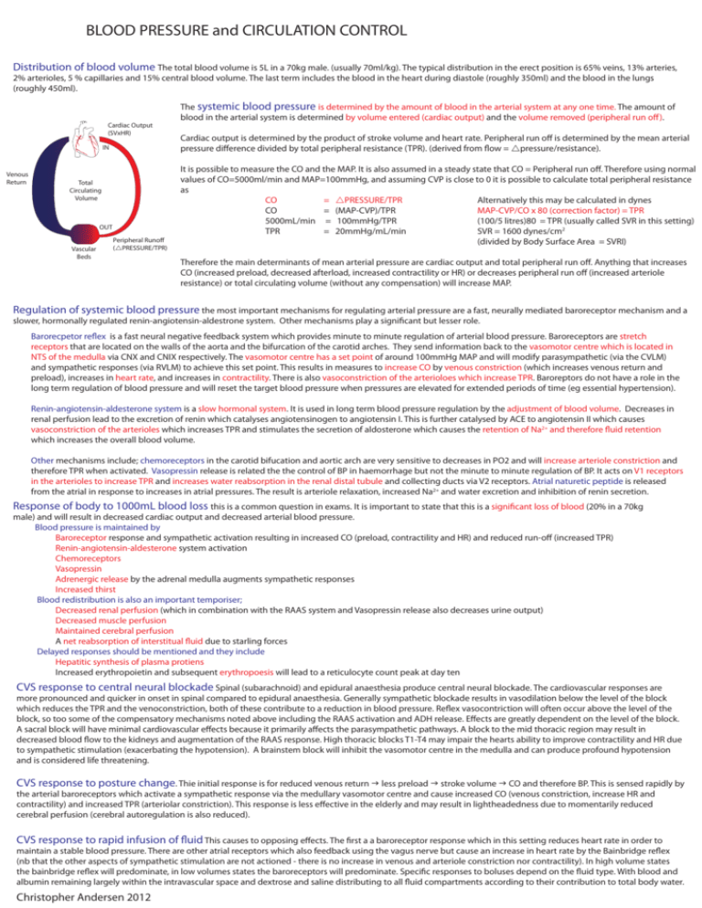
BLOOD PRESSURE and CIRCULATION CONTROL Distribution of blood volume The total blood volume is 5L in a 70kg male. (usually 70ml/kg). The typical distribution in the erect position is 65% veins, 13% arteries, 2% arterioles, 5 % capillaries and 15% central blood volume. The last term includes the blood in the heart during diastole (roughly 350ml) and the blood in the lungs (roughly 450ml). Cardiac Output (SVxHR) IN Venous Return Total Circulating Volume OUT Vascular Beds Peripheral Runoff (PRESSURE/TPR) The systemic blood pressure is determined by the amount of blood in the arterial system at any one time. The amount of blood in the arterial system is determined by volume entered (cardiac output) and the volume removed (peripheral run off ). Cardiac output is determined by the product of stroke volume and heart rate. Peripheral run off is determined by the mean arterial pressure difference divided by total peripheral resistance (TPR). (derived from flow = pressure/resistance). It is possible to measure the CO and the MAP. It is also assumed in a steady state that CO = Peripheral run off. Therefore using normal values of CO=5000ml/min and MAP=100mmHg, and assuming CVP is close to 0 it is possible to calculate total peripheral resistance as CO = PRESSURE/TPR Alternatively this may be calculated in dynes CO = (MAP-CVP)/TPR MAP-CVP/CO x 80 (correction factor) = TPR 5000mL/min = 100mmHg/TPR (100/5 litres)80 = TPR (usually called SVR in this setting) TPR = 20mmHg/mL/min SVR = 1600 dynes/cm2 (divided by Body Surface Area = SVRI) Therefore the main determinants of mean arterial pressure are cardiac output and total peripheral run off. Anything that increases CO (increased preload, decreased afterload, increased contractility or HR) or decreases peripheral run off (increased arteriole resistance) or total circulating volume (without any compensation) will increase MAP. Regulation of systemic blood pressure the most important mechanisms for regulating arterial pressure are a fast, neurally mediated baroreceptor mechanism and a slower, hormonally regulated renin-angiotensin-aldestrone system. Other mechanisms play a significant but lesser role. Barorecpetor reflex is a fast neural negative feedback system which provides minute to minute regulation of arterial blood pressure. Baroreceptors are stretch receptors that are located on the walls of the aorta and the bifurcation of the carotid arches. They send information back to the vasomotor centre which is located in NTS of the medulla via CNX and CNIX respectively. The vasomotor centre has a set point of around 100mmHg MAP and will modify parasympathetic (via the CVLM) and sympathetic responses (via RVLM) to achieve this set point. This results in measures to increase CO by venous constriction (which increases venous return and preload), increases in heart rate, and increases in contractility. There is also vasoconstriction of the arterioloes which increase TPR. Baroreptors do not have a role in the long term regulation of blood pressure and will reset the target blood pressure when pressures are elevated for extended periods of time (eg essential hypertension). Renin-angiotensin-aldesterone system is a slow hormonal system. It is used in long term blood pressure regulation by the adjustment of blood volume. Decreases in renal perfusion lead to the excretion of renin which catalyses angiotensinogen to angiotensin I. This is further catalysed by ACE to angiotensin II which causes vasoconstriction of the arterioles which increases TPR and stimulates the secretion of aldosterone which causes the retention of Na2+ and therefore fluid retention which increases the overall blood volume. Other mechanisms include; chemoreceptors in the carotid bifucation and aortic arch are very sensitive to decreases in PO2 and will increase arteriole constriction and therefore TPR when activated. Vasopressin release is related the the control of BP in haemorrhage but not the minute to minute regulation of BP. It acts on V1 receptors in the arterioles to increase TPR and increases water reabsorption in the renal distal tubule and collecting ducts via V2 receptors. Atrial naturetic peptide is released from the atrial in response to increases in atrial pressures. The result is arteriole relaxation, increased Na2+ and water excretion and inhibition of renin secretion. Response of body to 1000mL blood loss this is a common question in exams. It is important to state that this is a significant loss of blood (20% in a 70kg male) and will result in decreased cardiac output and decreased arterial blood pressure. Blood pressure is maintained by Baroreceptor response and sympathetic activation resulting in increased CO (preload, contractility and HR) and reduced run-off (increased TPR) Renin-angiotensin-aldesterone system activation Chemoreceptors Vasopressin Adrenergic release by the adrenal medulla augments sympathetic responses Increased thirst Blood redistribution is also an important temporiser; Decreased renal perfusion (which in combination with the RAAS system and Vasopressin release also decreases urine output) Decreased muscle perfusion Maintained cerebral perfusion A net reabsorption of interstitual fluid due to starling forces Delayed responses should be mentioned and they include Hepatitic synthesis of plasma protiens Increased erythropoietin and subsequent erythropoesis will lead to a reticulocyte count peak at day ten CVS response to central neural blockade Spinal (subarachnoid) and epidural anaesthesia produce central neural blockade. The cardiovascular responses are more pronounced and quicker in onset in spinal compared to epidural anaesthesia. Generally sympathetic blockade results in vasodilation below the level of the block which reduces the TPR and the venoconstriction, both of these contribute to a reduction in blood pressure. Reflex vasocontriction will often occur above the level of the block, so too some of the compensatory mechanisms noted above including the RAAS activation and ADH release. Effects are greatly dependent on the level of the block. A sacral block will have minimal cardiovascular effects because it primarily affects the parasympathetic pathways. A block to the mid thoracic region may result in decreased blood flow to the kidneys and augmentation of the RAAS response. High thoracic blocks T1-T4 may impair the hearts ability to improve contractility and HR due to sympathetic stimulation (exacerbating the hypotension). A brainstem block will inhibit the vasomotor centre in the medulla and can produce profound hypotension and is considered life threatening. CVS response to posture change. Thie initial response is for reduced venous return less preload stroke volume CO and therefore BP. This is sensed rapidly by the arterial baroreceptors which activate a sympathetic response via the medullary vasomotor centre and cause increased CO (venous constriction, increase HR and contractility) and increased TPR (arteriolar constriction). This response is less effective in the elderly and may result in lightheadedness due to momentarily reduced cerebral perfusion (cerebral autoregulation is also reduced). CVS response to rapid infusion of fluid This causes to opposing effects. The first a a baroreceptor response which in this setting reduces heart rate in order to maintain a stable blood pressure. There are other atrial receptors which also feedback using the vagus nerve but cause an increase in heart rate by the Bainbridge reflex (nb that the other aspects of sympathetic stimulation are not actioned - there is no increase in venous and arteriole constriction nor contractility). In high volume states the bainbridge reflex will predominate, in low volumes states the baroreceptors will predominate. Specific responses to boluses depend on the fluid type. With blood and albumin remaining largely within the intravascular space and dextrose and saline distributing to all fluid compartments according to their contribution to total body water. Christopher Andersen 2012
