COMMISSURAL FIBERS & ASSOCIATION FIBERS
advertisement
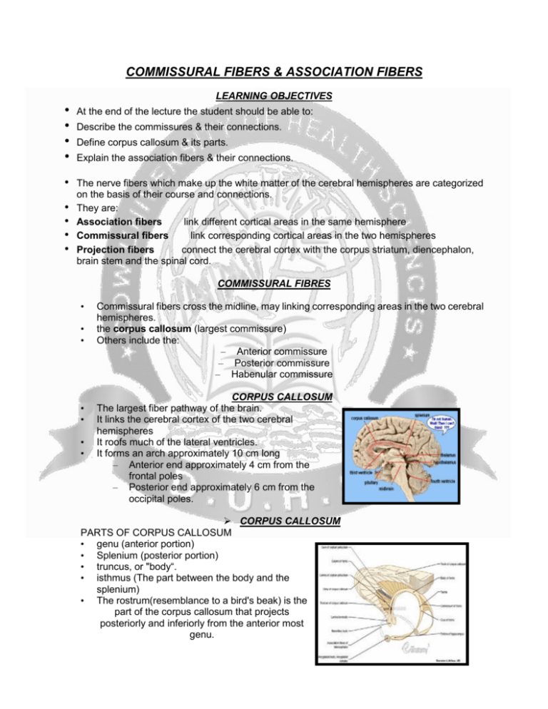
COMMISSURAL FIBERS & ASSOCIATION FIBERS LEARNING OBJECTIVES • • • • • • • • • At the end of the lecture the student should be able to: Describe the commissures & their connections. Define corpus callosum & its parts. Explain the association fibers & their connections. The nerve fibers which make up the white matter of the cerebral hemispheres are categorized on the basis of their course and connections. They are: Association fibers link different cortical areas in the same hemisphere Commissural fibers link corresponding cortical areas in the two hemispheres Projection fibers connect the cerebral cortex with the corpus striatum, diencephalon, brain stem and the spinal cord. COMMISSURAL FIBRES • • • • • • • Commissural fibers cross the midline, may linking corresponding areas in the two cerebral hemispheres. the corpus callosum (largest commissure) Others include the: – Anterior commissure – Posterior commissure – Habenular commissure CORPUS CALLOSUM The largest fiber pathway of the brain. It links the cerebral cortex of the two cerebral hemispheres It roofs much of the lateral ventricles. It forms an arch approximately 10 cm long – Anterior end approximately 4 cm from the frontal poles – Posterior end approximately 6 cm from the occipital poles. CORPUS CALLOSUM PARTS OF CORPUS CALLOSUM • genu (anterior portion) • Splenium (posterior portion) • truncus, or "body“. • isthmus (The part between the body and the splenium) • The rostrum(resemblance to a bird's beak) is the part of the corpus callosum that projects posteriorly and inferiorly from the anterior most genu. CORPUS CALLOSUM • • • Thinner axons in the genu connect the prefrontal cortex between the two halves of the brain. Thicker axons in the midbody of the corpus callosum and in the splenium interconnect areas of the premotor and supplementary motor regions and motor cortex. The posterior body of the corpus communicates somato sensory information between the two halves of the parietal lobe and visual center at the occipital lobe. ANTERIOR COMMISSURE • • • • • Also known as the precommissure Oval in shape Having a long vertical axis that measures about 5 mm. Bundle of nerve fibers connecting the two cerebral hemispheres across the midline Placed in front of the columns of the fornix CONNECTIONS • • • The fibers of the anterior commissure can be traced laterally and posteriorly on either side beneath the corpus striatum into the substance of the temporal lobe. It serves in this way to connect the two temporal lobes It also contains decussating fibers from the olfactory tracts and neospinothalamic tract for pain. POSTERIOR COMMISSURE • • • • also known as the epithalamic commissure a rounded band of white fibers crossing the middle line on the dorsal aspect of the upper end of the cerebral aqueduct. It is important in the bilateral pupillary light reflex Its fibers have their origin in a nucleus, the nucleus of the posterior commissure (nucleus of Darkschewitsch), CONNECTIONS • • • • nucleus lies in the central gray substance of the upper end of the cerebral aqueduct, in front of the nucleus of the oculomotor nerve. Some are probably derived from the thalamus and from the superior colliculus, others are believed to be continued downward into the medial longitudinal fasciculus. The posterior commissure interconnects the protectal nuclei, mediating the consensual pupillary light reflex. THE FORNIX • • Composed of myelinated nerve fibers Constitutes the efferent system of hippocampus passes to the mammillary bodies. PARTS OF THE FORNIX • • Alveus: – Thin layer of white matter covering the ventricular surface of hippocampus then converge to form the fimbria. Posterior commissure of fornix: – The fimbriae of two side increase in thickness, reach to the posterior end of hippocampus, arches above the thalamus below the corpus callosum PARTS OF THE FORNIX • • • • • • Body of fornix: – Formed by joining the two posterior columns in midline Commissure of the fornix: Consists of transverse fibers Across the midline From one column to another before formation of body Function: to connect the hippocampal formation of two side. CONNECTIONS • Fibers pass posterior to the anterior commissure to enter the mammillary body, end in medial nucleus • Fibers pass posterior to the anterior commissure to end in the anterior nuclei of the thalamus. • Fibers pass posterior to the anterior commissure to enter the tegmentum of midbrain. • Fibers pass anterior to the anterior commissure to end in: • Septal nuclei • Lateral preoptic area • Hypothalamus • Fibers join the stria medullaris thalami to reach the habenular nuclei HABENULAR COMMISSURE • • • A band of nerve fibers situated in front of the pineal gland. Connects the habenular nuclei on both sides of the diencephalon. Part of the trigonum habenulæ (a small depressed triangular area situated in front of the superior colliculus and on the lateral aspect of the posterior part of the tæni thalami. CONNECTIONS • • • The trigonum habenulæ also contains groups of nerve cells termed the ganglion habenulæ. Fibers enter the trigonum habenulæ from the stalk of the pineal gland, and the habenular commissure. Most of the trigonum habenulæ's fibers are, directed downward and form a bundle, the fasciculus retroflexus of Meynert, which passes medial to the red nucleus, and, after decussating with the corresponding fasciculus of the opposite side, ends in the interpeduncular ganglion. THE ASSOCIATION FIBERS • • unite different parts of the same cerebral hemisphere two kinds: (1) those connecting adjacent gyri, short association fibers; (2) those passing between more distant parts, long association fibers. THE ASSOCIATION FIBERS Short association fibers • also referred to as "U-fibers" • lie immediately beneath the gray substance of the cortex of the hemispheres. THE ASSOCIATION FIBERS Long association fibers include the following: Name From To uncinate fasciculus frontal lobe temporal lobe cingulum cingulate gyrus entorhinal cortex frontal lobe occipital lobe occipital lobe temporal lobe superior longitudinal fasciculus inferior longitudinal fasciculus perpendicular fasciculus inferior parietal lobule fusiform gyrus occipitofrontal fasciculus occipital lobe frontal lobe fornix hippocampus mammillary bodies Arcuate fasciculus frontal lobe temporal lobe -----------------------------THANK YOU--------------------------------
