internalization of a major group human rhinovirus does not require
advertisement
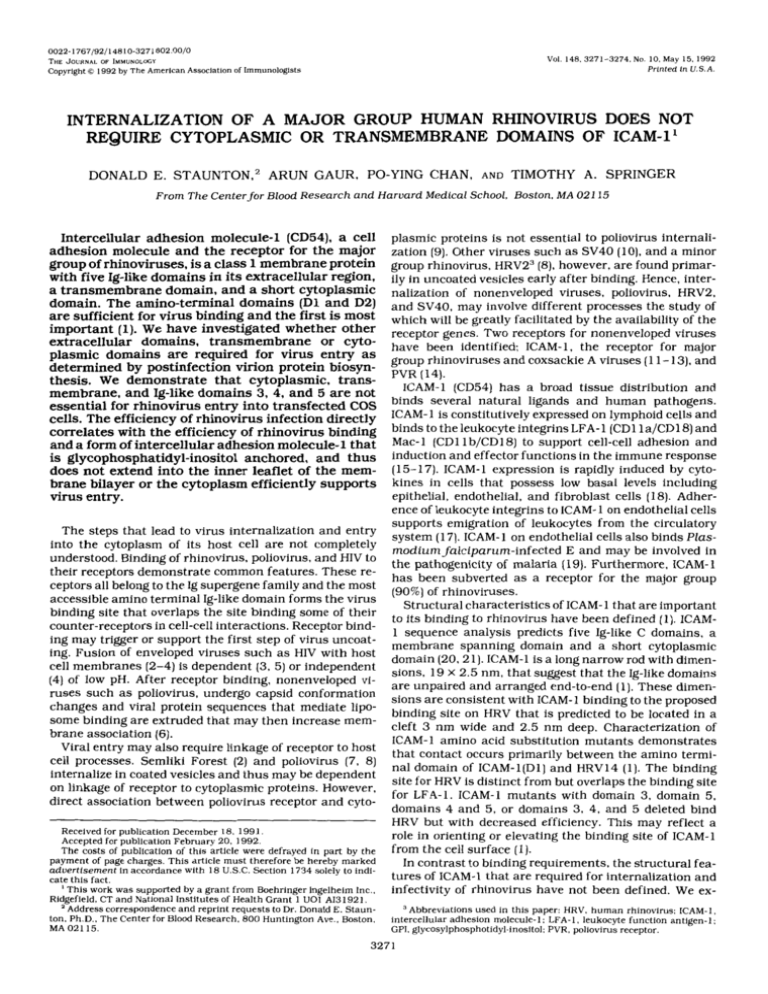
0022-1767/92/14810-3271$02.00/0 THEJOURNAL OF lMMUNOLoCY Copyright 0 1992 by The American Val. 148.3271-3274.No. IO.May 15. 1992 Prlnted In U.S.A. Associationof Immunologists INTERNALIZATION OF A MAJOR GROUP HUMAN RHINOVIRUS DOES NOT REQUIRE CYTOPLASMIC OR TRANSMEMBRANE DOMAINS OF ICAM-1' DONALD E. STAUNTON,2ARUNGAUR, PO-YING CHAN, AND TIMOTHY A. SPRINGER From The Center for Blood Research and Harvard Medical School, Boston. MA 021 15 plasmic proteins is not essential to poliovirus internaliIntercellularadhesionmolecule-1(CD54),acell zation (9).Other viruses such as SV40 (lo),and a minor adhesion molecule and the receptor for the major group ofrhinoviruses,is a class 1 membrane protein group rhinovirus, HRV23 (8), however, are found primarwith five Ig-like domains in its extracellular region, ily in uncoated vesicles early after binding. Hence, intera transmembrane domain, anda short cytoplasmic nalization of nonenveloped viruses, poliovirus, HRV2, domain. The amino-terminal domains (Dland D2) and SV40, may involve different processes the study of are sufficientfor virus binding and the firstis most which will be greatly facilitated by the availability of the important (1). We have investigated whether other receptor genes. Two receptors for nonenveloped viruses or cyto- have been identified: ICAM-1, the receptor for major extracellulardomains,transmembrane plasmicdomainsarerequired for virusentry as group rhinovirusesand Coxsackie A viruses (1 -113).and determined by postinfection virion protein biosynPVR (14). thesis. We demonstratethatcytoplasmic,transICAM-1 (CD54) has a broad tissuedistribution and membrane, and Ig-like domains 3, 4, and 5 are not binds several natural ligands andhuman pathogens. essential for rhinovirus entry into transfectedCOS cells. The efficiency of rhinovirus infection directly ICAM-1 is constitutively expressedon lymphoid cells and correlates with the efficiency of rhinovirus binding binds to the leukocyte integrins LFA- 1 (CD11a/CD18) and Mac-1 (CD1 lb/CD18) to support cell-cell adhesion and and a form of intercellular adhesion molecule-1 that is glycophosphatidyl-inositol anchored,and thus induction and effector functions in the immune response does not extend into the inner leaflet of the mem- (15- 17). ICAM- 1 expression is rapidly induced by cytobrane bilayer or the cytoplasm efficiently supports kines in cells that possess low basal levels including epithelial, endothelial, and fibroblast cells (18).Adhervirus entry. ence of leukocyte integrins to ICAM-1 on endothelial cells supports emigration of leukocytes from the circulatory The steps that lead to virus internalization and entry system (17). ICAM-1 on endothelial cells also bindsPlasinto the cytoplasm of its host cell are not completely modium falciparum-infected E and may be involved in understood. Binding of rhinovirus, poliovirus, and HIV to the pathogenicity of malaria (19). Furthermore. ICAM-1 their receptors demonstrate common features. These rehas been subverted as a receptor for the major group ceptors all belong to the Ig supergene family and themost (90%)of rhinoviruses. accessible amino terminalIg-like domain formsthe virus Structural characteristicsof ICAM- 1 that areimportant binding site that overlaps the site binding some of their to its binding to rhinovirus have been defined ( I ) . ICAMcounter-receptors incell-cell interactions. Receptor bind1 sequence analysis predicts five Ig-like C domains, a ing may trigger or support the first stepof virus uncoatmembrane spanning domain and a short cytoplasmic ing. Fusion of enveloped viruses such as HIV with host domain (20.21).ICAM-1 is a long narrow rod with dimencell membranes (2-4) is dependent ( 3 , 5) or independent sions, 19X 2.5 nm, thatsuggest that the Ig-like domains (4) of low pH. After receptor binding, nonenveloped viare unpaired and arranged end-to-end (1).These dimenruses such as poliovirus, undergo capsid conformation sions are consistentwith ICAM-I binding to the proposed changes and viral protein sequences that mediate lipobinding site on HRV that is predicted to be located in a some binding are extruded that may then increase memcleft 3 nm wide and 2.5 nm deep. Characterization of brane association (6). ICAM-I amino acid substitution mutants demonstrates Viral entry may also require linkageof receptor to host that contact occurs primarily between the amino termicell processes. Semliki Forest (2) and poliovirus (7, 8) nal domain of ICAM- 1( D l ) and HRVl4 (1). The binding internalize in coated vesicles and thusmay be dependent site for HRV is distinct from but overlapsthe binding site on linkage of receptor to cytoplasmic proteins. However, for LFA-1. ICAM- 1 mutants with domain 3 , domain 5, direct association between poliovirus receptor and cytodomains 4 and 5, or domains 3 , 4, and 5 deleted bind HRV but with decreased efficiency. This may reflect a Received for publication December 1 8 , 1991. role in orienting or elevating the binding site of ICAM-1 Accepted for publication February 20, 1992. from the cell surface (1). The costs of publication of this article were defrayed in part by the payment of page charges. This article must therefore be hereby marked In contrast to binding requirements,the structural feaaduertlsernent In accordance with 18 U.S.C.Section 1734 solely to inditures of ICAM-1 that arerequired for internalization and cate this fact. ' This work was supported by a grant from Boehringer Ingelhelm Inc.. infectivity of rhinovirus have not been defined. We exRidgefield, CT and National Institutes of Health Grant 1 UO1 AI31921. 'Address correspondence andreprint requests to Dr. Donald E. Staunton. Ph.D..The Center for Blood Research. 800 Huntington Ave.. Boston. MA 021 15. Abbreviations used in this paper: HRV. human rhinovirus: ICAM-1, intercellular adhesion molecule-1: LFA-1, leukocyte function antigen-I; GPI. glycosylphosphotidyl-inositol: PVR, poliovirus receptor. 327 1 3272 ICAM- 1 MUTANTS SUPPORTING HRV amine involvement ofICAM-1 features including potential interaction between cytoplasmic domain and cytoskeleton and distanceof binding site from the membrane. This characterization yields insight into the structural requirements of HRV internalization and what features may be common to other nonenveloped viruses. INTERNALIZATION A T N x LFA-3 * x sx P 0s G ACC GTG A A T GTC T T A AGC CCA AGC AGCGGT B MATERIALS AND METHODS v I PIL ICAM-1 "- Mock WT GPi pI.pLc: - + - + - .WW*mW + - kd Construction of CPI-anchored ICAM-1. A GPI-anchored form of ICAM-1 was generated by replacing ICAM-1 cDNA encoding trans- 91 membrane and cytoplasmic domains with the GPI signal sequence from LFA-3. ICAM-1 and LFA-3 expressionconstructswerede- 6R scribed previously (22. 23). A unique Aflll site was introduced by oligonucleotide-directed mutagenesis into ICAM-1 centered around sequence coding for residue L448 (silent mutation). amino-terminal - 43 to the transmembrane domain. TheAflII- BamHl fragment (unique BamHl site in theCDM8 vector) was then exchanged witha similar fragment from anLFA-3-CDM8 construct that contains an AflII site introduced into sequence centered around C177 (24). N-terminal to the predicted GPI-anchored attachment site at S180 (25) and the construct was designated ICl/GPIl.4. COS cell transfection a n d q u a n t i t a t i o nof H R V l 4 viral protein synthesis. COS cells a t 50% confluency were transfected as described previously by DEAE-dextran method using approximately 4 pg of plasmid per 10-cm dish (22). Two days posttransfection COS cells were suspended using trypsin-EDTA and reseeded into 6-cm dishes. Three days posttransfection confluent COS cells in 6-cm dishes were washed wlth RPMI 1640/10 mM MgCI2/25 mM HEPES pH 7.3 (binding buffer) and then incubated with HRV14 (multiplicity of Infection = 20) in binding buffer for 60 min at35°C wlth rotation. Binding buffer was aspirated and2 ml of complete media added and incubation continued for 2 h. Media was then aspirated and 120 pCi Flgure 1 . Constructlon and expression of a GPI-anchored form of of "S-methionine and 35S-cysteine was added in 1.5 ml of methio- ICAM- 1. A. Sequence of ICAM- 1/GPI at point of fuslon (AI11 site) between nine and cysteine-freeRPMI 1640 with 10% dialyzed FCS and gen- ICAM- 1 and LFA-3 mutant cDNA. The asterlsk indicates nucleotides that tamicin. After 6 h a t 35°C media was aspirated. cells were washed differ from wild-type. The predicted carboxyl-terminalresidue of the once, and lysed by three cycles of rapid freezing and thawing In 1 mature protein Is circled. E. ICAM-1 was immunoprecipitated from the supernatant of 13%] cystelne- and methionine-labeled mock- transfected ml of DMEM/40 mM HEPES. pH 7.3. Cell debris was removed by COS cells or COS cells expressingwild-type (WT) or GPI- anchored forms centrifugation in a mlcrofuge at 13.000 rpm for 10 min. Supernatants were layered on 30%sucrose with l M NaCl a n d 2 0 mM Trls- that had been treated wlth (+) or without (-) PI-PLC a s descrlbed (301. acetate. pH 7.5, and then centrifuged at35.000 rpm for 2 h a t 15°C Immunopreclpltates were subjected to SDS 10% PAGE and fluorography. (26).Theradiolabeledviruspelletwasresuspendedin 20 pl of DMEM/40 mM HEPES. pH 7.3. and an aliquot was subjected to reducing SDS 12% PAGE. Autoradiograms were scanned on a densltometer (Bio-Rad. McLean. VA). The area under VPl. VP2. and VP3 peaks was added and divided by the area under VP peaks from wild-type ICAM-1 -expressing cells. RESULTS A GPI-anchored form of ICAM-1 was generated by the exchange ofICAM- 1 transmembrane and cytoplasmic domain sequence for LFA-3 GPI signal sequence (Fig. 1A). When expressed in COS cells. ICAM-l/GPI but not wild-type (WT)ICAM-1 was specifically released into culture supernatant by phosphatidyl inositol-specific phospholipase Cdigestion (Fig. 1B). HRV 14 internalization was demonstratedby infecting COS cell transfectants andradiolabeling de novo synthesized viral proteins with [""SI cysteine and methionine. Virus was partially purified by sedimentation and the purified labeled viral capsid proteins(VP1,VP2, and VP3) visualized by SDS-PAGE (Fig. 2) migrated identically to viral proteins of highly purified HRV14 (data not presented). COS cells expressing human wild-type ICAM-1 clearly supported HRV14 VP synthesis. In contrast, no viral protein synthesis wasdetected in mock-transfected cells or cells expressing mouse ICAM-1, which does not bind HRV14 (1). In addition, a human-mouse chimera possessing the amino-terminalIg-like domains of human ICAM-1 (1) but not the reciprocal chimera, supports VP synthesis in COS cells. Thus, HRV14 entry intoCOS cells is dependent on expression of ICAM-1 and the presence of residues 1-1 68 of human ICAM-1. " VPI C VPZ+3 Flgure2. HRV14 viral protein synthesls In COS cells expressing ICAM-1 mutants. COS cells expresslng wild-type or mutant ICAM-1 a s indicated were Infected with H R V l 4 (Mol= 20)and de novo synthesized proteins labeled with [35S]methionine and cystelne. HRV was isolated from cells 9 h postinfection and subjected to SDS 12%PAGE and fiuorography. To determine if the cytoplasmic and transmembrane domains of ICAM- 1 are required for HRV14 internalization a GPI anchored form of ICAM-l(ICAM-l/GPI)and a cytoplasmic domain deletion mutant (1) were tested for their ability to support virus entry when expressed in COS cells. HRV14 internalization is supported by ICAM-1 with its 3273 INTERNALIZATION SUPPORTING MUTANTS HRV ICAM-1 FLgure3. Comparison of HRVl4 binding and viral proteinsynthesissupported by ICAM-1 mutants. ICAM-1 domaindeletion mutants (D3-. D5-. D4-5-, D3-4-5-, and Dcyt-), GPI anchored ICAM-1 (GPI-ICAM-1] andhuman-mousechimeric ICAM-1 (hm168) were expressed in COS cells. These cells were infected with HRV-14 and viral proteln synthesis was quantitatedby scanning densitometry of autoradiograms similar to thatpresentedinFigure2. Viral protein synthesis and binding (1) was normalized for percent expressing cells and is relative to wild-type. Binding to ICAM-1 mutants was previously published (1). except for GPI-ICAM-1. and is shown for comparison purposes. I VP synthesis HRV14 binding 1.o 05 nn ".- h W mWT entire cytoplasmic domain deleted (Dcyt-) as well as by ICAM-l/GPI (Fig. 2). The cytoplasmic andtransmembrane domains ofICAM-1 are therefore not essential to HRV14 infection. Previously, we havedemonstratedthat binding of HRV14 to ICAM-1 with Ig-like domains D3, D4, and D5 deleted was lessthan thatto wild-type (1). Thepossibility that deletion of these domains might restrict HRV internalization was tested inCOS cell transfectants. All ICAM1 domain deletion mutants conferred to COS cells the ability to internalize HRV14 (Fig. 2). Hence, these membrane proximal domains are not essential to the mechanism of HRV internalization. To compare the efficiency of HRV14 internalization supported by different ICAM-1 mutants, viral protein synthesis was quantitatedby densitometry and normalized for percent of transfected cells expressing ICAM-1. The level ofVP synthesis was decreased for domain deletion mutants, correlating with the previously determined efficiency of HRV14 binding of these mutants(Fig. 3) (1).In contrast, Dcyt- supported wild-type levels of VP synthesis andICAM-l/GPI resulted in greaterthan wildtype levels ofVP synthesis. This reflected the increased efficiency of HRV14 binding to these mutants(Fig. 3 ) . DISCUSSION We have demonstrated that the firsttwo amino terminal domains ofICAM-1 and amembraneanchorare sufficient to support HRV internalization and that ICAM1 Ig-like domains 3, 4, 5, and transmembrane and cytoplasmicdomains are not required. Truncation of the cytoplasmic domain and reanchoring on GPI did not decrease the efficiency of virus entry into the cytoplasm. GPI reanchoring results in replacement of the polypeptide membrane anchor with theGPI anchor, and this anchor is equivalent to aphospholipid and is present only in the outer leaflet of the membrane bilayer. The efficient internalization supported by the Dcyt- mutant and ICAMl/GPI demonstrates that interaction between the cytoplasmic domain of ICAM-1 and actin(27)or a cytoskeletal protein, e.g., a-actinin (0.Carpen, P. Pallai, D. E. Staunton, and T. A. Springer, manuscript submitted) is not required in this process. Poliovirus is a picornavirus that binds areceptor (PVR) possessing three Ig-like domains. The cytoplasmic domain ofPVR is also not necessary for poliovirus inter- hm-168 D5 D3 D4 5 D3 4 5 Dcyl - GPI-ICAM-1 nalization (9). Furthermore, CD4 with cytoplasmic domain deleted (28) or GPI anchored (29). supports HIV infection. Hence, surprisingly there appearsto be no role of the cytoplasmic domain in internalization of HRV. poliovirus or HIV. However, HIV possesses an envelope and cell entry occurs by, and may be entirely dependent on. direct fusion with the plasma membrane (4). Why are particular surfacemolecules subverted as virus receptors? This could be related to receptor cellular distribution or accessibility. Alternatively, receptor ligation by the virus might trigger a signal throughthe membrane or cytoplasmic domain of the receptor that mimics signalling by a biologic ligand and that is advantageous for the virus interaction with the host. We have only studied one aspect of this host interaction, entry into the cytoplasm, and it is possible that other aspects are important in vivo. REFERENCES 1. Staunton, D. E.. M. L. Dustin, H. P. Erickson. and T. A. Springer. 1990.Thearrangement of theimmunoglobulin-likedomains of ICAM-1 and the binding sites for LFA-1 and rhinovirus.Cell 61 :243. 2. Helenius, A., J. Kartenbeck, K. Simons, andE. Fries. 1980. On the entry of Semliki Forest virus intoBHK-21 cells. J . Cell Biol. 84:404. 3. White, J.. and A. Helenius. 1980. pH-dependent fusion between the Semliki Forest virus membrane and liposomes. Proc. Natl. Acad. Sci. USA 77:3273. 4. Stein. B. S . , S. D. Gowda, J. D. Lifson. R. C. Penhallow. K. G . Bensch. and E. G. Engleman. 1987. pH-independentHIV entry into CD4-positive T cells via virus envelope fusion to the plasma membrane. Cell 49:659. 5. Doms, R.W.. A. Helenius, and J. White. 1985. Membrane fusion activity of the influenza virus hemagglutinin: the low pH-induced conformational change. J . Biol. Chem. 260:2973. 6. Fricks, C.E., and J. M. Hogle. 1990. Cell-induced conformational change in poliovirus: externalization of the amino terminus of VP1 is responsible for liposome binding. J . Virol. 64: 1934. 7. Zeichhardt, H., K. Web, P. Willingmann, and K. -0. Habermehl. 1985. Entry of poliovirus type 1 and mouse elberfeld (ME)virus into HEp-2 cells: receptor-mediated endocytosis and endosomal or lysosoma1 uncoating. J . Gen. Virol. 66:483. 8. Madshus, I. H.,K. Sandvig, S . Olsnes, and B. Van Deurs. 1987. Effect of reduced endocytosis induced by hypotonic shock and potassium depletion on the infection of Hep 2 cells by picornaviruses, J . Cell. Physiol. 131:14. 9. Koike, S . , I. Ise, and A. Nomoto. 1991. Functional domains of the poliovirus receptor. Proc. Natl. Acad. Sci. USA 88:4104. 10. Kartenbeck, J..H. Stukenbrok, andA. Helenius. 1989. Endocytosis of simian virus 40 into the endoplasmic reticulum. J . CellBiol. 109:2721. 11. Greve, J. M., G . Davis, A. M. Meyer. C. P. Forte, S . C. Yost, C. W. Marlor, M. E. Kamarck. and A. McClelland. 1989. Themajor human rhinovirus receptoris ICAM-1. Cell 56:839. 12. Staunton. D. E.. V. J.Merluzzi. R. Rothlein. R. Barton, S . D. Marlin, and T.A. Springer. 1989. A cell adhesion molecule, ICAM-1, is the major surface receptorfor rhinoviruses. Cell 56:849. 3274 ICAM- 1 MUTANTS INTERNALIZATION SUPPORTING HRV 13. Tomassini, J. E., D. Graham, C. M. DeWitt. D.W. Lineberger, J. A, 22. Staunton, D. E., M. L. Dustin, andT. A. Springer. 1989. Functional cloning of ICAM-2, a cell adhesion ligand for LFA-1 homologous to Rodkey, and R. J. Colonno. 1989. cDNA cloning reveals that the major group rhinovirus receptoron HeLa cells is intercellular adheICAM-1. Nature 339331. sion molecule 1. Proc. Natl. Acad. Sci. USA 86:4907. 23. Seed, B. 1987. An LFA-3 cDNA encodes a phosphoiipld-linked mem14. Mendelsohn, C. L.,E. Wimmer, and R. Racaniello. 1989. Cellular brane protein homologous to its receptorCD2. Nature 329:840. receptor for poliovirus: Molecular cloning, nucleotide sequence. and 24. Wallner, B. P.. A. 2.Frey. R. Tizard. R. J. Mattaliano. C. Hession, expression of a new member of the immunoglobulin superfamily. M. E. Sanders. M. L. Dustin, and T.A. Springer. 1987. Primary Cell 56:855. structure of lymphocyte function associated antigen-3 (LFA-3):the ligand of the T-lymphocyteCD2 glycoprotein. J. Exp. Med. 1661923. 15. Marlin, S. 0.. and T. A. Springer. 1987. Purified intercellular adhesion molecule-I (ICAM-1) is a ligand for lymphocyte function-asso- 25. Ferguson, M. A. J.. and A. F. Williams. 1988. Cell surface anchoring dated antigen 1 (LFA-1).Cell 51:813. of proteins via glycosyl-phophatidylinositolstructures. ARRU. Rev. Biochem. 57:285. 16. Diamond, M. S., D. E. Staunton, A. R. d e Fougerolles, S . A. Stacker, J. Garcia-Aguilar, M. L. Hibbs, and T. A. Springer. 1990. ICAM-1 26. Sherry, B.. A. G . Mosser, R. J. Colonno. and R. R. Rueckert. 1986. (CD54)-a counter-receptor for Mac-I (CDllb/CD18). J. Cell Blol. Use of monoclonal antibodies to identifyfour neutralization immu11 1~3129. nogens on a common cold picornavirus, human rhinovirus 14. J. Vtrol. 571246. 17. Springer, T.A. 1990. Adhesion receptors of the immune system. Nature 346:425. 27.Vogetseder, W.. and M. P. Dierich.1991.Intercellularadhesion 18. Dustin, M. L., R. Rothlein, A. K. Bhan, C.A. Dinarello, and T. A. molecule- 1 (ICAM-1. CD 54) is associated with actin-filaments. Immunobiology 182: 143. Springer. 1986. Induction by IL-1 and interferon, tissue distribution, biochemistry, and function of a natural adherence molecule (ICAM- 28. Bedinger, p., A. Moriarty, R. C. von Borstel11. N. J. Donovan, K.S. Steimer, and D.R. Littman. 1988. Internalization of the human 1).J. Immunol. 137:245. immunodeficiency virus does not require the cytoplasmic domain of 19. Berendt, A. R.,D. L. Simmons, J. Tansey, C. I. Newbold, and K. Marsh. 1989. Intercellular adhesionmolecule-I is a n endothelial cell CD4. Nature 334:162. adhesion receptor for Plasmodium falciparum. Nature 341~57. 29. Diamond. D. C., R. Finberg, S. Chaudhuri, B. P. Sleckman, and S . 20. Simmons, D.. M. W. Makgoba, and B. Seed. 1988. ICAM. a n adhesion J. Burakoff. 1990. Human immunodeficiency virus infection is efligand of LFA- 1, is homologous to the neuralcell adhesion molecule ficiently mediatedby a glycolipid-anchored form of CD4. Proc. Natl. NCAM. Nature 3313324. Acad. Sci. USA 87:5001. 21. Staunton, D. E., S. D. Marlin, C. Stratowa, M. L. Dustin, and T. A. 30. Selvaraj, P.,M. L.Plunkett, M. Dustin. M. E. Sanders, S . Shaw, and Springer. 1988. Primary structure of intercellular adhesionmolecule T. A. Springer. 1987. The T lymphocyte glycoprotein CD2 jLFA-2/ T1 1/E-Rosette receptor] binds the cell surface ligand LFA-3. Nature 1 (ICAM-I]demonstrates interaction between members of the im326:400. munoglobulin and integrin supergene families.Cell 521925.
