LASER HAIR REMOVAL: SCIENTIFIC PRINCIPLES AND
PRACTICAL ASPECTS
Christine C. Dierickx, M.D.
Visiting Research Scientist
Wellman Laboratories of Photomedicine
Harvard Medical School
ABSTRACT
The use of lasers for hair removal has been studied for a number of years. In this procedure,
laser light is absorbed by melanin in the hair shaft, damaging the follicular epithelium. A clinical study evaluated the use of the LightSheer™ Diode Laser for hair removal. Of 92 patients,
all had temporary hair loss and 89% had permanent hair reduction. Regrowing hairs were
shown to be thinner and lighter than previously. Extensive clinical use of this high-power,
pulsed diode laser has resulted in recommendations for patient selection and proper use of the
laser. Appropriate fluence settings have been shown to cause permanent hair reduction without
damaging the epidermis, regardless of skin type.
BACKGROUND
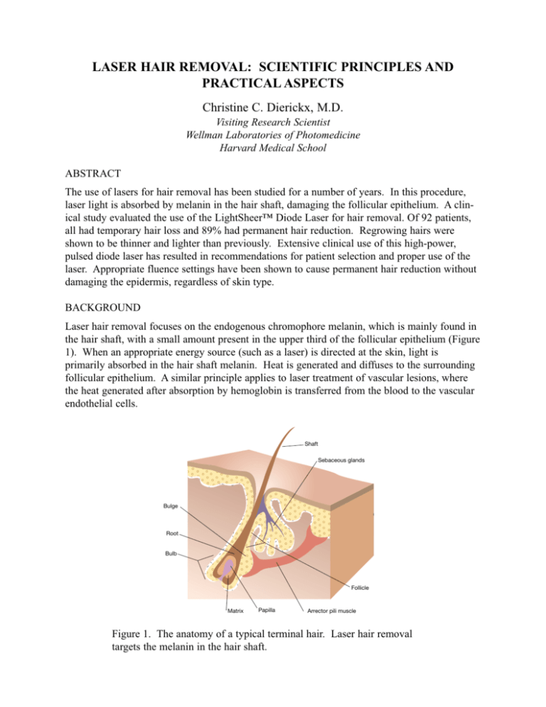
Laser hair removal focuses on the endogenous chromophore melanin, which is mainly found in
the hair shaft, with a small amount present in the upper third of the follicular epithelium (Figure
1). When an appropriate energy source (such as a laser) is directed at the skin, light is
primarily absorbed in the hair shaft melanin. Heat is generated and diffuses to the surrounding
follicular epithelium. A similar principle applies to laser treatment of vascular lesions, where
the heat generated after absorption by hemoglobin is transferred from the blood to the vascular
endothelial cells.
Shaft
Sebaceous glands
Bulge
Root
Bulb
Follicle
Matrix
Papilla
Arrector pili muscle
Figure 1. The anatomy of a typical terminal hair. Laser hair removal
targets the melanin in the hair shaft.
Laser hair removal is based on the principles of selective photothermolysis: a combination of
the appropriate laser wavelength, pulse duration, and fluence.
• Wavelengths between approximately 700 and 1000 nanometers (nm) are selectively
absorbed by melanin; the competing chromophores (oxyhemoglobin and water)
absorb less energy at these wavelengths. Figure 2 shows the absorption of different
chromophores in the skin. Therefore, any light source that operates between 700
and 1000 nm is appropriate for targeting melanin in the hair shaft.
Diode ~ 800 nm
Alexandrite ~ 755 nm
Nd:YAG ~ 1064 nm
Ruby ~ 694 nm
Absorption (log scale)
Melanin
Oxyhemoglobin
Water
300
500
700
1000
2000
Wavelength (nm)
Figure 2. The absorption of various chromophores as a function of wavelength. Ruby
lasers operate at 694 nm, alexandrite lasers at 755 nm, diode lasers at 800 nm and
Nd:YAG lasers at 1064 nm. (Adapted from Boulnois JL. Photophysical processes in recent
medical laser developments: a review. Published in Lasers in Medical Science, Vol 1, 1986.)
• Pulse duration (or pulse width) must be equal to or shorter than the thermal relaxation time of the target to confine thermal damage. The thermal relaxation time of
the whole follicular structure depends on its diameter and is on the order of tens of
milliseconds. Consequently, the laser source must have a range of pulse widths to
selectively damage different size follicles.
• Pulse width must be matched with the appropriate amount of fluence (energy per
unit area) necessary to cause follicular damage.
Hair removal devices available today include 694 nm ruby lasers, 755 nm alexandrite lasers,
800 nm diode lasers, 1064 nm Nd:YAG lasers, and filtered xenon flashlamps. This paper
focuses on an 800 nm diode laser (LightSheer Diode Laser, Lumenis Inc., Santa Clara, CA).
This wavelength effectively targets the melanin while deeply penetrating the dermis.
HAIR LOSS AND REGROWTH
One hundred patients were treated in a clinical study with the high-power, pulsed diode laser.
The study evaluated different combinations of fluence and pulse width in eight test sites. The
patients were followed-up at one, three, six, nine, and 12 months following the last treatment.
Ninety-two patients completed the study. Hair loss was assessed from hair counts using digital
photographs before treatment and at each follow-up visit. Tattoos identified the location of
each test site.
The study showed that the high-power diode laser induces two separate effects: temporary hair
loss and permanent hair reduction.
Temporary hair loss occurs in all patients, for all hair colors and at all laser fluences. It usually
lasts from one to three months.
Permanent hair reduction is defined as a significant reduction in the number of terminal hairs at
a given body site that is stable for a period of time longer than the follicles’ complete growth
cycle (Figure 3, Table I). Test sites were mainly given on the back and thighs, where complete
Anagen
Catagen
Telogen
Figure 3. Hair growth cycle. Anagen is the active growth phase, catagen
is the regression phase, telogen is a resting phase.
3
Location
Telogen
(months)
Anagen
(months)
Total
(months)
Back
Thigh
Arm
Calf
Axilla
Upper Lip
3-6
3-6
3-5
3-4
2-3
1-2
3-6
3-6
1-2
4-5
3-4
3-4
6-12
6-12
4-7
7-9
5-7
4-6
Bikini
3-4
2-3
5-7
Table I. Duration of hair growth cycles.
hair growth cycles vary between six months and a year. A one year follow-up allowed time for
one to two complete growth cycles at these anatomic sites.
There is a difference between permanent hair reduction and complete hair loss. Complete hair
loss implies that there are no regrowing hairs. This can be a temporary or permanent phenomenon. The LightSheer Diode Laser usually produces complete but temporary hair loss,
followed by a partial but permanent hair reduction. This is an important distinction to make
when setting patient expectations.
With this laser, 100% of the patients experienced temporary hair loss, while 89% of the patients
had permanent hair reduction at one year follow-up. Of the 11% of patients who did not have
long-term hair loss, most had blond hair. Because blond hair contains less melanin than darker
hair, there is less chromophore for the laser to target, and the response is less. However, these
patients experienced temporary hair loss.
Numbers cited for hair loss only take into account the absolute number of hairs. They do not
reflect the fact that the regrowing hairs are lighter and thinner than before, which also adds to
apparent clinical hair loss. Hair color was measured by calculating the absorption coefficient
from the hairs’ transmission of 700 nm light. Hair diameter was measured from digital
photographs. The study showed that the regrowing hairs appeared lighter (with a transmission
coefficient 1.41 times higher than the value before treatment) and were thinner (with a decrease
in the mean hair diameter by 19.9%) than the original hairs.
Histologic observations support two mechanisms for permanent hair reduction: miniaturization
of coarse hair follicles to vellus-like hair follicles, and destruction of the hair follicle with granulomatous degeneration, leaving a fibrotic remnant. Clinically, both of these mechanisms
produced reduction in hair.
The study design used a fixed set of fluence-pulse-width combinations in each patient, regardless of skin type. If skin type and color had been matched to appropriate fluences, the incidence of side effects could have been reduced. Epidermal damage was seen in 6% of cases.
Textural change occurred in 3% of cases, where triple pulsing was used at the highest fluence.
These changes disappeared after three months. Transient pigment changes were seen in about
4
10% of cases and usually occurred in the darker skin types or in patients who had tans at the
time of treatment.
DIODE LASER CHARACTERISTICS
The characteristics of the LightSheer Diode Laser are seen in Table II. The ChillTip™ handpiece directs the laser onto the skin through an integrated, cold (approximately 5 degrees C)
sapphire window.
Laser Wavelength
800 nm
Pulse Duration
5 to 100 milliseconds
Spot Size
9 by 9 millimeters*
Fluence
10 to 40 Joules/cm2 *
Repetition Rate
1 pulse per second*
Table II. LightSheer Diode Laser characteristics.
*Other LightSheer models have expanded
capabilities for these specifications.
The laser has a range of pulse widths from 5 to 100 milliseconds, which is longer than the
thermal relaxation time of the epidermis and comparable to that of the follicle. This pulse
width range can effectively damage the follicle. However, the epidermis also contains some
melanin and must be protected. A sapphire window (ChillTip) with high thermal conductivity is
put in direct contact with the skin. It cools the epidermis before, during, and after each laser
pulse. Because of index matching, it also reduces internal reflection of back-scattered light.
These combined thermal and optical cooling effects protect the epidermis from damage.
Besides preserving the epidermis, compressing the skin with the ChillTip has two other advantages. The pressure removes oxyhemoglobin, a chromophore that competes with melanin. It
also flattens the epidermis, bringing the hair roots closer to the surface. Hair roots closer to the
surface have a greater probability of absorbing the laser light.
CLINICAL GUIDELINES
Patient Selection
By studying hair color and skin type it is easy to determine which patients will have the best
results with laser hair removal. Patients with red, gray, or blond hair can be advised that they
should not expect permanent hair reduction. It is especially important to see if the patient has a
tan or not. If patients have a tan they should be instructed to stay out of the sun, use a
bleaching cream and sun block, and return for treatment when the tan is gone or start treatment
with the 100ms pulse width.
5
Because the hair shaft is the chromophore, it is essential that the hair shaft is present in the hair
follicle at the time of treatment. Patients are therefore not allowed to pluck, wax, or have electrolysis for at least six weeks before the laser treatment. Shaving and depilatory creams are allowed because they leave the hair shaft in the follicle. It is important to take a history, including
an endocrine history. Female patients with hirsutism can be treated regardless of the cause.
Patients with a history of herpes simplex 1 or 2 should be put on oral antiviral drugs (Zovirax®
or Famvir®) beginning the day before treatment. This is important when treating an upper lip or
even a bikini line because reactivation of herpes simplex 1 or 2 has been reported after laser
treatment.
There is no consensus on how long Accutane® should be stopped before treatment. The general
rule is to stop Accutane® treatments for six months before laser hair removal.
Treatment Methods
It is important to shave before beginning the treatment. If the external hair shaft is present the
laser will burn it, in turn burning the skin. Depilatory creams can be used with patients who
object to shaving.
Anesthesia is usually not required; however, this depends on the patient and body area. When
treating the upper lip some kind of anesthesia is recommended.
There is a high risk for eye damage with the laser because the retina has a very high concentration of melanin. For this reason treatment must not be carried out inside the bony area of the
eye. It is important that the patient, nurse, and doctor all wear goggles.
During treatments it is important to regularly clean the handpiece. When the hair shaft
carbonizes, it leaves debris on the sapphire window. This build up can make it hot and can
make it difficult for the laser light to penetrate. Cleaning the ChillTip handpiece with alcohol
prevents this barrier from forming. There is a small but real risk of infection because the handpiece is in direct contact with the skin. Therefore, between patients the handpiece should be
disinfected with a liquid disinfectant such as Virex.
Fluence Selection
Hair color and skin color determine the best fluence to use. Darker skin types IV to VI (Table
III) can be treated between 10 and 20 J/cm . Fair skin types I to III can take the highest
fluences, from 25 to 40 J/cm .
2
2
Type I
Always burns, never tans
Type II
Always burns, sometimes tans
Type III
Sometimes burns, always tans
Type IV
Rarely burns, always tans
Type V
Moderately pigmented
Type VI
Black skin
Table III. Fitzpatrick classification of skin types.
6
Treatment should be performed with the highest fluence the skin can tolerate. Studies have
shown that the percentage hair loss is fluence-dependent, with higher percentages of hair loss at
higher fluences.
Each skin type has its own threshold fluence at which pigmentation changes occur. To minimize hypo- or hyperpigmentation, lower fluences than those suggested above should be used
while gaining clinical experience. With multiple pulsing the incidence of pigment changes
increases without an increase in efficacy. For this reason, double and triple pulsing are not
recommended. If hypo- or hyperpigmentation occurs, it is transient. The duration of these
pigment changes, however, depends on the anatomic area.
The ChillTip handpiece must be in firm contact with the skin. A single pulse should be placed
at test sites within or near the treatment area. If epidermal damage is present (blistering, ablation, graying or whitening of the epidermis, or a positive Nikolski sign) the fluence should be
lowered by 5 to 10 J/cm .
2
Several pulses should then be placed next to one another while looking for the epidermal
response. An effective fluence is one where the hair carbonizes, followed by very selective
follicular swelling and redness (Figure 4).
Figure 4. Immediately after laser treatment of the bikini area on a Fitzpatrick skin type II;
treatment at 40 J/cm fluence and 20 ms pulse duration.
2
Some areas may be missed during treatment because the redness and swelling may become
confluent, and it may be difficult to distinguish the treated areas. A template or other skin
marking method can be helpful. A polarized light source with a magnifying loop (Syris
Scientific LLC, Gray, ME) allows visualization of individual follicles, helping to define the
treated area.
Additionally, within several days of treatment there is a phenomenon in which hair casts,
7
carbonized by the laser, will be shed from the hair follicle. Patients may believe that their hair
is regrowing. These hair casts can be pulled out easily with tweezers.
There is an additive effect for a second treatment. Second treatments should be given when the
hair begins to regrow. This will occur at different times for different anatomical areas. For the
face, armpit, and bikini it is usually after one to two months. On other sites such as the back
and legs, the growth delay is usually two to three months.
Follow-up
Perifollicular swelling and redness are desired clinical endpoints. They indicate that the patient
has been treated with an appropriate fluence. The sunburned feeling and swelling usually last
one to three hours. Applying ice will give relief and reduce the swelling duration. A topical
cortisone cream can also be used. Redness can last for a few days but can by easily covered by
applying makeup. If there are signs of epidermal damage, the patient should use an antibiotic
ointment or call if there are problems. Patients should avoid sun exposure.
CONCLUSION
Both temporary and permanent hair reduction can be achieved safely and effectively with the
LightSheer Diode Laser.
By matching pulse duration and fluence to specific hair color, skin color and type, the laser can
effectively treat a broad range of patients with excellent results. Eighty-nine percent of patients
studied experienced permanent hair reduction, and 100% had short-term hair loss. These results
were achieved with few, if any, adverse side effects.
Lumenis Inc.
2400 Condensa Street
Santa Clara, CA 95051 USA
Tel 408 764 3000
800 635 1313
Fax 408 764 3999
800 505 1133
Lumenis, its logo, LightSheer, OptiPulse, and ChillTip are trademarks or registered trademarks of the Lumenis Group of
Companies. Copyright © 2002 the Lumenis Group of Companies. All rights reserved.
internet: www.lumenis.com
Federal law restricts this device to sale by or on the order of a physician. Product specifications are subject to change
without notice.
email: information@lumenis.com
Printed in the U.S.A.
A1003C 0701 10M
PB0000173


