Ch. 46 Sensory Receptors, Neuronal Circuits for
advertisement
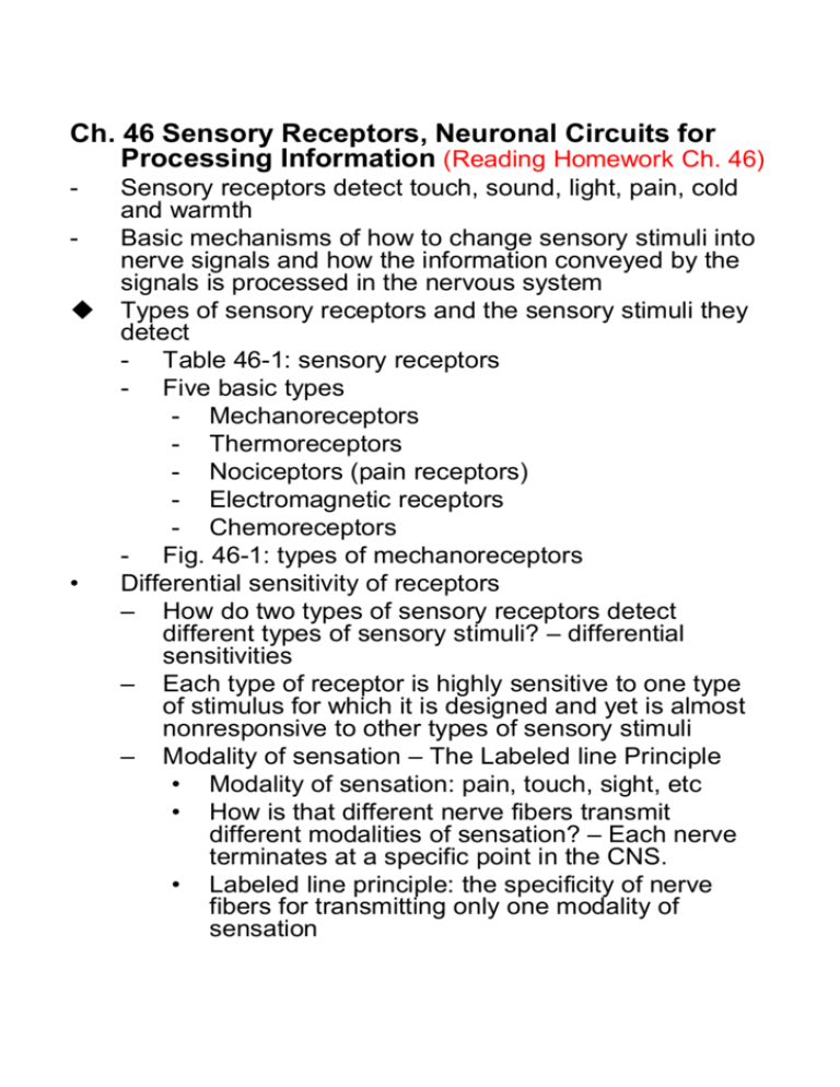
Ch. 46 Sensory Receptors, Neuronal Circuits for Processing Information (Reading Homework Ch. 46) u • Sensory receptors detect touch, sound, light, pain, cold and warmth Basic mechanisms of how to change sensory stimuli into nerve signals and how the information conveyed by the signals is processed in the nervous system Types of sensory receptors and the sensory stimuli they detect - Table 46-1: sensory receptors - Five basic types - Mechanoreceptors - Thermoreceptors - Nociceptors (pain receptors) - Electromagnetic receptors - Chemoreceptors - Fig. 46-1: types of mechanoreceptors Differential sensitivity of receptors – How do two types of sensory receptors detect different types of sensory stimuli? – differential sensitivities – Each type of receptor is highly sensitive to one type of stimulus for which it is designed and yet is almost nonresponsive to other types of sensory stimuli – Modality of sensation – The Labeled line Principle • Modality of sensation: pain, touch, sight, etc • How is that different nerve fibers transmit different modalities of sensation? – Each nerve terminates at a specific point in the CNS. • Labeled line principle: the specificity of nerve fibers for transmitting only one modality of sensation u • Transduction of sensory stimuli into nerve impulses Receptor potentials: the change in potential at a receptor – Mechanisms of receptor potentials: excitation by • Mechanical deformation of the receptor: stretch and opening of ion channels • Application of chemical to the membrane • Change of the temperature • Electromagnetic radiation – Maximum receptor potential amplitude: 100mV – Relation of the receptor potential to action potentials: Fig. 46-2 – Fig. 46-3 – Fig. 46-4: relation between stimulus intensity and receptor potential • Amplitude increases rapidly at first, but then progressively less rapidly at high stimulus strength • It allows the receptor to be sensitive to very weak sensory experience and reach a maximum firing rate until the sensory experience is maximum. • The receptor have an extreme range of response • • • Adaptation of receptors – Sensory receptors adapt either partially or completely to any constant stimulus after a period of time. – Fig. 46-5: Typical adaptation of certain types of receptors – Mechanisms by which receptors adapt • Receptor potential appears at the onset of stimuli, not after • Accommodation – Tonic receptors: slowly adapting receptors can continue to transmit information for many hours – Rate receptors (movement receptors or phasic receptors): Rapidly adapting receptors detect changes in stimulus strength – Importance of rate receptors – predictive function Physiological classification and functions of nerve fibers (Fig. 46-6) Spatial and Temporal Summation – Spatial summation • Receptor field • Fig. 46-7 • Stronger stimulus, more fibers – Temporal summation • Fig. 46-8 u Transmission and processing of signals in neuronal pools – Neuronal pools – Each pool has its own special characteristics – Relaying of signals through neuronal pools • Stimulatory field • Fig. 46-9 • Excitatory stimulus – suprathreshold stimulus • Facilitated – subthreshold • Inhibitory zone • Fig. 46-10 – Divergence of signals: Fig. 46-11 – Convergence of signals: Fig. 46-12 – Inhibitory circuits: Fig. 46-13 – Reverberatory (Oscillatory) circuits: Fig. 46-14 Instruments: EEG Ÿ Hans Berger (1929) Ÿ Reasonably low-cost Ÿ Widely used in clinical practice + Neuropsychology research units EEG Instrumentation n Electrode Board = load plug-in box or input box n Electrode Selectors => montage n Differential Amplifiers n Filters n Penmotors n Chart Drive n Power Supply n Calibrator n Electrodes (Sensors) n Electrolytes, Gels, and Pastes * WWW Neuro Scan Labs Instruments: EEG MicroMed Electrical Geodesics NeuroScan Spontaneous Brain Activity Oscillations of electrical activity which are thought to be the average across thousands of cells • Alpha Rhythm: 8-13Hz • Relax & meditation • Beta Activity: 13-35Hz • Alert or anxious • Theta Activity: 3-7Hz • Abnormal in awake adults • Delta Activity: <3Hz Event-related Evoked Potentials An evoked potential is essentially the same kind of recording, but with larger change of electrical activity which are triggered by a stimulus. An electroencephalogram (EEG) records much smaller oscillations of electrical activity which are thought to be the average across thousands of cells. An evoked potential is essentially the same kind of recording, but with larger changes of electrical activity which are triggered by a stimulus. Brain Mapping with EEG and fMRI
