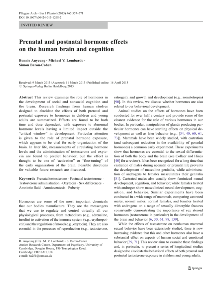
Pflugers Arch - Eur J Physiol (2013) 465:557–571
DOI 10.1007/s00424-013-1268-2
INVITED REVIEW
Prenatal and postnatal hormone effects
on the human brain and cognition
Bonnie Auyeung & Michael V. Lombardo &
Simon Baron-Cohen
Received: 9 March 2013 / Accepted: 11 March 2013 / Published online: 16 April 2013
# Springer-Verlag Berlin Heidelberg 2013
Abstract This review examines the role of hormones in
the development of social and nonsocial cognition and
the brain. Research findings from human studies
designed to elucidate the effects of both prenatal and
postnatal exposure to hormones in children and young
adults are summarized. Effects are found to be both
time and dose dependent, with exposure to abnormal
hormone levels having a limited impact outside the
“critical window” in development. Particular attention
is given to the role of prenatal hormone exposure,
which appears to be vital for early organization of the
brain. In later life, measurements of circulating hormone
levels and the administration of testosterone and oxytocin are found to predict behavior, but the effect is
thought to be one of “activation” or “fine-tuning” of
the early organization of the brain. Possible directions
for valuable future research are discussed.
Keywords Prenatal testosterone . Postnatal testosterone .
Testosterone administration . Oxytocin . Sex differences .
Amniotic fluid . Amniocentesis . Puberty
Hormones are some of the most important chemicals
that our bodies manufacture. They are the messengers
that we use to regulate and control virtually all our
physiological processes, from metabolism (e.g., adrenaline,
insulin) to activation of the immune system (e.g., erythropoietin) and the regulation of mood (e.g., oxytocin). They are also
essential in the processes of reproduction (e.g., testosterone,
B. Auyeung (*) : M. V. Lombardo : S. Baron-Cohen
Autism Research Centre, Department of Psychiatry, University of
Cambridge, Douglas House, 18b Trumpington Road,
Cambridge CB2 8AH, UK
e-mail: ba251@cam.ac.uk
estrogen), and growth and development (e.g., somatotropin)
[90]. In this review, we discuss whether hormones are also
related to our behavioral development.
Animal studies on the effects of hormones have been
conducted for over half a century and provide some of the
clearest evidence for the role of various hormones in our
bodies. In particular, manipulation of glands producing particular hormones can have startling effects on physical development as well as later behavior (e.g., [34, 40, 60, 61,
73]). Mammals have been widely studied, with castration
(and subsequent reduction in the availability of gonadal
hormones) a common early experiment. These experiments
show that hormones are essential to the sexual differentiation of both the body and the brain (see Collaer and Hines
[40] for a review). It has been recognized for a long time that
castration of males during neonatal or prenatal life prevents
the development of masculine genitalia, while administration of androgens to females masculinizes their genitalia
[81]. Castrated males also usually show feminized neural
development, cognition, and behavior; while females treated
with androgen show masculinized neural development, cognition, and behavior. Similar experiments have been
conducted in a wide range of mammals, comparing castrated
males, normal males, normal females, and females treated
with androgens on a range of sexually dimorphic features
consistently demonstrating the importance of sex steroid
hormones (testosterone in particular) in the development of
the brain and behavior [6, 30, 61, 98, 139].
While the effects of testosterone on nonhuman mammal
sexual behavior have been extensively studied, there is now
increasing evidence that this and other hormones also have a
substantial effect on aspects of human social and emotional
behavior [39, 71]. This review aims to examine these findings
and, in particular, to present a series of longitudinal studies
designed to elucidate the behavioral effects of both prenatal and
postnatal testosterone exposure in children and young adults.
558
Pflugers Arch - Eur J Physiol (2013) 465:557–571
1000
The timing of hormonal effects is crucial when studying lasting
effects on development. There are generally thought to be two
types of hormonal effects: organizational and activational
[109]. Organizational effects are most likely to occur during
early development when most neural structures are being
established, producing permanent changes in the brain [109],
whereas activational effects are short term and are dependent on
current hormone levels. It is widely thought that organizational
effects are maximal during certain critical periods of development. These are hypothetical windows of time in which a tissue
can be formed [71]. Outside the sensitive period, the effect of
the hormone will be limited, protecting the animal from disruptive influences. This means, for example, that circulating sex
hormones necessary for adult sexual functioning do not cause
unwanted alterations to tissues, even though the same hormones might have been essential in laying down cellular organization during the initial development of those tissues.
Animal research has indicated that the critical period for
sexual differentiation of the brain occurs when differences in
serum testosterone are highest between sexes [40]. Therefore,
it is likely that this is an important period for sexual differentiation of the human brain as well. It is difficult to get accurate
measurements of hormone levels for humans, but studies that
have sampled fetal serum, plasma, and amniotic fluid during
pregnancy have indicated that for typical human males, there
is a surge in fetal testosterone (FT) levels between weeks 8 and
24 of gestation, peaking around week 16 [2, 36, 115, 116,
126]. During this period, male fetuses produce more than 2.5
times the levels observed in females [20]. There is then a
decline to barely detectable levels from the end of this period
until birth. As a result, it is expected that the most significant
effects of FT on development are likely to occur within this
window. For typical human females, levels are generally very
low throughout pregnancy and childhood [71].
In addition to the fetal surge, two other periods of elevated testosterone have been observed in typical males. The
first takes place shortly after birth and reaches a peak when
the child reaches approximately 3–4 months [126], and the
second occurs around puberty. Figure 1 shows the typical
sex differences in circulating testosterone levels during the
prenatal and neonatal period.
Prenatal hormone effects in humans
Studies in clinical conditions
Some naturally occurring clinical conditions render the human hormone environment abnormal. As we consider artificial manipulation of the hormone environment during
critical periods of development to be unethical, such
Testosterone (ng/dl)
Timing and critical periods
Amniocentesis
Window
500
males
Critical
Period
females
0
10
20
30
birth 10
Weeks
20
30
40
50
Fig. 1 Circulating levels of testosterone in the human fetus and neonate. Males (blue line) have been shown to have higher levels of
testosterone than females (red dashed line), particularly from about
week 8 to 24 of gestation and week 2 to 26 of postnatal life. (Figure
adapted from Hines [71]. Brain gender. New York, NY: Oxford University Press, Inc.)
conditions are a natural starting point for evaluating the
impact of androgens and other hormones on development.
One such condition is congenital adrenal hyperplasia
(CAH), a genetic disorder which causes excess adrenal
production of androgen hormones (including testosterone
and other hormones thought to be responsible for the development of masculinizing features) beginning prenatally in
both males and females [103].
Studies of individuals with CAH have generally found that
girls with the condition show masculinization of behavioral
performance in activities such as spatial orientation, visualization, targeting, personality, cognitive abilities, and sexuality
[64, 74, 114]. Several research groups have reported that girls
with CAH show increased male-typical toy, playmate, and
activity preferences [49, 70, 71, 106]. The determining role of
prenatal steroids in sex-role identity appears to be supported
by studies of females with CAH who demonstrate a masculine
bias on various personality inventories (e.g., Detachment and
Indirect Aggression Scales, Aggression and Stress Reaction
Scales, Reinish’s Aggression Inventory) [40].
While CAH provides an opportunity to investigate the
effects of additional androgen exposure, the relatively rare
occurrence of CAH makes it difficult to obtain large-enough
groups for generalization of research findings to the wider
population. Researchers have also suggested that CAHrelated disease characteristics, rather than prenatal androgen
exposure, could be responsible for the atypical cognitive
profiles observed in this population [51, 112].
Polycystic ovary syndrome (PCOS) is the most common
endocrine disorder in women and affects an estimated 1 in
15 women worldwide. It is a heterogeneous disorder generally characterized by disruption of the ovulation cycle, a
number of small cysts around the edge of their ovaries
(polycystic ovaries), and excessive production and/or secretion of androgens (masculinizing hormones) referred to as
Pflugers Arch - Eur J Physiol (2013) 465:557–571
“hyperandrogenism” [104]. A study of children of women
with PCOS found that daughters of these women showed
lower Empathy Quotient (EQ) scores, a measure where girls
generally show higher scores than boys [105]. In the same
study, daughters of women with PCOS also showed higher
Systemizing Quotient (SQ) scores, a measure where boys
generally score higher than girls. These findings are consistent with the idea that PCOS increases androgen exposure in
the womb and that this increased exposure leads to more
masculinized behavior in later life.
Studies using amniotic fluid measurements
Amniocentesis is the process of extracting a sample of
amniotic fluid during the second trimester of pregnancy to
detect clinical abnormalities in mothers thought to be at risk.
Amniocentesis is typically performed during a relatively
narrow time window which is thought to coincide with the
hypothesized critical period for human sexual differentiation
(between approximately weeks 8 and 24 of gestation, see
Fig. 1) [71]. Samples taken this way indicate that both male
and female human fetuses produce testosterone, with male
fetuses producing on average 2.5 times the levels observed
in females; see Fig. 2.
Males are exposed to testosterone from the fetal adrenals
and testes. The female fetus is also exposed to androgens,
but at much lower levels. In early prenatal life, this testosterone enters the amniotic fluid via diffusion through the
fetal skin and later enters the fluid via fetal urination [118].
Testosterone levels are also affected by other processes—the
underproduction of aromatase may result in higher FT levels
by impairing conversion of testosterone to estrogen [1].
Similarly, dihydrotestosterone is produced from testosterone
559
and may be a stronger activator of the androgen receptor
than testosterone itself [90]. While these processes limit the
conclusions we can draw from a snapshot measurement of
FT level in the amniotic fluid, it is a useful starting point
from which to develop our understanding.
A number of studies have linked elevated levels of FT in
the amniotic fluid with the masculinization of certain behaviors, beginning shortly after birth. In particular, the
Cambridge Child Development Project is an ongoing longitudinal study investigating the relationship between prenatal
hormone levels and the development of later behaviors [15,
85]. Mothers of participating children had all undergone
amniocentesis for clinical reasons. To date, these children
have been tested postnatally at several time points.
Eye contact at 12 months
Reduced eye contact is a characteristic common in children
with autism [96, 129]. The first study aimed to measure FT
and estradiol levels in relation to eye contact in a sample of
70 typically developing 12-month-old children [96]. Frequency and duration of eye contact were measured using
videotaped sessions. Sex differences were found, with girls
making significantly more eye contact than boys. The
amount of eye contact varied with FT levels when the sexes
were combined and also within the boys’ group [96]. No
relationships were observed between the outcome and estradiol levels. Results were taken to indicate that FT may
play a role in shaping the neural mechanisms underlying
social development [96].
Vocabulary size at 18–24 months
Another study of 87 children focused on the relationship
between vocabulary size in relation to FT and estradiol
levels from amniocentesis. Vocabulary size was measured
at both 18 and 24 months of age using the Communicative
Development Inventory, a self-administered checklist of
words for parents to complete [63]. Girls were found to
have significantly larger vocabularies than boys at both time
points [97], and results showed that levels of FT inversely
predicted the rate of vocabulary development [97].
Autistic traits in toddlers at 18–24 months
Fig. 2 Sex differences in amniotic fluid measurements. (Data obtained
from the Cambridge Child Development Project). Circles indicate a small
deviation from the mean. Stars indicate a large deviation from the mean
Autism spectrum conditions (ASCs) are a group of related
conditions characterized by impairments in reciprocal social
interaction and communication, alongside strongly repetitive behaviors and unusually narrow interests [5]. It has
been well documented that autism is much more prevalent
in males than females [32, 58], so the possibility that androgens may have a role to play in the etiology of these
conditions was explored.
560
Pflugers Arch - Eur J Physiol (2013) 465:557–571
Studies have examined the effects of FT on the later
development of autism and autistic traits. In the first of these
studies, autistic traits were measured using the Quantitative
Checklist for Autism in Toddlers (Q-CHAT) [4]. This questionnaire has been used to measure autistic traits elsewhere
and has been validated by following children throughout
their early development (Fig. 3 shows an example question).
The Q-CHAT questionnaire was administered to mothers
who had also undergone amniocentesis, providing measurements of FT level and fetal estradiol (FE)—a second hormone which forms prenatally from testosterone and is
considered to be the most biologically active estrogen
[40]. Samples of postnatal testosterone (PT) levels were also
taken from saliva at 3–4 months of age in a small sample of
these children. The study revealed a significant sex difference in autistic traits, with boys scoring higher (indicating
more autistic traits) than girls. Q-CHAT scores were predicted by FT levels only, with both sex and the FT/sex interaction excluded from the model [13].
The relationship between FT and Q-CHAT score was also
visible within the subset of children who participated in the
follow-up study measuring PT levels at 3–4 months. However, no relationships between FE, PT levels, and Q-CHAT
scores were observed. In addition, FE and PT levels showed
no sex differences or relationships with FT levels [7].
Use of mental and affective language at 4 years
The Cambridge Child Development study has also completed much longer-term studies, with participants recruited at
amniocentesis being followed up 4–5 years after birth. This
has provided the opportunity to establish a much greater
understanding of how FT levels could affect behavioral
development in later life.
Thirty-eight children completed a “moving geometric
shapes” task at age 4. The children were asked to describe
cartoons with two moving triangles whose interaction with
each other suggested social relationships and psychological
motivations [87]. Sex differences were observed, with girls
using more mental and affective state terms to describe the
• Does your child point to share interest with you (e.g. pointing at an
interesting sight)?
many times a day
a few times a day
a few times a week
less than once a week
never
• How easy is it for you to get eye contact with your child?
very easy
quite easy
quite difficult
very difficult
impossible
Fig. 3 Example items from the Q-CHAT
cartoons compared to boys; however, no relationships between FT levels and mental or affective state terms were
observed. Girls were found to use more intentional propositions than males, and a negative relationship between FT
levels and frequency of intentional propositions was observed when the sexes were combined and in boys. Boys
used more neutral propositions than females. FT was related
to the frequency of neutral propositions when the sexes were
combined.
Social relationships and narrow interests at age 4 years
A separate follow-up study at 4 years of age utilized the
Children’s Communication Checklist—a questionnaire
designed to screen for communication difficulties in children 4–16 years of age [24]. A quality of social relationships
subscale demonstrated an association between higher FT
levels and poorer quality of social relationships for both
sexes combined (but not within each sex). A lack of significant correlations within each sex was thought to be a result
of the small sample size (n=58).
Levels of FT were also associated with a subscale for
narrower interests when the sexes were combined and also
within boys [86]. This within-sex result is interesting since it
suggests that this subscale of the measurement is sensitive to
even moderate changes in FT level. Sex differences were
also reported, with males scoring higher (i.e., having more
narrow interests) than females [86].
Gender-typical play at 6–9 years
At 6–9 years of age, the children from the Cambridge Child
Development Project were followed up using the PreSchool Activities Inventory (PSAI). This is a standardized
questionnaire measure of gender-typical play in both boys
and girls. The PSAI includes 24 items and is completed by a
parent to describe the child’s behavior. Higher scores reflect
more male-typical behavior, and females with CAH obtain
elevated (more male-typical) scores in the PSAI [72],
suggesting sensitivity to the effects of prenatal androgen
exposure. A significant relationship was found between FT
levels and sexually differentiated play behavior in both girls
and boys [9].
Gender-role behavior at 6–9 years
The Bem Sex Role Inventory (BSRI) is a questionnaire
developed to measure feminine and masculine personality
traits on the basis of cultural definitions of sex-typed social
desirability [21]. This is a 60-item (20 feminine, 20 masculine, and 20 non-gender-related items) questionnaire. Examination of scores in this measure indicated that higher FT
levels are associated with higher masculinity scores in the
Pflugers Arch - Eur J Physiol (2013) 465:557–571
561
BSRI when boys and girls were examined together and
when girls were examined alone. No relationships were
found between FT levels and scores in the femininity scale.
Within-sex results suggest that girls exposed to higher testosterone levels in utero are perceived as exhibiting more
masculinized behavior [7].
Mind reading at 6–9 years
Mind reading is the ability to put oneself into the mind of
another person and infer what the person is thinking or feeling.
It is also referred to as theory of mind [91] or mentalizing [55].
One method of measuring an individual’s capability for
mentalizing is the child version of the “Reading the Mind in
the Eyes” test. This measure consists of 28 pictures from the
eye region of the face, with each depicting a mental state—
some including subtle emotions [16]. Figure 4 shows an
example item, with four emotions for the participant to choose
from (the correct answer is “a bit worried”).
Results from this study revealed a significant negative
correlation with FT—with higher levels predicting lower
mind reading capability. Within-sex analyses revealed a significant negative correlation between FT and the eyes test
within both the boys’ and girls’ groups [33]. The significance
within each sex points to a much more sensitive dependency
on FT level than in the entire population, where boys have
much higher levels of FT.
Empathy and systemizing at 6–9 years
Empathy has been described as the drive to identify another
person’s emotions and thoughts and to respond to these
appropriately [14]. This is an aspect of social interaction
where there is usually a strong advantage for females. Sex
differences in the precursors of empathy are seen from birth,
with female babies showing a stronger preference for
looking at social stimuli (faces) 24 h after birth [41] and
more eye contact at 12 months of age [96]. Girls also tend to
show more comforting, sad expressions or sympathetic vocalizations than boys when witnessing another’s distress as
early as 1 year of age [76].
angry
friendly
Systemizing has been defined as the drive to analyze,
explore, and construct a system [14]. Systemizing allows
one to predict the behavior of a system and to control it. A
system is defined as something that takes inputs, which can
then be operated on in variable ways, to deliver different
outputs in a rule-governed way. A growing body of evidence
suggests that, on average, males spontaneously systemize to a
greater degree than females. Boys, on average, engage in more
mechanical and constructional play than girls [22, 92]. This
sex difference in toy choice has been observed in humans as
early as the first year of life [125], as well as in nonhuman
primates [3], suggesting the possibility of a biological basis
for these preferences. Boys are better than girls at using
directional cues in map reading and map making [19, 56,
82]. They are also more accurate on the Mental Rotation Test
[80, 100] and the Rod and Frame Test [23, 141]. All of these
can be seen as involving systemizing since they involve
relating input to output via a lawful operation.
The Cambridge Child Development study has developed
parental questionnaires to assess a child’s capability in the
above dimensions: the Empathy Quotient (EQ-C) and Systemizing Quotient (SQ-C). These questionnaires are based
on similar versions for adolescents and adults, which have
consistently identified significant differences between males
and females [8, 138]. Although limited by the questionnaire
format, these measures have the advantage that parents
observe their child’s behavior in a wide number of contexts.
Girls generally scored higher than boys on the EQ-C at
ages 6–8 years. When scores were compared with prenatal
measurements of FT, a significant negative correlation between FT levels and EQ-C score was observed when the
sexes were combined and also within boys [33].
Results for the SQ-C showed that boys scored significantly
higher than girls on this scale. A significant positive association was found between SQ-C and FT levels when boys and
girls were examined together. When sexes were examined
together, the only significant predictor was FT. Sex was not
included in the final regression model, suggesting that FT
levels could play a greater role than the child’s sex in terms
of differences in systemizing preference [10].
The dimensions of Empathizing and Systemizing are also
useful to our understanding of autism. Administration of a
wide variety of tasks measuring the ability to empathize has
demonstrated that individuals with an ASC are much weaker in this area. Conversely, there is some evidence that some
individuals with an ASC are better at tasks that involve
systemizing.
Autistic traits at 6–9 years of age
unkind
a bit worried
Fig. 4 Example item from the children’s Reading the Mind in the Eyes
Test. The child is asked to indicate which of the four choices best
describes what the person in the picture is feeling
In order to further evaluate the potential effects of prenatal
exposure to testosterone on the development of autistic
behaviors in later life, FT measurements were directly
562
Pflugers Arch - Eur J Physiol (2013) 465:557–571
evaluated against a child’s score on the Childhood Autism
Spectrum Test (CAST) [123, 140] and the Child Autism
Spectrum Quotient (AQ-Child) [12]. The CAST is a validated and widely used autism screening measure used to
detect who is at risk for ASC. The AQ-Child is a measure
that quantifies autistic traits and has been used widely in
research.
FT levels were positively associated with higher scores
(indicating greater number of autistic traits) on both the
CAST and the AQ-Child. For the AQ-Child, this relationship was seen within both the male and female groups as
well as when the sexes were combined, suggesting that this
is an effect of FT rather than an effect of sex. The relationship between CAST scores and FT was also seen within
boys, but not girls [11].
Summary of the Cambridge Child Development Project
Table 1 describes the measures used in the Cambridge Child
Development Project to identify sex differences in behavior
and the links with FT for boys and girls together. For each
measure, we describe the direction of the sex differences (if
present). The final columns indicate whether FT levels
(independent of sex) were a significant predictor in the
associated regression analyses.
FT and the brain
Results from the above studies suggest that higher prenatal
hormone levels contribute to greater masculinization of
behavior. In order to understand some of the mechanisms
by which this could take place, we have recently
extended our investigation into how FT may affect brain
development.
Increased FT levels have been shown to affect brain
morphology, showing a significant relationship with increased rightward asymmetry (e.g., right > left) of a posterior subsection of the callosum [35]. FT also influences later
cortical gray matter volume, which has been observed to be
sexually dimorphic [94]. Higher FT predicted increased
gray matter in the right temporo-parietal junction, and this
brain region shows a male > female pattern of sexual dimorphism. The right temporo-parietal junction is one region
that is associated with tasks requiring one to think about
other people’s thoughts and mental states [121], suggesting
a link between FT exposure and the neural development of
mentalizing. Similarly, the gray matter in the planum
temporale and posterior lateral orbitofrontal cortex is inversely
related to FT levels and also show a female > male
pattern of sexual dimorphism. Thus, FT predicts development
of gray matter in directions that are congruent with observed
sexual dimorphism and is indicative of the organizational
nature of its influence on sexually dimorphic brain
development; see Fig. 5.
Recent studies on functional brain response have also
indicated that higher levels of FT predict enhanced sensitivity to positive (happy faces) compared to negatively
valenced information (fear faces) in reward-related structures within the striatum. Furthermore, the effect FT has
on influencing sensitivity to positive over negatively
valenced information mediates the relationship FT has on
predicting later behavioral approach tendencies (e.g., When
my child sees an opportunity for something, he/she gets
excited right away) [93].
Table 1 Cambridge Child Development Project results
Characteristic
Measure
Child age
Sex difference
FT sig. predictor
Eye contact
Vocabulary size
Mental and affective language
Restricted interests
Social relationships
Gender-typical play
Gender-role behavior
12 months
18–24 months
4 years
4 years
4 years
6–9 years
6–9 years
6–9 years
6–9 years
Yes
Yes
Yes
Yes
Yes
Yes
Yes
Yes
No
Yes
(F >
(F >
(F >
(F <
(F >
(F <
(F >
(F <
Mind Reading
Empathy
Frequency
Communicative Development Inventory
Intentional Propositions
Children’s Communication Checklist
Children’s Communication Checklist
Pre-School Activities Inventory
Bem Sex Role Inventory Femininity Total
Bem Sex Role Inventory Masculinity Total
Reading the Mind in the Eyes
Empathy Quotient
(F > M)
Yes
Yes
Yes
Yes
Yes
Yes
–a
Yes
Yes
No
Systemizing
Autistic traits
Autistic traits
Autistic traits
Systemizing Quotient
Q-CHAT
AQ-Child
CAST
6–9 years
18–24 months
6–10 years
6–10 years
Yes
Yes
Yes
Yes
(F <
(F <
(F <
(F <
Yes
Yes
Yes
Yes
a
A regression analysis was not conducted
M)
M)
M)
M)
M)
M)
M)
M)
M)
M)
M)
M)
Pflugers Arch - Eur J Physiol (2013) 465:557–571
563
Fig. 5 FT correlations with
gray matter volume. a Areas
where FT predicts local gray
matter volume. Red/orange
voxels denote positive
correlations; blue voxels denote
negative correlations. b Areas
of sexual dimorphism in local
GM volume. Red/orange voxels
denote a male > female pattern;
blue voxels denote a female >
male pattern. (Figure from
Lombardo et al., [94])
Limitations of measuring prenatal exposure to hormones
in amniotic fluid
The findings presented in Table 1 make use of testosterone levels in amniotic fluid (via amniocentesis). The
benefit of this method is that it provides a sample
which is close to the fetus and which is collected as
part of normal clinical practice for mothers thought to
be at risk of complications during pregnancy or birth.
Amniocentesis is generally also conducted in a fairly
narrow time window, aiding repeatability of measurements. Ideally, it would be best to make direct measurements of testosterone at regular intervals throughout
gestation and into postnatal life. Even for amniocentesis,
it is not currently possible to obtain repeated samples of
FT because the procedure carries a risk of causing
miscarriage (of about 1 %) [42, 120]. It is also known
that hormones fluctuate during the day and between
days, even in fetuses [124, 137].
Given the estimated timeline for testosterone secretion,
the most promising time to measure FT is probably at
prenatal weeks 8 to 24 [126], but this is still a relatively
wide range. Research on nonhuman primates has also
shown that androgens masculinize different behaviors at
different times during gestation, suggesting that different
behaviors may also have different sensitive periods for
development [60].
For all these reasons, the inferences we can therefore
draw about the single measurement of FT are necessarily
limited. At the same time, a significant correlation between
amniotic FT and a behavior should represent a conservative
estimate of the potential effect of FT exposure on that
behavior.
Human behavior is complex, and biological, social, or
cultural factors are continuously interacting, making it challenging to investigate the causes of behavior. To the extent
that social factors have been considered within the experiments presented so far, these have been restricted to certain
564
demographic variables (such as maternal age, parental education, and number of siblings), and behaviors and traits are
likely to be influenced by a range of social factors that have
not been measured in these studies.
Postnatal hormone effects in humans
Studies of current (activational) hormones
Studies of postnatal hormone exposure have examined the
effects of current (or activational) hormones. The most
obvious example of postnatal hormone exposure is during
puberty—a major period of hormonal, physical, and behavioral change and development.
Studies in nonhuman mammals have investigated whether changes during puberty indicate a critical period for the
effects of steroid hormones. One study showed that gonadectomy in male ferrets before puberty but after the early
critical period did not affect sexual development when these
animals were treated with testosterone in adulthood [17]. A
study in rats showed that early steroid hormone deprivation
resulted in systemic reduction in sensitivity to later androgen effects [59]. More recent findings suggest that steroid
hormones during puberty have an activational affect on
brain development [122]. These results indicate that although the critical window during perinatal development is
vitally important for early sexual differentiation of the brain,
the pubertal period also plays a large role in “fine-tuning”
the organizational effects of steroid hormones [119].
In the area of physical development, it is well known that
during puberty changes in adrenal androgens, rapid growth in
body size, changes in fat composition, and the development of
secondary sex characteristics occur [54]. In contrast, the studies examining the relationships between puberty, hormone
changes, and the effects on social and emotional behavior
have been relatively few. This is because the onset of puberty
varies greatly between individuals as well as between sexes,
so recruitment of appropriate age groups can be difficult. In
addition, there is little research on this age group due to the
discomfort and embarrassment that may occur when trying to
obtain reliable and accurate information on sexual maturation.
Other studies have relied on parental or self-report measures
of puberty, which can also have difficulties [107]. It is also
very difficult to disentangle the many physical aspects of
maturation from the co-occurring changes in social and cultural status that are associated with this age group.
Much of what is known about adolescent development in
humans comes from studies that do not specifically include
biological measures of pubertal development (such as hormone levels). For example, there is evidence suggesting that
brain regions such as the rostral prefrontal cortex which are
involved in certain executive functions are still developing
Pflugers Arch - Eur J Physiol (2013) 465:557–571
during adolescence [48]. A study that used a narrow age
range and measures of pubertal development showed a
positive correlation between pubertal development and an
increased tendency towards sensation seeking [99]. The
increase in sensation seeking during puberty may relate to
the increase in risk taking observed in adolescents, which
seems to decline in adulthood [145]. However, how the
development of these systems might be related to the effect
of puberty or changes in steroid hormones is not known.
Efforts have also been made to examine the links between both prenatal as well as activational hormone effects
on aggressive behavior in same-sex and opposite-sex twins,
with the assumption that girls from pairs of opposite-sex
twins are exposed to higher levels of prenatal testosterone
compared to same-sex twin girls. The researchers hoped to
control for postnatal environmental effects by comparing
data with similar measurements of same-sex female twins.
In this study, the Dutch translation of the Reinisch Aggression Inventory (RAI) [113] and the Dutch translation of a
modified version of the Olweus Multifaceted Aggression
Inventory (OMAI) [52] were used to measure aggression
in 74 opposite-sex and 55 same-sex 13-year-old twin pairs.
Opposite-sex twin girls scored in the masculine direction on
the withdrawal and verbal aggression subscales of the RAI,
whereas no differences were observed between same-sex
and opposite-sex twin girls on the OMAI. These differences
may have existed because the RAI measures how prone an
individual is to aggressive behavior, whereas the OMAI
focuses on overt aggressive behavior [38]. The activational
effects of testosterone were assessed using salivary testosterone measures in addition to a measure of pubertal status
using the Tanner drawings [130]. Although there was some
evidence of associations between free testosterone levels
and personality traits (such as aggressive impulses and
boredom susceptibility in boys, and experience seeking
and extraversion in girls), the authors concluded that at this
age, no clear associations between circulating testosterone
levels and behavioral traits were apparent [38].
More recently, sex differences were observed in the relationship between circulating testosterone levels using bloodspot
samples and thickness in areas of the brain which are associated
with high androgen receptor density (including the left inferior
parietal lobule, middle temporal gyrus, calcarine sulcus, and
right lingual gyrus) [29]. These findings provide new evidence
for the role of testosterone in pubertal structural brain development and sexual differentiation; however, further work is
needed to ascertain how these changes may relate to social,
cognitive, and emotional development.
Studies of testosterone administration
The majority of findings discussed so far have relied on
observations in clinical conditions characterized by atypical
Pflugers Arch - Eur J Physiol (2013) 465:557–571
exposure to hormones or by obtaining samples of amniotic
fluid, blood, or saliva to measure hormone levels and relating these to measurements of interest. In some cases, it is
also possible to study the effects of directly altering circulating hormone levels (though prenatal manipulation of hormone levels is considered too dangerous and unethical).
Recent studies in adult women have used a sublingual
administration of testosterone, leading to a short-term large
increase in circulating testosterone. Using this method, a
series of studies have examined the effects of a single dose
of testosterone versus placebo on social and emotional behavior (see Bos et al. [27] for a review).
Administration studies have shown that testosterone decreases theory of mind and facial emotion recognition in
these women. Using the Reading the Mind in the Eyes test, a
measure examining subtle emotion and mental states from
pictures of the eye region, testosterone administration led to
lower scores compared to placebo [134]. Interestingly, the
2D:4D digit length ratios (thought to be a proxy for prenatal
hormone exposure) of the women tested in this study predicted approximately 50 % of the variance in the effect of
testosterone on task performance. The authors suggest that
the testosterone administration effect may be primed by
prenatal exposure to testosterone [134].
Testosterone administration has also been shown to decrease recognition of angry expressions, and the authors
hypothesize that testosterone may reduce the recognition
of social threat, which may point towards a role for testosterone in social aggression [133]. Angry faces may be an
implicit signal of threat or competition, and testosterone
administration has also been shown to increase gaze to the
eye region of threatening faces that are viewed unconsciously,
suggesting a role for testosterone in implicit social dominance
[131]. Testosterone has also been shown to reduce empathic
facial imitation [68]. In a functional MRI (fMRI) study, testosterone administration activated areas such as the orbitofrontal
cortex and amygdala (both considered to be emotion processing regions) when looking at angry-versus-happy facial expressions, again suggesting a role for testosterone in social
threat [69]. A recent fMRI study also suggests that administration of testosterone alters functional connectivity between
brain regions when looking at social stimuli. Testosterone
(vs. placebo) decreases connectivity between the amygdala
and orbitofrontal cortex (OFC) [26], and amygdala activation
shifts away from the OFC towards the thalamus [136].
Testosterone is also related to trust. Administration of testosterone is related to rating pictures as being less trustworthy
compared to placebo, even when baseline testosterone levels
do not differ [28]. Administration of testosterone increases the
responsiveness of the amygdala to untrustworthy faces,
perhaps due to heightened social vigilance [26].
Testosterone decreases the amount of collaboration between two participants by increasing the egocentricity of the
565
individual’s choices [143] and decreases generosity [144].
However, another study found that testosterone administration increases social cooperation in individuals with low
levels of prenatal testosterone exposure (measured using
2D:4D ratio) [132], which provides some evidence that responses following testosterone administration may be in
part dependent on early organizational effects. Following
testosterone (vs. placebo) administration, women have increased activation in the thalamo-cingulate region, insula,
and the cerebellum in response to infant crying, indicating
testosterone may have a role in modulating parental care
[25].
Testosterone also affects responsivity to reward. Using
the Iowa gambling task, women show an increase in risk
taking after testosterone administration [135]. Using a monetary incentive delay task, testosterone administration increases ventral striatum activation, associated with reward
anticipation, in individuals with low appetitive motivation
(behavior directed toward goals that are usually associated
with reward processes) [67].
Participants who believe that they received testosterone,
regardless of whether they actually received it or not, behave more unfairly than those who believed that they were
treated with placebo. In fact, testosterone administration
increases the frequency of fair bargaining [50].
Although these studies provide interesting and novel
evidence for testosterone administration effects, the sample
sizes are small and further replication of the results is
needed. These studies also include mainly females, and
while they do control for the phase of the menstrual cycle,
which itself predicts emotion recognition [43] and brain
function (e.g., amygdala response) [44], many of the women
are also using oral contraceptives which suppress ovarian
hormone production [53]. The effects of how all these
factors interact and the effects of social and emotional
behavior need further investigation.
Studies of oxytocin administration
Oxytocin is another hormone that has been shown to be
essential to our social functioning. Interestingly, research
examining the administration of oxytocin has shown seemingly “opposite” results to those found for testosterone when
examining its effect on aspects of human social behavior.
Oxytocin production is unique to mammals, and a great
deal of research has investigated the critical role it plays in
the social behavior of nonhuman mammals. Studies have
shown that oxytocin is associated with social memory [111],
affiliation [79, 142], and pair bonding in animals [31, 77,
78], and some researchers have suggested that oxytocin may
also play a largely social role in human behavior [66].
Studies in humans are at an early stage but are gradually
revealing some potentially useful results. In one of the
566
earlier studies, duration and pattern of social gaze (towards
the eye region) in men were increased by administration of
an intranasal dose of oxytocin [62]. Gaze is generally
thought to be predictive of the ability to interpret the meaning of social situations and the intentions of others [84].
Research in social behavior has shown that a dose of
intranasal oxytocin increases trust in social situations,
suggesting that it might serve an affiliative purpose in
humans, as well as animals [88] (especially among ingroup members). For this reason, oxytocin is sometimes
dubbed the “trust hormone”. However, later work has
shown that oxytocin modulates much more in social cognition than just interpersonal trust. fMRI studies have shown
that oxytocin exerts influence on important neural circuits
for a wide range of social–cognitive abilities such as eye
gaze, mentalizing, emotion recognition, and learning [18,
46, 47, 57, 83, 89, 110, 117]. Regions in these studies which
are affected by oxytocin, such as the amygdala, fusiform
gyrus, ventromedial prefrontal cortex, insula, superior temporal gyrus, and inferior frontal gyrus [108], are consistently
atypical in conditions where difficulties in social cognition
are a defining feature as in ASC [45, 95]. Extensive reviews
on oxytocin can be found elsewhere (e.g., [66, 128]).
Testosterone versus oxytocin administration
Due to the seemingly disparate effects of administering
testosterone and oxytocin, it has recently been proposed that
steroids and neuropeptides are important in different environments [27]. For example, testosterone may increase vigilance and motivation for action and may reduce social
cognition in environments that demand action (such as in
emergencies or high stress situations). Neuropeptides such
as oxytocin may increase social cognition in environments
that are safe or that do not demand action. The subtleties of
these interactions are numerous and varied and need further
testing [27]; however, it will be important to consider the
environment and situational contexts when interpreting the
findings from research of this kind, where the vast majority
of studies are conducted in laboratory settings.
Further, unlike the testosterone studies that mainly include
females, the vast majority of the studies on the effects of
oxytocin have only included males. These samples were
mainly chosen as a result of the practicalities of the side effects
associated with each hormone. The generalizability of results
from these studies has not yet been thoroughly tested, and
possible sex-dependent outcomes have not been ruled out.
Future directions
In all the research described above, the role of the social
environment has largely not been considered in depth.
Pflugers Arch - Eur J Physiol (2013) 465:557–571
Social interactions undoubtedly play an important role in
the development of social and emotional behavior. For
example, research on gender-based expectations may cause
parents, teachers, or caregivers to elicit and reinforce
expected behavior from children [127], thus shaping the
child’s behavior. Further work on the role of the environment and how it interacts with hormone levels and behavior
would be highly informative.
The relationships between hormones and behaviors in
humans are likely to be dependent on many factors, and in
the main report correlations with hormone levels measured
at a single time point. Research in animals has generally
shown that hormonal effects on behavior may be dose and
time dependent [39, 71], and these issues need to be clarified.
The replication of the results in larger sample sizes would also
help to increase the range of hormone levels observed in these
studies and assist in identifying any factors that are linked with
levels in the extreme ranges.
It would also be valuable to further establish the relationships between direct measures of hormones (e.g., amniotic
fluid or serum measures) and physical characteristics (e.g.,
2D:4D ratio or dermatoglyphics) which have been used as
proxy measures of hormone exposure. The benefit of using
these types of measurements is that they are easy to obtain
and have also been linked to multiple areas of human
development. However, limited evidence exists for a relationship between these proxy measures and exposure to
prenatal hormones. If such a link was confirmed using direct
measures of hormones, it could simplify future investigations of hormone effects.
In studies of puberty, it would be beneficial for the field
to include more in-depth studies that investigate the contribution of pubertal development, hormone levels, and social
influences on development. The degree to which genetic
variation is coupled with changes in hormone exposure is
also unknown, and it may be that changes in hormone levels
are simply a manifestation of a genetic influence. This
would be an interesting area for future research since investigations of current testosterone levels have shown rates
of heritability between 50and 66 % [65, 75]. Sex hormones
also have an epigenetic role in changing gene expression
throughout development and likely interact with sex chromosome effects on sexual differentiation [101, 102], and
further exploration of applications to social behavior would
be important.
With regards to the administration studies, it is worth
noting that the majority of studies that have used this methodology have restricted their samples to either a female sample when using testosterone or a male sample when using
oxytocin. As a result, the findings of the abovementioned
administration studies may not necessarily be generalized to
samples of the opposite sex. Future studies should compare
the responses of males and females to ascertain whether there
Pflugers Arch - Eur J Physiol (2013) 465:557–571
may be any sex-dependent effects. The testosterone administration studies also include those who are using oral contraceptives, which itself is a hormone manipulation. It would be
important for this area for studies to investigate how oral or
hormone contraception may interact with the testosterone
administration.
Conclusions
Research suggests that human social and emotional behaviors may be affected by gonadal hormones, in particular
exposure to testosterone. The role of prenatal testosterone
appears to be vital for early organization of the brain and, in
particular, in the programming of sexual differentiation during critical periods of development. In humans, the most
important period appears to be early–mid pregnancy. This
finding has been repeated in studies looking at a number of
behavioral measures and is also indicated by tendencies in
those who are naturally exposed to elevated levels of the
hormone through clinical conditions. It is also generally
supported by studies in nonhuman mammals.
In later life, the effects of hormones such as testosterone
during puberty have been shown to also predict behavior,
but it is thought that these effects activate or fine-tune the
early organization of the brain, although the exact relationships between these two time periods are far from clear. To
some extent, the activational effect of hormones during
puberty appears to be dependent on exposure during the
organizational period of early development, when key tissues are first formed.
While the above conclusions appear to generally hold, it
is apparent that there is still much work to be done to further
understand the subtle effects that specific changes to hormone levels may have. Administration of hormones to an
individual can provide some further clues. Generally speaking, such studies have concluded that increased testosterone
levels seem to be involved with decreased social and emotional behavior, whereas administration of oxytocin increases social and emotional behavior.
More recent studies are beginning to identify the physical
processes which may be involved in the effects of hormones
on development and behavior. This research is generally at
an early stage, though there is an indication that specific
areas of the brain are more developed in those with higher
prenatal testosterone levels. Functional MRI studies involving administration of particular hormones also indicate
greater or reduced response from individual areas of the
brain due to changes in testosterone or oxytocin levels. Such
experiments are useful because they do not require a longitudinal design but at the same time cannot easily examine
organizational effects. The ways in which steroids interact
with neuropeptides and other hormones, as well as the cause
567
of natural variation of sex steroids in general, is still not well
understood.
The investigation of both organizational and activational
hormone exposure on behavioral development remains an
area needing much more detailed research. In addition to
helping us map the process of human development, findings
in this area could have major implications for clinical conditions characterized by social and emotional difficulties
such as autism. Such conditions have a very major impact
on an individual’s quality of life.
Although it does not yet provide a clear way to help, the
science examining hormonal effects on social and emotional
development has come a long way and continues to evolve
at a rapid pace. This remains a vibrant area of research
where long-term commitment to research goals can greatly
increase our understanding of human physical and behavioral development. Many exciting studies are underway, and
we look forward to disentangling some of the many biological and environmental factors that contribute to shaping
human social and emotional behavior.
Acknowledgments BA, MVL, and SBC were supported by grants
from the Nancy Lurie Marks Family Foundation, the Shirley Foundation, the MRC, and the Wellcome Trust during the period of this work.
We are grateful to our colleagues and the families who have taken part
in the research over the years. Parts of this review have been updated
from Baron-Cohen, S., Tager-Flusberg, H., Lombardo, MV. (eds) Understanding Other Minds (Oxford University Press).
References
1. Abramovich DR (1974) Human sexual differentiation—in utero
influences. J Obstet Gynecol 81:448–453
2. Abramovich DR, Rowe P (1973) Foetal plasma testosterone
levels at mid-pregnancy and at term: relationship to foetal sex. J
Endocrinol 56(3):621–622
3. Alexander GM, Hines M (1994) Gender labels and play styles:
their relative contribution to children’s selection of playmates.
Child Dev 65:869–879
4. Allison C, Baron-Cohen S, Wheelwright S, Charman T, Richler J,
Pasco G, Brayne C (2008) The Q-CHAT (Quantitative Checklist
for Autism in Toddlers): a normally distributed quantitative measure of autistic traits at 18–24 months of age: preliminary report. J
Autism Dev Disord 38:1414–1425
5. APA (1994) DSM-IV Diagnostic and statistical manual of mental
disorders, 4th edn. American Psychiatric Association,
Washington
6. Arnold AP, Gorski RA (1984) Gonadal steroid induction of
structural sex differences in the central nervous system. Annu
Rev Neurosci 7:413–442
7. Auyeung B (2008) Foetal testosterone, cognitive sex differences
and autistic traits. University of Cambridge, Cambridge
8. Auyeung B, Allison C, Wheelwright S, Baron-Cohen S (2012)
Brief report: development of the adolescent empathy and systemizing quotients. J Autism Dev Disord 42(10):2225–2235.
doi:10.1007/s10803-012-1454-7
568
9. Auyeung B, Baron-Cohen S, Ashwin E, Knickmeyer R, Taylor
K, Hackett G, Hines M (2009) Fetal testosterone predicts sexually
differentiated childhood behavior in girls and in boys. Psychol
Sci 20:144–148
10. Auyeung B, Baron-Cohen S, Chapman E, Knickmeyer R, Taylor
K, Hackett G (2006) Foetal testosterone and the child systemizing
quotient. Eur J Endocrinol 155(suppl 1):S123–S130
11. Auyeung B, Baron-Cohen S, Chapman E, Knickmeyer R, Taylor
K, Hackett G (2009) Fetal testosterone and autistic traits. Brit J
Psychol 100:1–22
12. Auyeung B, Baron-Cohen S, Wheelwright S, Allison C (2008)
The Autism Spectrum Quotient: children’s version (AQ-Child). J
Autism Dev Disord 38:1230–1240
13. Auyeung B, Taylor K, Hackett G, Baron-Cohen S (2010) Foetal
testosterone and autistic traits in 18 to 24-month-old children.
Mol Autism 1(1):11
14. Baron-Cohen S (1999) The extreme male-brain theory of autism.
In: Tager Flusberg H (ed) Neurodevelopmental disorders. MIT
Press, Cambridge
15. Baron-Cohen S, Lutchmaya S, Knickmeyer R (2004) Prenatal
testosterone in mind. MIT Press, Cambridge
16. Baron-Cohen S, Wheelwright S, Spong A, Scahill L, Lawson J
(2001) Are intuitive physics and intuitive psychology independent?
A test with children with Asperger syndrome. J Dev Learn Disord
5:47–78
17. Baum MJ, Erskine MS (1984) Effect of neonatal gonadectomy
and administration of testosterone on coital masculinization in the
ferret. Endocrinology 115(6):2440–2444
18. Baumgartner T, Heinrichs M, Vonlanthen A, Fischbacher U, Fehr
E (2008) Oxytocin shapes the neural circuitry of trust and trust
adaptation in humans. Neuron 58(4):639–650. doi:10.1016/
j.neuron.2008.04.009
19. Beatty WW, Tröster AI (1987) Gender differences in geographical knowledge. Sex Roles 16(11–12):565–590
20. Beck-Peccoz P, Padmanabhan V, Baggiani AM, Cortelazzi D,
Buscaglia M, Medri G, Marconi AM, Pardi G, Beitins IZ
(1991) Maturation of hypothalamic-pituitary-gonadal function
in normal human fetuses: circulating levels of gonadotropins,
their common alpha-subunit and free testosterone, and discrepancy between immunological and biological activities of
circulating follicle-stimulating hormone. J Clin Endocr Metab
73:525–532
21. Bem SL (1974) The measurement of psychological androgyny. J
Consult Clin Psychol 42(2):155–162
22. Berenbaum SA, Hines M (1992) Early androgens are related to
childhood sex-typed toy preferences. Psychol Sci 3:203–206
23. Berlin DF, Languis ML (1981) Hemispheric correlates of the rodand-frame test. Percept Mot Ski 52(1):35–41
24. Bishop DVM (1998) Development of the children’s communication checklist (CCC): a method for assessing qualitative aspects
of communicative impairment in children. J Child Psychol
Psychiatry 6:879–891
25. Bos PA, Hermans EJ, Montoya ER, Ramsey NF, van Honk J
(2010) Testosterone administration modulates neural responses to
crying infants in young females. Psychoneuroendocrinology
35(1):114–121. doi:10.1016/j.psyneuen.2009.09.013
26. Bos PA, Hermans EJ, Ramsey NF, van Honk J (2012) The neural
mechanisms by which testosterone acts on interpersonal trust.
NeuroImage 61(3):730–737. doi:10.1016/j.neuroimage.2012.04.002
27. Bos PA, Panksepp J, Bluthe RM, van Honk J (2012) Acute effects
of steroid hormones and neuropeptides on human social-emotional
behavior: a review of single administration studies. Front
Neuroendocrinol 33(1):17–35. doi:10.1016/j.yfrne.2011.01.002
28. Bos PA, Terburg D, van Honk J (2010) Testosterone decreases
trust in socially naive humans. Proc Natl Acad Sci U S A
107(22):9991–9995. doi:10.1073/pnas.0911700107
Pflugers Arch - Eur J Physiol (2013) 465:557–571
29. Bramen JE, Hranilovich JA, Dahl RE, Chen J, Rosso C, Forbes
EE, Dinov ID, Worthman CM, Sowell ER (2012) Sex matters
during adolescence: testosterone-related cortical thickness maturation differs between boys and girls. PLoS One 7(3):e33850.
doi:10.1371/journal.pone.0033850
30. Breedlove SM (1994) Sexual differentiation of the human nervous system. Ann RevPsychol 45:389–418
31. Carter CS, Williams JR, Witt DM, Insel TR (1992) Oxytocin and
social bonding. Ann N Y Acad Sci 652:204–211
32. Chakrabarti S, Fombonne E (2005) Pervasive developmental
disorders in preschool children: confirmation of high prevalence.
Am J Psychiatry 162(6):1133–1141
33. Chapman E, Baron-Cohen S, Auyeung B, Knickmeyer R, Taylor
K, Hackett G (2006) Fetal testosterone and empathy: evidence
from the Empathy Quotient (EQ) and the ‘Reading the Mind in
the Eyes’ test. Soc Neurosci 1:135–148
34. Christensen LW, Gorski RA (1978) Independent masculinization
of neuroendocrine systems by intracerebral implants of testosterone or estradiol in the neonatal female rat. Brain Res 146(2):325–
340. doi:10.1016/0006-8993(78)90977-0
35. Chura LR, Lombardo MV, Ashwin E, Auyeung B, Chakrabarti B,
Bullmore ET, Baron-Cohen S (2010) Organizational effects of
fetal testosterone on human corpus callosum size and asymmetry.
Psychoneuroendocrinology 35:122–132
36. Clements JA, Reyes FI, Winter JS, Faiman C (1976) Studies on
human sexual development. III. Fetal pituitary and serum, and
amniotic fluid concentrations of LH, CG, and FSH. J Clin
Endocrinol Metab 42(1):9–19
37. Cohen J (1988) Statistical power analysis for the behavioral
sciences, 2nd edn. Lawrence Erlbaum Associates, Hillsdale
38. Cohen-Bendahan CCC, Buitelaar JK, van Goozen SHM,
Orlebeke JF, Cohen-Kettenis PT (2005) Is there an effect of
prenatal testosterone on aggression and other behavioral traits?
A study comparing same-sex and opposite-sex twin girls. Horm
Behav 47:230–237
39. Cohen-Bendahan CC, van de Beek C, Berenbaum SA (2005)
Prenatal sex hormone effects on child and adult sex-typed
behavior: methods and findings. Neurosci Biobehav R
29(2):353–384
40. Collaer ML, Hines M (1995) Human behavioural sex differences:
a role for gonadal hormones during early development? Psychol
Bull 118:55–107
41. Connellan J, Baron-Cohen S, Wheelwright S, Batki A, Ahluwalia
J (2000) Sex differences in human neonatal social perception.
Infant Behav Dev 23(1):113–118
42. d’Ercole C, Shojai R, Desbriere R, Chau C, Bretelle F, Piechon L,
Boubli L (2003) Prenatal screening: invasive diagnostic approaches. Child’s Nerv Syst 19(7–8):444–447
43. Derntl B, Kryspin-Exner I, Fernbach E, Moser E, Habel U (2008)
Emotion recognition accuracy in healthy young females is associated with cycle phase. Horm Behav 53(1):90–95. doi:10.1016/
j.yhbeh.2007.09.006
44. Derntl B, Windischberger C, Robinson S, Lamplmayr E,
Kryspin-Exner I, Gur RC, Moser E, Habel U (2008) Facial
emotion recognition and amygdala activation are associated with
menstrual cycle phase. Psychoneuroendocrinology 33(8):1031–
1040. doi:10.1016/j.psyneuen.2008.04.014
45. Di Martino A, Ross K, Uddin LQ, Sklar AB, Castellanos FX,
Milham MP (2009) Functional brain correlates of social and
nonsocial processes in autism spectrum disorders: an activation
likelihood estimation meta-analysis. Biol Psychiatry 65(1):63–
74. doi:10.1016/j.biopsych.2008.09.022
46. Domes G, Heinrichs M, Glascher J, Buchel C, Braus DF,
Herpertz SC (2007) Oxytocin attenuates amygdala responses to
emotional faces regardless of valence. Biol Psychiatry 62:1187–
1190
Pflugers Arch - Eur J Physiol (2013) 465:557–571
47. Domes G, Lischke A, Berger C, Grossmann A, Hauenstein K,
Heinrichs M, Herpertz SC (2010) Effects of intranasal oxytocin on
emotional face processing in women. Psychoneuroendocrinology
35(1):83–93. doi:10.1016/j.psyneuen.2009.06.016
48. Dumontheil I, Burgess PW, Blakemore SJ (2008) Development
of rostral prefrontal cortex and cognitive and behavioural disorders. Dev Med Child Neurol 50(3):168–181. doi:10.1111/j.14698749.2008.02026.x
49. Ehrhardt AA, Meyer-Bahlburg HF (1981) Effects of prenatal sex
hormones on gender-related behavior. Science 211(4488):1312–1318
50. Eisenegger C, Naef M, Snozzi R, Heinrichs M, Fehr E (2010)
Prejudice and truth about the effect of testosterone on human
bargaining behaviour. Nature 463(7279):356–359. doi:10.1038/
nature08711
51. Fausto-Sterling A (1992) Myths of gender. Basic Books, New
York
52. Finkelstein JW, Susman EJ, Chinchilli VM, Kunselman SJ,
D’Arcangelo MR, Schwab J, Demers LM, Liben LS,
Lookingbill G, Kulin HE (1997) Estrogen or testosterone increases self-reported aggressive behaviors in hypogonadal adolescents. J Clin Endocrinol Metab 82(8):2433–2438
53. Fleischman DS, Navarrete CD, Fessler DM (2010) Oral contraceptives suppress ovarian hormone production. Psychol Sci
21(5):750–752. doi:10.1177/0956797610368062, author reply
753
54. Forbes EE, Dahl RE (2010) Pubertal development and behavior:
hormonal activation of social and motivational tendencies. Brain
Cogn 72(1):66–72. doi:10.1016/j.bandc.2009.10.007
55. Frith U, Morton J, Leslie AM (1991) The cognitive basis of a
biological disorder: autism. Trends Neurosci 14(10):433–438
56. Galea LA, Kimura D (1993) Sex differences in route-learning.
Personal Individ Differ 14(1):53–65
57. Gamer M, Zurowski B, Buchel C (2010) Different amygdala
subregions mediate valence-related and attentional effects of
oxytocin in humans. Proc Natl Acad Sci U S A 107(20):9400–
9405. doi:10.1073/pnas.1000985107
58. Gillberg C, Cederlund M, Lamberg K, Zeijlon L (2006) Brief
report: “the autism epidemic”. The registered prevalence of autism
in a Swedish urban area. J Autism Dev Disord 36(3):429–435
59. Gotz F, Dorner G (1976) Sex hormone-dependent brain maturation and sexual behaviour in rats. Endokrinologie 68(3):275–282
60. Goy RW, Bercovitch FB, McBrair MC (1988) Behavioral masculinization is independent of genital masculinization in prenatally androgenized female rhesus macaques. Horm Behav
22:552–571
61. Goy RW, McEwen BS (1980) Sexual differentiation of the brain.
MIT Press, Cambridge
62. Guastella AJ, Mitchell PB, Dadds MR (2008) Oxytocin increases
gaze to the eye region of human faces. Biol Psychiatry 63(1):3–5
63. Hamilton A, Plunkett K, Shafer G (2000) Infant vocabulary development assessed with a British communicative inventory: lower
scores in the UK than the USA. J Child Lang 27(3):689–705
64. Hampson E, Rovet JF, Altmann D (1998) Spatial reasoning in
children with congenital adrenal hyperplasia due to 21hydroxylase deficiency. Dev Neuropsychol 14(2):299–320
65. Harris JA, Vernon PA, Boomsma DI (1998) The heritability of
testosterone: a study of Dutch adolescent twins and their parents.
Behav Genet 28:165–171
66. Heinrichs M, von Dawans B, Domes G (2009) Oxytocin, vasopressin, and human social behavior. Front Neuroendocrinol
30(4):548–557. doi:10.1016/j.yfrne.2009.05.005
67. Hermans EJ, Bos PA, Ossewaarde L, Ramsey NF, Fernandez G,
van Honk J (2010) Effects of exogenous testosterone on the
ventral striatal BOLD response during reward anticipation in
healthy women. NeuroImage 52(1):277–283. doi:10.1016/
j.neuroimage.2010.04.019
569
68. Hermans EJ, Putman P, van Honk J (2006) Testosterone administration reduces empathetic behavior: a facial mimicry study.
Psychoneuroendocrinology 31(7):859–866
69. Hermans EJ, Ramsey NF, van Honk J (2008) Exogenous testosterone enhances responsiveness to social threat in the neural
circuitry of social aggression in humans. Biol Psychiatry
63(3):263–270. doi:10.1016/j.biopsych.2007.05.013
70. Hines M (2003) Sex steroids and human behavior: prenatal
androgen exposure and sex-typical play behavior in children.
Ann N Y Acad Sci 1007:272–282
71. Hines M (2004) Brain gender. Oxford University Press, New York
72. Hines M, Brook C, Conway GS (2004) Androgen and psychosexual development: core gender identity, sexual orientation and
recalled childhood gender role behavior in women and men with
congenital adrenal hyperplasia (CAH). J Sex Res 41(1):75–81
73. Hines M, Davis FC, Coquelin A, Goy RW, Gorski RA (1985)
Sexually dimorphic regions in the medial preoptic area and the
bed nucleus of the stria terminalis of the guinea pig brain: a
description and an investigation of their relationship to gonadal
steroids in adulthood. J Neurosci 5(1):40–47
74. Hines M, Fane BA, Pasterski VL, Matthews GA, Conway GS,
Brook C (2003) Spatial abilities following prenatal androgen
abnormality: targeting and mental rotations performance in individuals with congenital adrenal hyperplasia.
Psychoneuroendocrinology 28:1010–1026
75. Hoekstra R, Bartels M, Boomsma DI (2006) Heritability of
testosterone levels in 12-year-old twins and its relation to pubertal
development. Twin Res Hum Genet 9:558–565
76. Hoffman ML (1977) Sex differences in empathy and related
behaviors. Psychol Bull 84(4):712–722
77. Insel TR, Hulihan TJ (1995) A gender-specific mechanism for
pair bonding: oxytocin and partner preference formation in monogamous voles. Behav Neurosci 109(4):782–789
78. Insel TR, Winslow JT, Wang ZX, Young L, Hulihan TJ (1995)
Oxytocin and the molecular basis of monogamy. Adv Exp Med
Biol 395:227–234
79. Insel TR, Winslow JT, Witt DM (1992) Homologous regulation
of brain oxytocin receptors. Endocrinology 130(5):2602–2608
80. Johnson ES, Meade AC (1987) Developmental patterns of spatial
ability: an early sex difference. Child Dev 58(3):725–740
81. Jost A (1970) Hormonal factors in the sex differentiation of the
mammalian foetus. Philos Trans R Soc Lon: B Biol Sci
259(828):119–130
82. Kimura D (1999) Sex and cognition. MIT Press, Cambridge
83. Kirsch P, Esslinger C, Chen Q, Mier D, Lis S, Siddhanti S,
Gruppe H, Mattay VS, Gallhofer B, Meyer-Lindenberg A
(2005) Oxytocin modulates neural circuitry for social cognition
and fear in humans. J Neurosci 25(49):11489–11493
84. Klin A, Jones W, Schultz R, Volkmar F, Cohen D (2002) Visual
fixation patterns during viewing of naturalistic social situations as
predictors of social competence in individuals with autism. Arch
Gen Psychiatry 59(9):809–816
85. Knickmeyer RC, Baron-Cohen S (2006) Fetal testosterone and
sex differences. Early Human Dev 82(12):755–760
86. Knickmeyer R, Baron-Cohen S, Raggatt P, Taylor K (2005)
Foetal testosterone, social relationships, and restricted interests
in children. J Child Psychol Psychiatry 46(2):198–210
87. Knickmeyer R, Baron-Cohen S, Raggatt P, Taylor K, Hackett
G (2006) Fetal testosterone and empathy. Horm Behav
49:282–292
88. Kosfeld M, Heinrichs M, Zak PJ, Fischbacher U, Fehr E (2005)
Oxytocin increases trust in humans. Nature 435(7042):673–676
89. Labuschagne I, Phan KL, Wood A, Angstadt M, Chua P,
Heinrichs M, Stout JC, Nathan PJ (2010) Oxytocin attenuates
amygdala reactivity to fear in generalized social anxiety disorder.
Neuropsychopharmacology 35(12):2403–2413
570
90. Larsen PR, Kronenberg HM, Melmed S, Polonsky KS (eds)
(2002) Williams textbook of endocrinology, 10th edn.
Philadelphia, Saunders
91. Leslie AM (1987) Pretence and representation: the origins of
“theory of mind”. Psychol Rev 94:412–426
92. Liss MB (1979) Variables influencing modeling and sex-typed
play. Psychol Rep 44:1107–1115
93. Lombardo MV, Ashwin E, Auyeung B, Chakrabarti B, Lai MC,
Taylor K, Hackett G, Bullmore ET, Baron-Cohen S (2012) Fetal
programming effects of testosterone on the reward system and
behavioral approach tendencies in humans. Biol Psychiatry.
doi:10.1016/j.biopsych.2012.05.027
94. Lombardo MV, Ashwin E, Auyeung B, Chakrabarti B, Taylor K,
Hackett G, Bullmore ET, Baron-Cohen S (2012) Fetal testosterone
influences sexually dimorphic gray matter in the human brain. J
Neurosci 32(2):674–680. doi:10.1523/JNEUROSCI.4389-11.2012
95. Lombardo MV, Baron-Cohen S, Belmonte MK, Chakrabarti B
(2011) Neural endophenotypes of social behaviour in autism spectrum conditions. In: Decety J, Cacioppo J (eds) Oxford handbook of
social neuroscience. Oxford University Press, Oxford
96. Lutchmaya S, Baron-Cohen S, Raggatt P (2002) Foetal testosterone and eye contact in 12 month old infants. Infant Behav Dev
25:327–335
97. Lutchmaya S, Baron-Cohen S, Raggatt P (2002) Foetal testosterone and vocabulary size in 18- and 24-month-old infants. Infant
Behav Dev 24(4):418–424
98. MacLusky N, Naftolin F (1981) Sexual differentiation of the
central nervous system. Science 211:1294–1303
99. Martin CA, Kelly TH, Rayens MK, Brogli BR, Brenzel A, Smith
WJ, Omar HA (2002) Sensation seeking, puberty, and nicotine,
alcohol, and marijuana use in adolescence. J Am Acad Child
Adolesc Psychiatry 41(12):1495–1502. doi:10.1097/00004583200212000-00022
100. Masters MS, Sanders B (1993) Is the gender difference in mental
rotation disappearing? Behav Genet 23(4):337–341
101. McCarthy MM, Arnold AP (2011) Reframing sexual differentiation of the brain. Nat Neurosci 14(6):677–683
102. McCarthy MM, Auger AP, Bale TL, De Vries GJ, Dunn GA,
Forger NG, Murray EK, Nugent BM, Schwarz JM, Wilson ME
(2009) The epigenetics of sex differences in the brain. J Neurosci
29(41):12815–12823
103. New MI (1998) Diagnosis and management of congenital adrenal
hyperplasia. Ann Rev Med 49:311–328
104. Norman RJ, Dewailly D, Legro RS, Hickey TE (2007) Polycystic
ovary syndrome. Lancet 370(9588):685–697
105. Palomba S, Marotta A, Di Cello A, Russo T, Falbo A, Orio F, Tolino
A, Zullo F, Esposito R, La Sala GB (2012) Pervasive developmental
disorders in children of hyperandrogenic women with polycystic
ovary syndrome: a longitudinal case–control study. Clin Endocrinol
(Oxf). doi:10.1111/j.1365-2265.2012.04443.x
106. Pasterski VL, Geffner ME, Brain C, Hindmarsh P, Brook C,
Hines M (2005) Prenatal hormones and postnatal socialization
by parents as determinants of male-typical toy play in girls with
congenital adrenal hyperplasia. Child Dev 76(1):264–278
107. Petersen AC, Crockett L, Richards M, Boxer A (1988) A selfreport measure of pubertal status: reliability, validity, and initial
norms. J Youth Adolesc 17:117–133
108. Petrovic P, Kalisch R, Singer T, Dolan RJ (2008) Oxytocin
attenuates affective evaluations of conditioned faces and amygdala activity. J Neurosci 28(26):6607–6615
109. Phoenix CH, Goy RW, Gerall AA, Young WC (1959) Organizing
action of prenatally administered testosterone propionate on the
tissues mediating mating behavior in the female guinea pig.
Endocrinology 65:369–382
110. Pincus D, Kose S, Arana A, Johnson K, Morgan PS, Borckardt J,
Herbsman T, Hardaway F, George MS, Panksepp J, Nahas Z (2010)
Pflugers Arch - Eur J Physiol (2013) 465:557–571
Inverse effects of oxytocin on attributing mental activity to others in
depressed and healthy subjects: a double-blind placebo controlled
FMRI study. Front Psychiatry 1:134. doi:10.3389/fpsyt.2010.00134
111. Popik P, Vos PE, Van Ree JM (1992) Neurohypophyseal hormone
receptors in the septum are implicated in social recognition in the
rat. Behav Pharmacol 3(4):351–358
112. Quadagno DM, Briscoe R, Quadagno JS (1977) Effects of perinatal gonadal hormones on selected nonsexual behavior patterns:
a critical assessment of the nonhuman and human literature.
Psychol Bull 84:62–80
113. Reinisch JM, Sanders SA (1986) A test of sex differences in
aggressive response to hypothetical conflict situations. J
Personal Soc Psychol 50(5):1045–1049
114. Resnick SM, Berenbaum SA, Gottesman II, Bouchard TJ (1986)
Early hormonal influences on cognitive functioning in congenital
adrenal hyperplasia. Dev Psychol 22(2):191–198
115. Reyes FI, Boroditsky RS, Winter JS, Faiman C (1974) Studies on
human sexual development. II. Fetal and maternal serum gonadotropin
and sex steroid concentrations. J Clin Endocrinol Metab 38(4):612–617
116. Reyes FI, Winter JS, Faiman C (1973) Studies on human sexual
development. I. Fetal gonadal and adrenal sex steroids. J Clin
Endocrinol Metab 37(1):74–78
117. Riem MM, Bakermans-Kranenburg MJ, Pieper S, Tops M,
Boksem MA, Vermeiren RR, van Ijzendoorn MH, Rombouts
SA (2011) Oxytocin modulates amygdala, insula, and inferior
frontal gyrus responses to infant crying: a randomized controlled
trial. Biol Psychiatry. doi:10.1016/j.biopsych.2011.02.006
118. Robinson J, Judd H, Young P, Jones D, Yen S (1977) Amniotic fluid
androgens and estrogens in midgestation. J Clin Endocrinol
45:755–761
119. Romeo RD, Richardson HN, Sisk CL (2002) Puberty and the
maturation of the male brain and sexual behavior: recasting a
behavioral potential. Neurosci Biobehav Rev 26(3):381–391
120. Sangalli M, Langdana F, Thurlow C (2004) Pregnancy loss rate
following routine genetic amniocentesis at Wellington Hospital.
N Z Med J 117(1191):U818
121. Saxe R, Kanwisher N (2003) People thinking about thinking
people. The role of the temporo-parietal junction in “theory of
mind”. NeuroImage 19(4):1835–1842
122. Schulz KM, Molenda-Figueira HA, Sisk CL (2009) Back to the
future: the organizational-activational hypothesis adapted to puberty and adolescence. Horm Behav 55(5):597–604. doi:10.1016/
j.yhbeh.2009.03.010
123. Scott FJ, Baron-Cohen S, Bolton P, Brayne C (2002) The CAST
(Childhood Asperger Syndrome Test): preliminary development
of a UK screen for mainstream primary-school-age children.
Autism 6(1):9–13
124. Seron-Ferre M, Ducsay CA, Valenzuela GJ (1993) Circadian
rhythms during pregnancy. Endocr Rev 14(5):594–609
125. Servin A, Bohlin G, Berlin D (1999) Sex differences in 1-, 3-, and
5-year-olds’ toy-choice in a structured play session. Scand J
Psychol 40:43–48
126. Smail PJ, Reyes FI, Winter JSD, Faiman C (1981) The fetal
hormonal environment and its effect on the morphogenesis of
the genital system. In: Kogan SJ, Hafez ESE (eds) Pediatric
andrology. Martinus Nijhoff, Boston, pp 9–19
127. Stern M, Karraker KH (1989) Sex stereotyping of infants: a
review of gender labeling studies. Sex Roles 20:501–522
128. Striepens N, Kendrick KM, Maier W, Hurlemann R (2011)
Prosocial effects of oxytocin and clinical evidence for its therapeutic potential. Front Neuroendocrinol 32(4):426–450.
doi:10.1016/j.yfrne.2011.07.001
129. Swettenham J, Baron-Cohen S, Charman T, Cox A, Baird G,
Drew A, Rees L, Wheelwright S (1998) The frequency and
distribution of spontaneous attention shifts between social and
non-social stimuli in autistic, typically developing, and non-
Pflugers Arch - Eur J Physiol (2013) 465:557–571
130.
131.
132.
133.
134.
135.
136.
autistic developmentally delayed infants. J Child Psychol
Psychiatry 9:747–753
Tanner JM (1962) Growth at adolescence: with a general consideration of the effects of hereditary and environmental factors
upon growth and maturity from birth to maturity, 2nd edn.
Blackwell, Oxford
Terburg D, Aarts H, van Honk J (2012) Testosterone affects
gaze aversion from angry faces outside of conscious awareness.
Psychol Sci 23(5):459–463. doi:10.1177/0956797611433336
van Honk J, Montoya ER, Bos PA, van Vugt M, Terburg D
(2012) New evidence on testosterone and cooperation. Nature
485(7399):E4–E5. doi:10.1038/nature11136, discussion E5-6
van Honk J, Schutter DJ (2007) Testosterone reduces conscious
detection of signals serving social correction: implications for
antisocial behavior. Psychol Sci 18(8):663–667. doi:10.1111/
j.1467-9280.2007.01955.x
van Honk J, Schutter DJ, Bos PA, Kruijt AW, Lentjes EG, BaronCohen S (2011) Testosterone administration impairs cognitive empathy in women depending on second-to-fourth digit ratio. Proc Natl
Acad Sci U S A 108(8):3448–3452. doi:10.1073/pnas.1011891108
van Honk J, Schutter DJ, Hermans EJ, Putman P, Tuiten A,
Koppeschaar H (2004) Testosterone shifts the balance between
sensitivity for punishment and reward in healthy young women.
Psychoneuroendocrinology 29(7):937–943. doi:10.1016/
j.psyneuen.2003.08.007
van Wingen G, Mattern C, Verkes RJ, Buitelaar J, Fernandez G
(2010) Testosterone reduces amygdala-orbitofrontal cortex coupling. Psychoneuroendocrinology 35(1):105–113. doi:10.1016/
j.psyneuen.2009.09.007
571
137. Walsh SW, Ducsay CA, Novy MJ (1984) Circadian hormonal
interactions among the mother, fetus, and amniotic fluid. Am J
Obstet Gynecol 150(6):745–753
138. Wheelwright S, Baron-Cohen S, Goldenfeld N, Delaney J, Fine
D, Smith R, Weil L, Wakabayashi A (2006) Predicting Autism
Spectrum Quotient (AQ) from the Systemizing Quotient-Revised
(SQ-R) and Empathy Quotient (EQ). Brain Res 1079(1):47–56
139. Williams CL, Meck WH (1991) The organizational effects of
gonadal steroids on sexually dimorphic spatial ability.
Psychoneuroendocrinology 16(1–3):155–176
140. Williams J, Scott F, Stott C, Allison C, Bolton P, Baron-Cohen S,
Brayne C (2005) The CAST (Childhood Asperger Syndrome
Test): test accuracy. Autism 9(1):45–68
141. Witkin HA, Dyk RB, Fattuson HF, Goodenough DR, Karp SA
(1962) Psychological differentiation: studies of development.
Wiley, Oxford, p 418
142. Witt DM, Winslow JT, Insel TR (1992) Enhanced social interactions in rats following chronic, centrally infused oxytocin.
Pharmacol Biochem Behav 43(3):855–861
143. Wright ND, Bahrami B, Johnson E, Di Malta G, Rees G, Frith
CD, Dolan RJ (2012) Testosterone disrupts human collaboration
by increasing egocentric choices. Proc Biol Sci 279(1736):2275–
2280. doi:10.1098/rspb.2011.2523
144. Zak PJ, Kurzban R, Ahmadi S, Swerdloff RS, Park J, Efremidze
L, Redwine K, Morgan K, Matzner W (2009) Testosterone administration decreases generosity in the ultimatum game. PLoS
One 4(12):e8330. doi:10.1371/journal.pone.0008330
145. Zuckerman M (1971) Dimensions of sensation seeking. J Consult
Clin Psychol 36:45–52

