Previews
1125
transfer affected the dynamics. The result confirmed
experimental observation that the T89V mutation has
no effect on the capacity of the mutant to pump protons,
and the calculations indicated that the OH moiety of T89
is not essential for proton transfer reaction.
The success of Fischer and coworkers extends far
beyond the special problem of the initial proton transfer
step of Bacteriorhodopsin. Once the methodology has
passed the test and can reliably reproduce the experimental observations, it can be used as a research tool
for dissecting the mechanism and predicting the sequence of events in other enzyme systems.
Menachem Gutman
Laser Laboratory for Fast Reactions in Biology
Department of Biochemistry
Tel Aviv University
Tel Aviv
Israel
Selected Reading
Bondar, A.-N., Elstner, M., Suhai, S., Smith, J.C., and Fischer, S.
(2004). Structure 12, this issue, 1281–1288.
Cleland, W.W. (2000). Arch. Biochem. Biophys. 382, 1–5.
Cukeir, R.I. (2004). Biochim. Biophys. Acta 1656, 189–202.
Edman, K., Royant, A., Larsson, G., Jacobson, F., Taylor, T., van
der Spoel, D., Landau, E.M., Pebay-Peyroula, E., and Neutze, R.
(2004). J. Biol. Chem. 279, 2147–2158.
Lanyi, J.K., and Schobert, B. (2003). J. Mol. Biol. 328, 439–450.
Luecke, H., Schobert, B., Richter, H.T., Cartailler, J.P., and Lanyi,
J.K. (1999). J. Mol. Biol. 291, 899–911.
Mitchell, P. (1966). Biol. Rev. Camb. Philos. Soc. 41, 445–502.
Sacks, V., Marantz, Y., Aagaard, A., Checover, S., Nachliel, E., and
Gutman, M. (1998). Bioch. Bioph. Acta 1365, 232–240.
Scheiner, S., and Hillenbrand, E.A. (1985). Proc. Natl. Acad. Sci. USA
82, 2741–2745.
Warshel, A. (2002). Acc. Chem. Res. 35, 385–395.
Structure, Vol. 12, July, 2004, 2004 Elsevier Ltd. All rights reserved.
DOI 10.1016/j.str.2004.06.004
Structurally Analogous
Proteins Do Exist!
adopting similar structures because of the general packing and folding rules (Lesk et al., 1989). However, with
the development of more sophisticated methods of sequence and structure analysis, it became clear that most
TIM barrels are likely to share a common origin and
thus are homologous (Nagano et al., 2002). Nowadays,
homology has become the default explanation for the
majority of structural similarities. Are we pushing homology too far?
The main problem here is that evolutionary concepts
are difficult if not impossible to probe experimentally.
Many researchers would argue that evolution happened
once and thus by definition is not subject to experiment.
However, it is possible to experiment with evolutionary
rules and to see what is likely and what is not. For
example, it is possible to demonstrate experimentally
that analogous structures exist. In fact, this has been
done as an unintentional by-product of the recent study
by Lo Surdo et al. (Lo Surdo et al., 2004). Lo Surdo
et al. report the crystal structure of a protein domain
selected from a large pool of random sequences and
optimized by function-directed in vitro evolution (Keefe
and Szostak, 2001). Although the artificial nucleotide
binding protein (ANBP) was selected for ATP binding
only, in addition to ADP, the crystal structure also revealed a zinc ion bound to four cysteine residues (Lo
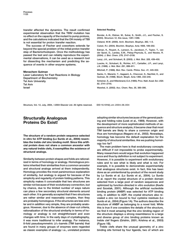
Surdo et al., 2004) (Figure 1A). The authors describe the
structure of ANBP as belonging to a novel fold. While
this is true if one considers the details of the structure,
we find it most amazing that the zinc binding region of
the structure displays a strong resemblance to a large
and diverse group of zinc binding proteins known as
treble clef fingers (Grishin, 2001; Krishna et al., 2003)
(Figure 1B).
Treble clefs share the unusual geometry of a zinc
binding site formed by four ligands, two of which are
The structure of a random protein sequence selected
in vitro for ATP binding (Lo Surdo et al., 2004) resembles the treble clef zinc binding motif. Since this artificial protein does not share a common ancestor with
any natural treble clefs, it exemplifies the existence of
structural analogs.
Similarity between protein shapes and folds are rationalized in terms of homology or analogy. Homologous proteins inherited their similarities from a common ancestor
and structural analogs arrived at them independently.
Homology provides the most parsimonious explanation
of similarity, but analogy is argued for because of the
simplicity and regularity of protein folding patterns. This
simplicity makes it conceivable that two structures are
similar not because of their evolutionary connection, but
by chance, due to the limited number of ways nature
can place a few secondary structural elements around
each other. It is generally accepted that if two structures
are rather similar and reasonably complex, then they
are probably homologous. If the structures are less similar and in addition very simple, they are probably analogous. However, due to the absence of clear-cut criteria,
rationalization of the structural similarity in terms of homology or analogy is not straightforward and even
changes with time. In the early days of crystallography,
it was more traditional to infer analogy. For instance,
8-fold pseudosymmetric /␣-barrels (TIM barrels) that
are found in many groups of enzymes were regarded
as classic examples of analogy: i.e., unrelated proteins
Structure
1126
Figure 1. Diagrams of ANBP and ARF-GAP
Domain
(A and B) The structural diagrams of (A) ANBP
(1uw1) and (B) Pyk2-associated protein 
ARF-GAP domain (1dcq) are shown. 1uw1 is
an isolated folding unit and 1dcq is a domain
within a larger structure with which it has stabilizing intramolecular interactions. In both
figures, the N-terminal -hairpin is purple, the
zinc knuckle is red, and the C-terminal helix
is cyan. The -hairpin between the N-terminal
zinc knuckle and the C-terminal helix is yellow. All other secondary structure elements
that do not contribute to the core of the treble
clef finger are white. The side chains of the
zinc-chelating residues are shown in balland-stick representation, and the zinc atom
(orange) is shown as a ball.
(C) Stereo diagram of the structural superposition of ANBP (1uw1, red) and the ARF-GAP
domain (1dcq, black). The structures were superimposed using the program insightII by
manually defining equivalent residue pairs
(1dcq_A: 262–268, 282–294; 1uw1_A: 21–27,
44–56, shown as thick lines). All figures were
made using the program BOBSCRIPT (Esnouf, 1999).
contributed by an N-terminal -hairpin (knuckle) and the
other two from the first turn of an ␣ helix. The knuckle
(purple) is connected to the ␣ helix (cyan) by a -hairpin
(yellow) (Figure 1B). The structural similarity of ANBP to
treble clef fingers is remarkable and residues from the
zinc binding region of ANBP superimpose with a RMSD
of 1.21 Å (160 backbone atoms from 20 residues) to the
treble clef finger (Pyk2-associated protein  ARF-GAP
domain) shown in Figures 1B and 1C. The side chain
orientation of the zinc-chelating residues and the geometry of the zinc binding site of ANBP are strikingly similar
to those of natural treble clef fingers. It is likely that
the zinc ion in ANBP plays a structural rather than a
functional role (Lo Surdo et al., 2004) similar to those of
treble clef fingers (Grishin, 2001; Krishna et al., 2003).
Also, treble clef fingers generally develop the functional
site around the ␣ helix (cyan) (Grishin, 2001). Interestingly, the key residues of the nucleotide binding site in
ANBP are contributed by the C-terminal ␣ helix as well
(Lo Surdo et al., 2004) (Figure 1A). Thus, there is a significant structural similarity between ANBP and naturally
occurring treble clef finger proteins. Since in vitro generation of ANBP is independent of natural evolutionary
processes, ANBP cannot be homologous to treble clef
proteins and thus represents a structural analog of any
treble clef finger.
It is important to have experimental proof of analogy.
The structure of ANBP shows that the possibilities in
stabilizing small (ⵑ80 residues) proteins are rather limited and have been probed thoroughly by nature. It is
remarkable that a small randomly synthesized protein
selected for ATP binding developed a core of a treble
clef finger, which is commonly found in many proteins.
The in vitro translation of the evolving proteins took
place in the presence of rabbit reticulocyte lysate, which
contains zinc at micromolar concentration (Keefe and
Szostak, 2001). However, as far as we can tell, there
was no selection force in the experiment by Keefe and
Szostak that would specifically mold the zinc binding
region of ANBP into a treble clef; i.e., the protein was
selected for ATP binding and not for zinc binding. An
unexpected and convergent appearance of the treble
clef motif suggests that there are a limited number of
ways to chelate the zinc ion. The remarkable structural
similarity between ANBP and treble clef fingers that extends over the ligand binding site urges caution in inferring homology based only on structural similarity and
the presence of similar ligand binding sites. The whole
structure of ANBP forms an isolated folding unit, and it
is not known whether the treble clef motif constitutes
its folding nucleus (Lo Surdo et al., 2004). Generally, the
task of identifying remote homologs in the protein world
remains difficult and controversial. Homology is typically
inferred by sequence, structural, and functional similarities (Murzin, 1998). On the other hand, analogy cannot
be directly argued for and thus is inferred by the lack
Previews
1127
of homology. A number of studies (Matsuo and Bryant,
1999) have explored different discriminators between
homologs and analogs. However, the largest difficulty
here is to assemble a set of analogs, since it is difficult
to distinguish analogs from remote homologs. Proteins
like ANBP could provide first examples of true structural
analogs.
S. Sri Krishna2 and Nick V. Grishin1,2
1
Howard Hughes Medical Institute and
2
Department of Biochemistry
University of Texas Southwestern Medical Center
5323 Harry Hines Boulevard
Dallas, Texas 75390
Selected Reading
Esnouf, R.M. (1999). Acta Crystallogr. D Biol. Crystallogr. 55,
938–940.
Grishin, N.V. (2001). Nucleic Acids Res. 29, 1703–1714.
Keefe, A.D., and Szostak, J.W. (2001). Nature 410, 715–718.
Krishna, S.S., Majumdar, I., and Grishin, N.V. (2003). Nucleic Acids
Res. 31, 532–550.
Lesk, A.M., Branden, C.I., and Chothia, C. (1989). Proteins 5,
139–148.
Lo Surdo, P., Walsh, M.A., and Sollazzo, M. (2004). Nat. Struct. Mol.
Biol. 11, 382–383.
Matsuo, Y., and Bryant, S.H. (1999). Proteins 35, 70–79.
Murzin, A.G. (1998). Curr. Opin. Struct. Biol. 8, 380–387.
Nagano, N., Orengo, C.A., and Thornton, J.M. (2002). J. Mol. Biol.
321, 741–765.
Acknowledgments
We are grateful to Dr. Lo Surdo for helpful comments on our manuscript. This work was supported by NIH grant GM67165 to N.V.G.
Structure, Vol. 12, July, 2004, 2004 Elsevier Ltd. All rights reserved.
DOI 10.1016/j.str.2004.06.010
A Novel Approach to HighThroughput Screening: A Solution
for Structural Genomics?
diffusion method is the most popular and mostly employed by the automated crystallization trials. In fact,
current crystallization procedures can be broken down
into two stages: “screening,” in which various experimental conditions are tried, and “optimization,” wherein
the quality and size of the crystals are improved (Chayen
and Helliwell, 1998). To address the first stage, structural
genomic and proteomic projects have been leading the
development of high-throughput automated crystallization stations that can prepare hundreds of experiments.
These “brute force” crystallization stations are usually
equipped with automated microscopes as their only diagnostic tool and, although many times successful, reliable production of diffraction quality crystals that can
yield atomic resolution structural information has been
limited.
The automated analysis of vapor diffusion crystallization drops with X-rays proposed by Jean-Luc Ferrer’s
group is a novel approach to the diagnostic problem
and the production of diffraction quality crystals. The
authors demonstrated that it is possible using the vapor
diffusion method and standard crystallization plates
(see http://www.hamptonresearch.com/support/pdf101/
greiner.pdf) to screen for best crystallization conditions
and even determine the 3D structure with X-ray diffraction without removing the crystals from the crystallization drop. One of the main disadvantages in the current
configuration of the method is that the plates must be
moved from their usual horizontal position into a vertical
configuration for the X-ray diffraction analysis. Although
the authors used small drops and small reservoir volumes to help with the stability of the drop during the
rotation process, the actual effect of the rotation on the
shape of the drop and the crystals is not clear. It is also
possible that if the crystals are floating in the drop they
will move during data collection and it would be impossi-
A quasi in situ technique for screening of diffraction
quality biomolecular crystals presents itself to revolutionize the crystallogenesis field.
For decades structural biology has been key to the understanding of the role of biological molecules in the
living cell. To date 85.3% of structures deposited in
the Protein Data Bank have been determined by X-ray
diffraction crystallography. As the name crystallography
suggests, this method absolutely requires the preparation of crystals, a task that is challenging and time consuming at times. Today much of the effort that goes into
the determination of the 3D structure of a biological
molecule actually goes in finding the ideal growth conditions and obtaining the crystals.
Unlike inorganic or small organic molecules, biological molecules are quite complex and present distinctive
physicochemical properties. Therefore, the crystallization process of these molecules depends on a much
larger number of parameters including pH, temperature,
protein concentration, nature of the solvent and precipitant used, and purity, not to mention biological contaminants (for a detailed discussion, see Ducruix and Giege
[1991]). There are few rules and little guidance available
to explain how to crystallize a new macromolecule. With
rare exceptions the phase diagram of these complex
biological molecules is unknown. Several methods,
batch, vapor, counter diffusion, and their variations, are
available to the crystallographer. Among these the vapor

