Cowan L et al.
Caution: when combining topical wound treatments, more is not always better
Caution: when combining topical wound
treatments, more is not always better
Cowan L, Phillips P, Liesenfeld B, Mikhaylova A, Moore D, Stechmiller J & Schultz G
Abstract
Most wound care providers are aware of the principles embodied in the concept of 'wound bed preparation', which is an
integrated approach that seeks to enhance healing of acute and chronic wounds by optimising four key aspects of a wound bed:
1) tissue debridement; 2) inflammation/infection; 3) moisture levels; and 4) epithelial edge healing. Ideally, treatment strategies
should simultaneously address each of these four aspects. This often leads to combining advanced topical and/or systemic
therapies that stimulate healing or remove barriers to endogenous healing. While some laboratory data and clinical trial results
suggest specific combinations of treatments may synergistically enhance healing, other combinations may destructively interact
and reduce the effectiveness of the treatment components. This brief review presents some examples of constructive, destructive
and neutral interactions of combinations of wound treatments and emphasises the need for clinicians to carefully consider how
combinations of wound treatments may interact in the wound bed.
Keywords: combined therapies, dressings, debriding enzymes, silver
Introduction
Wound bed preparation is a comprehensive approach to
wound management which focuses on optimising conditions
in the wound bed to encourage normal endogenous processes
of healing1. It is based on an understanding of cellular
and molecular processes that underlie and regulate normal
wound healing, and it has been applied to both chronic
wounds and acute wounds, such as thermal burns2-4. The
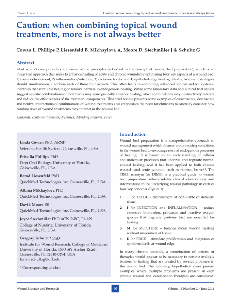
TIME acronym (or DIME) is a practical guide to wound
bed preparation, which relates clinical observations and
interventions to the underlying wound pathology in each of
four key concepts (Figure 1):
Linda Cowan PhD, ARNP
Veterans Health System, Gainesville, FL, USA
Priscilla Phillips PhD
Dept Oral Biology, University of Florida,
Gainesville, FL, USA
Bernd Liesenfeld PhD
QuickMed Technologies Inc, Gainesville, FL, USA
Albina Mikhaylova PhD
QuickMed Technologies Inc, Gainesville, FL, USA
1. T for TISSUE – debridement of non-viable or deficient
tissue.
David Moore BS
2. I for INFECTION and INFLAMMATION – reduce
excessive bioburden, proteases and reactive oxygen
species that degrade proteins that are essential for
healing.
QuickMed Technologies Inc, Gainesville, FL, USA
Joyce Stechmiller PhD ACN P-BC, FAAN
College of Nursing, University of Florida,
Gainesville, FL, USA
3. M for MOISTURE – balance moist wound healing
without maceration of tissue.
Gregory Schultz * PhD
4. E for EDGE – stimulate proliferation and migration of
epidermal cells at wound edge.
Institute for Wound Research, College of Medicine,
University of Florida, 1600 SW Archer Road,
Gainesville, FL 32610-0294, USA
Email schultzg@ufl.edu
In many chronic wounds, a combination of actions or
therapies would appear to be necessary to remove multiple
barriers to healing that are created by several problems in
the wound bed. The following hypothetical cases present
examples where multiple problems are present in each
chronic wound and combination therapies are considered.
* Corresponding author
Wound Practice and Research
60
Volume 19 Number 2 – June 2011
Cowan L et al.
Caution: when combining topical wound treatments, more is not always better
Figure 1. Wound bed preparation. Wound bed preparation is a comprehensive approach to wound management which focuses on optimising
four key concepts for healing: TISSUE – debridement of non-viable or deficient tissue; INFECTION and INFLAMMATION – reduce excessive
bioburden proteases, reactive oxygen species that degrade proteins that are essential for healing; MOISTURE – balance moist wound healing
without maceration of tissue; EDGE – stimulate proliferation and migration of epidermal cells at wound edge.
The potential constructive and destructive interactions of the
combination therapies are discussed
surrounding oedema but slight localised erythema noted
up to 1.0 cm from the callus edges. It was judged to not
be clinically infected but was considered to be moderately
inflamed. Importantly, there was minimal granulation tissue
over about two-thirds of the wound bed. This portion of
the wound bed appeared pale pink. The wound care team
decided to utilise an integrated combination of therapies to
reduce the inflammation and rapidly stimulate formation of
granulation tissue in the wound bed of such long duration.
The patient was not on any type of anticoagulation therapy
and had no known history of coagulopathy. The wound care
team recommended sharp paring down of the peri-wound
callus and another sharp wound debridement of the slough
in the wound bed. They also recommended treatment with
recombinant platelet-derived growth factor (Regranex®) to
promote more rapid development of granulation tissue. They
decided to replace the simple gauze dressing with a protease
inhibiting dressing (Promogran®) covered by a semi-occlusive
dressing. The patient was required to use a specially-fitted
Hypothetical Case #1. A 60-year-old man with poorly
controlled type II diabetes and a documented history of
peripheral neuropathy presented to the wound clinic with
a chronic ulcer of one year duration on the plantar surface
of his left foot. The ulcer had been treated in the past
with occasional debridement and gauze packing with an
off-loading custom orthotic shoe. The patient’s last Ankle
Brachial Index (ABI) was 0.8, and the patient demonstrated
palpable pedal and posterior tibial pulses (1–2+). The wound
measured 2.0 cm length x 2.0 cm width x 0.5 cm depth. The
wound edge was not macerated but there was moderate
periwound callus noted and moderate yellow exudate (no
foul odour detected) with thick yellow slough noted over
one-third of the wound base. Bacterial swab culture of the
wound bed after debridement indicated moderate growth
(<1,000,000 cfu) of several typical bacteria species, but
without MRSA. The client denied pain and there was no
Wound Practice and Research
61
Volume 19 Number 2 – June 2011
Cowan L et al.
Caution: when combining topical wound treatments, more is not always better
off-loading orthotic at all times and return to clinic in one
week for follow-up.
the wound clinic with stage III pressure ulcer on her sacrum,
of reportedly five weeks' duration with no improvement.
The ulcer measured 5.0 cm length x 6.0 cm width x 0.5 cm
depth and had moderate serosanguinous exudate and yellow
slough over two-thirds of the wound base and bacterial swab
culture indicated moderate growth (<1,000,000 cfu) of several
typical bacteria species, including MRSA. It was judged to
not be clinically infected but was considered to be inflamed,
with a high bioburden. There was minimal granulation
tissue over about one-third of the wound bed, but there
was substantial proteinaceous wound slough covering the
wound bed. The surrounding skin showed mild erythema
at immediate wound edges (up to 0.5 cm from wound
edge) but no appreciable warmth, fluctuance or oedema.
The wound was being treated with an alginate dressing
(changed daily) as well as interventions such as pressure
redistributing support surface with frequent turning but
healing had not progressed in four weeks. The wound team
considered using an integrated combination of therapies to
first debride the wound bed with an enzymatic debriding
ointment containing bacterially derived collagenase (Santyl®)
while simultaneously using a silver-releasing dressing to help
reduce the level of planktonic MRSA.
From the perspective of wound bed preparation, this treatment
plan simultaneously addressed several key issues of the
TIME paradigm. Firstly, the surgical debridement would
remove defective tissue (peri-wound callus) and reduce the
levels of planktonic (free-floating) and sessile biofilm bacteria
(stationary, mature polymicrobial colony), which would
reduce the level of inflammation and should reduce the level
of wound exudate. The wound team suspected the level of
proteases would be highly elevated in the wound bed and
they understood that both endogenous and exogenous growth
factors would be degraded by high levels of proteases like the
matrix metalloproteinases (MMPs) and neutrophil elastase
(NE)5,6. Therefore, they decided to combine the PDGF growth
factor therapy with a dressing that contains collagen and
oxidised regenerated cellulose (ORC). These two components
of Promogran® act as substrate sinks for MMPs and NE
and can reduce the levels of protease activities in chronic
wound fluids to values that are more similar to the low levels
of proteases found in acute healing wounds7. In addition,
laboratory data show that protease-inhibiting dressings can
substantially reduce the breakdown of PDGF by proteases
present in chronic wound fluids8. Importantly, each of these
topical treatments (Regranex® and Promogran®) has been
shown in randomised controlled trials (RCT) to separately
improve healing of chronic wounds9-11. Thus, it was reasonable
to consider combining these two topical treatments (based on
biochemical knowledge of both) with the expectation that they
would enhance the effects of the other treatment and improve
healing better than either treatment alone.
While this combination of treatments would appear to address
two important components of wound bed preparation,
specifically, debridement of necrotic or defective tissue and
reduction of inflammation/infection, one of the wound team
members pointed out that laboratory data included with the
Santyl® product insert indicate that ionic silver and iodine both
reduce the enzymatic activity of collagenase contained in the
debriding ointment (Table 1). Thus, this combination of two
active topical treatments would destructively interfere with the
actions of the enzymatic debriding agent and this combined
treatment strategy was rejected by the wound care team.
Hypothetical Case #2. A very frail, 70-year-old female who
is a resident in an extended care nursing home presented to
Table 1. Effect of wound dressings on Clostridium collagenase enzyme activity.
Dressing type
Selected product
Inhibitory effect
Nanocrystalline silver dressing
Acticoat [Smith & Nephew, Largo, FL]
>50%13
Carboxymethyl cellulose hydrofibre dressing
with ionic silver
Aquacel Ag [ConvaTec, Skillman, NJ]
Low inhibition14
Alginate dressing with ionic silver
Algicell AG [Derma Sciences, Princeton, NJ]
Contraindicated+
Pigment-complexed polyvinyl alcohol dressing
Hydrofera Blue [Healthpoint Ltd, Fort Worth, TX]
None13
Iodine dressings
Iodoflex [Smith & Nephew, Largo, FL]
>90%13
PHMB gauze
small cationic monomer
Kerlix AMD™ [Covidien, Mansfield, MA]
>80%*
pDADMAC gauze
large cationic polymer
Bioguard™ [Derma Sciences, Princeton, NJ]
None*
Collagen dressing plus alginate
Fibracol Plus [Johnson & Johnson, Minneapolis, MN]
None13,14
* QuickMed Technologies, Inc, internal data; + www.woundcareresources.net
Wound Practice and Research
62
Volume 19 Number 2 – June 2011
Cowan L et al.
Caution: when combining topical wound treatments, more is not always better
Another treatment approach the wound team considered for
this patient was to combine the enzymatic debriding agent with
a bacterial barrier gauze dressing, Kerlix AMD®, that contains
the microbicidal agent polyhexanide (or polyhexamethylene
biguanide, PHMB). However, laboratory data (Table 1)
show this bacterial barrier dressing also inactivates the
bacterial collagenase, probably due to interaction with the
PHMB that elutes from the dressing. In contrast, a different
microbicidal bacterial barrier dressing, BioGuard®, which
contains a bound microbicidal polyquat polymer (polydiallyldimethylammonium chloride, pDADMAC) does not
inhibit bacterial collagenase (Table 1). Thus, combinations
of dressings with collagenase debriding agents can have
very different effects on the enzyme’s activity. The wound
team also considered the enzymatic debriding agent requires
additional moisture to activate the enzymatic activity. Since
the patient was having a moderate amount of wound
exudate, this would provide the adequate moisture. After
cleansing the wound with saline, the enzymatic ointment was
applied to the slough in the wound bed in a recommended
thickness of 2 mm. It was recognised that if some of the
collagenase enzymatic debriding ointment contacted the
granulation tissue it would cause no harm. The microbial
barrier dressing was applied over this with orders to change
the entire dressing daily (using saline as a wound cleanser)
and schedule a wound follow-up visit in two weeks.
Hypothetical Case #3. A 65-year-old female with chronic
venous insufficiency developed an ulcer located on the
lateral surface near her left ankle that was improving with
standard compression therapy dressing, but after several
weeks of compression therapy, the patient presented with
acute, severe pain in the ulcer bed. The wound measured 3.0
cm length x 2.0 cm width x 0.3 cm depth. Macerated wound
edges, erythema and warmth were noted in the surrounding
skin. After examining the wound bed, the wound team
noted the ulcer had developed signs of an acute infection,
consistent with Pseudomonas aeruginosa (light-green sheen
and a musty, earthy odour). The wound team decided to
prescribe systemic antibiotics (gentamicin) combined with
topical wound cleansing with Dakin’s solution followed by
gauze dressing soaked with dilute (1/4 strength) Dakin’s
solution (0.125% sodium hypochlorite buffered with 0.04%
boric acid). The wound team initially thought about using the
bacterial barrier gauze dressing, Kerlix AMD®, that contains
the microbicidal agent polyhexanide, but one team member
remarked that Kerlix AMD® gauze dressing inactivated
sodium hypochlorite (Table 1), so the team decided to use
the BioGuard® bacterial barrier gauze since it does not
COVIDIEN 2011 Infection Control Scholarship
holarship
Applications for the COVIDIEN 2011 Infection Control
Scholarship are now open, with total funding of $40,000
to promote excellence in Infection Control being awarded
across four categories!
For further information or an application form:
Email: Aust.Infection.Control@Covidien.com
Visit: www.aica.org.au or contact your local Covidien
Product Specialist
Applications close 30th June 2011
COVIDIEN, COVIDIEN with Logo and ™ marked brands
are trademarks of Covidien AG or its affiliate. © 2010
Covidien AG or its affiliate. All rights reserved.
WC 125-02-11
Wound Practice and Research
63
Volume 19 Number 2 – June 2011
Cowan L et al.
Caution: when combining topical wound treatments, more is not always better
In conclusion, the purpose of this article is to alert wound
care providers to potentially detrimental interactions
between certain wound products. Caution is advised when
considering combining products where there may be an
unknown interaction, or there exists laboratory data (such
as those in Table 1) to demonstrate interactive effects. The
hypothetical scenarios portrayed in this article are not meant
in any way to be clinical advice for treating specific wounds,
but rather an illustration of common reasoning wound
providers may use in making treatment decisions. Please
consult evidence-based wound treatment guidelines for
specific wound treatment recommendations.
inactivate the sodium hypochlorite solution. The wound team
anticipates that treatment with dilute Dakin’s solution will be
short term (<four weeks) because they recognise that sodium
hypochlorite solutions >0.01% or 0.025% are cytotoxic to
fibroblasts15-16. However, the infection is far more detrimental
to the wound bed and fibroblasts are not expected to
proliferate in the presence of acute infection in a wound that is
deteriorating. The wound team decided they would address
the bacterial infection first. The treatment was ordered for
two weeks and included continued compression stockings
to bilateral lower extremities daily. A wound follow-up
appointment was scheduled for two weeks. After the acute
infection is resolved, a non-cytotoxic wound dressing will be
ordered and wound progression toward healing is expected.
References
Summary
1. Schultz GS, Sibbald RG, Falanga V et al. Wound bed preparation: a
systematic approach to wound management. Wound Repair Regen 2003;
11(Suppl 1):S1–S28.
These hypothetical cases illustrate several key concepts
that clinicians should consider when designing optimised
treatment strategies for individual patients. Firstly, most
chronic wounds first present with several aspects that need to
be addressed within the concepts of wound bed preparation.
This typically leads to combining several therapeutic
interventions to correct the molecular and microbial problems
in the wound bed. However, the clinician must recognise the
potential for positive, negative or neutral interactions that
can occur between the different agents and dressings. Some
of these negative interactions are straightforward and rather
well known. For example, certain silver dressings should be
hydrated only with sterile water and not with solutions that
contain substantial concentrations of anions like chloride
(isotonic saline) or phosphate (phosphate buffered saline)
or protein (plasma) because these solutions precipitate and
inactivate silver anion (Acticoat® package insert). Another
destructive combination is the use of silver dressings with
Tegederm Matrix® dressing, which contains a mixture of
four cations (calcium, zinc, potassium and rubidium) with
chlorine counter anion (3M FAQs Wound Management).
Other interactions may not be as widely recognised, such as
the inactivation of debriding enzymes by reactive metal ions
like silver or iodine or other microbicides like PHMB (Table
1). Another potential negative interaction may occur between
collagenase debriding enzymes and systemic antibiotics
of the tetracycline family (tetracycline, doxycycline,
minocycline) because all the tetracycline family of antibiotics
are competitive inhibitors of MMPs12. Of equal importance
for clinicians, however, is to know when there is no known
detrimental effect of combining different therapies. Examples
of neutral interactions between different topical wound
treatments include collagenase debriding ointment used
with dressings such as saline moistened gauze, BioGuard®
microbicidal gauze dressing, Hydrofera Blue® PVA dressing
or Fibrocol Plus® collagen plus alginate dressing (Table 1).
Wound Practice and Research
2. Schultz G, Mozingo D, Romanelli M & Claxton K. Wound healing and
TIME; new concepts and scientific applications. Wound Repair Regen
2005; 13(4 Suppl):S1–S11.
3. Ayello EA & Cuddigan JE. Conquer chronic wounds with wound bed
preparation. Nurse Pract 2004; 29(3):8–25.
4. Sibbald RG, Orsted H, Schultz GS, Coutts P & Keast D. Preparing the
wound bed 2003: focus on infection and inflammation. Ostomy Wound
Manage 2003; 49(11):23–51.
5. Ladwig GP, Robson MC, Liu R, Kuhn MA, Muir DF & Schultz GS. Ratios
of activated matrix metalloproteinase-9 to tissue inhibitor of matrix
metalloproteinase-1 in wound fluids are inversely correlated with healing
of pressure ulcers. Wound Repair Regen 2002; 10(1):26–37.
6. Yager DR, Chen SM, Ward SI, Olutoye OO, Diegelmann RF & Cohen
IK. Ability of chronic wound fluids to degrade peptide growth factors is
associated with increased levels of elastase activity and diminished levels
of proteinase inhibitors. Wound Repair Regen 1997; 5(1):23–32.
7. Cullen B, Smith R, McCulloch E, Silcock D & Morrison L. Mechanism of
action of PROMOGRAN, a protease modulating matrix, for the treatment
of diabetic foot ulcers. Wound Repair Regen 2002; 10(1):16–25.
8. Cullen B, Watt PW, Lundqvist C et al. The role of oxidised regenerated
cellulose/collagen in chronic wound repair and its potential mechanism
of action. Int J Biochem Cell Biol 2002; 34(12):1544–56.
9. Veves A, Sheehan P & Pham HT. A Randomized, Controlled Trial of
Promogran (a Collagen/Oxidized Regenerated Cellulose Dressing) vs
Standard Treatment in the Management of Diabetic Foot Ulcers. Arch Surg
2002; 137(7):822–7.
10. Vin F, Teot L & Meaume S. The healing properties of Promogran in venous
leg ulcers. J Wound Care 2002; 11(9):335–41.
11. Steed DL. Clinical evaluation of recombinant human platelet-derived
growth factor for the treatment of lower extremity diabetic ulcers. Diabetic
Ulcer Study Group. J Vasc Surg 1995; 21(1):71–8.
12. Chin GA, Thigpin TG, Perrin KJ, Moldawer LL & Schultz GS. Treatment
of Chronic Ulcers in Diabetic Patients with a Topical Metalloproteinase
Inhibitor, Doxycycline. Wounds 2003; 15(10):315–23.
13. Shi L, Ermis R, Kiedaisch B & Carson D. The effect of various wound
dressings on the activity of debriding enzymes. Adv Skin Wound Care
2010; 23(10):456–62.
14. Shi L & Carson D. Collagenase Santyl ointment: a selective agent for
wound debridement. J Wound Ostomy Continence Nurs 2009; 36(6
Suppl):S12–S16.
15. Heling et al. Bacteriocidal and Cytotoxic Effects of Sodium Hypochlorite
and Sodium Dichloroisocyanurate Solutions In Vitro. J Endod 2001 Apr;
27(4).
16. Heggers JP, Sazy JA, Stenberg BD et al. Bactericidal and wound-healing
properties of sodium hypochlorite solutions: the 1991 Lindberg Award. J
Burn Care Rehabil 1991 Sep-Oct; 12(5):420–4.
64
Volume 19 Number 2 – June 2011

