Norrie Disease
advertisement
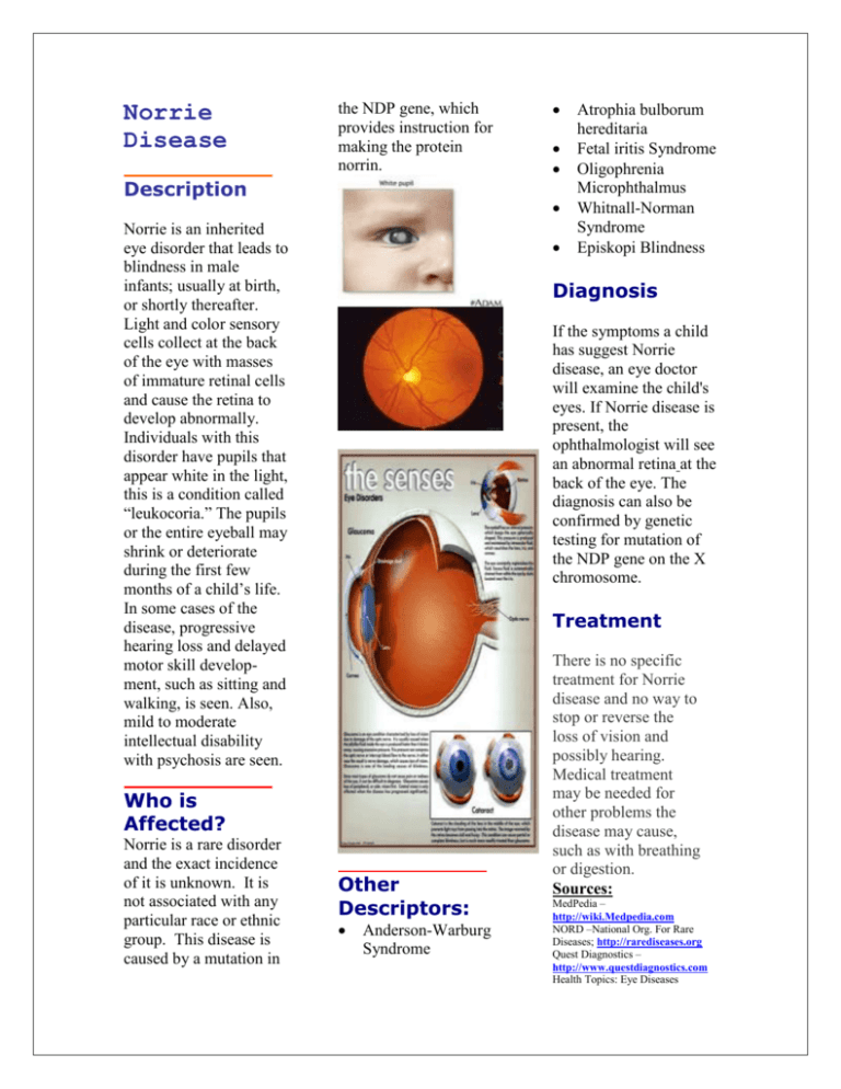
Norrie Disease the NDP gene, which provides instruction for making the protein norrin. Description Norrie is an inherited eye disorder that leads to blindness in male infants; usually at birth, or shortly thereafter. Light and color sensory cells collect at the back of the eye with masses of immature retinal cells and cause the retina to develop abnormally. Individuals with this disorder have pupils that appear white in the light, this is a condition called “leukocoria.” The pupils or the entire eyeball may shrink or deteriorate during the first few months of a child’s life. In some cases of the disease, progressive hearing loss and delayed motor skill development, such as sitting and walking, is seen. Also, mild to moderate intellectual disability with psychosis are seen. Atrophia bulborum hereditaria Fetal iritis Syndrome Oligophrenia Microphthalmus Whitnall-Norman Syndrome Episkopi Blindness Diagnosis If the symptoms a child has suggest Norrie disease, an eye doctor will examine the child's eyes. If Norrie disease is present, the ophthalmologist will see an abnormal retina at the back of the eye. The diagnosis can also be confirmed by genetic testing for mutation of the NDP gene on the X chromosome. Treatment Who is Affected? Norrie is a rare disorder and the exact incidence of it is unknown. It is not associated with any particular race or ethnic group. This disease is caused by a mutation in Other Descriptors: Anderson-Warburg Syndrome There is no specific treatment for Norrie disease and no way to stop or reverse the loss of vision and possibly hearing. Medical treatment may be needed for other problems the disease may cause, such as with breathing or digestion. Sources: MedPedia – http://wiki.Medpedia.com NORD –National Org. For Rare Diseases; http://rarediseases.org Quest Diagnostics – http://www.questdiagnostics.com Health Topics: Eye Diseases http://www.nlm.nih.gov Prepared By: Gwendolyn Jones Description Retinal detachment (Knobloch Syndrome, Type I) is a separation of the light-sensitive membrane in the back of the eye, the retina, from the supporting layers. The retina is a clear tissue positioned at the back of the eye. It helps a person see images that are focused on by the eye. When retinal detachment occurs, bleeding from small retinal blood vessels may cloud the interior of the eye, this area is normally filled with a fluid called the vitreous fluid. The central vision will become severely affected if the macula, the part of the retina responsible for fine vision becomes detached. Causes Retinal detachment is associated with a tear or hole in the retina that results in fluids leaking. This condition results in a separation of the retina from the underlying tissues. Retinal detachment sometimes happens without any underlying cause. It may also be caused by trauma, diabetes, an inflammatory disorder, or by a related condition called posterior vitreous detachment. Symptoms Symptoms of Retinal detachment are: Bright flashes of light, especially in peripheral vision Blurred vision Floaters in the eye Shadow or blindness in a part of the visual field of one eye Treatment Most patients who experience a retinal detachment will need immediate surgery, at times within 24-hours of the first symptoms. If there are no symptoms or if you have had the detachment for a while, surgery may not be needed. However unsuccessful reattachments results in vision loss. Most retinal detachments can be successfully repaired. Prevention Always use protective eye wear to prevent eye trauma; control your blood sugar if diabetic. Have annual eye exam. Sources: 1. Genetics Home Reference, http://ghr.nlm.nih.gov/conditi 2. MedlinePlus – Health information Encyclopedia: Health Topics: Usher Syndrome http://www.nlmnih.gov/medli neplus/ushersyndrome.html 3.Rare Diseases Information Center http://rarediseases.info.nih.go v/GARD/ 4.MadisonsFoundation http://www.madisonsfoundati on.org/index 5.National Institutes of Health http://www.usersyndrome.nih .gov/ Prepared by: Gwendolyn Jones


