Visual Pathways & Function
advertisement
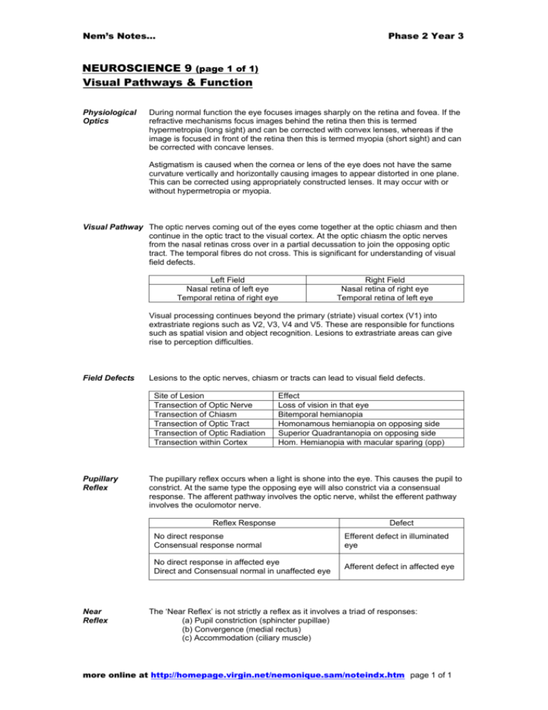
Nem’s Notes… Phase 2 Year 3 NEUROSCIENCE 9 (page 1 of 1) Visual Pathways & Function Physiological Optics During normal function the eye focuses images sharply on the retina and fovea. If the refractive mechanisms focus images behind the retina then this is termed hypermetropia (long sight) and can be corrected with convex lenses, whereas if the image is focused in front of the retina then this is termed myopia (short sight) and can be corrected with concave lenses. Astigmatism is caused when the cornea or lens of the eye does not have the same curvature vertically and horizontally causing images to appear distorted in one plane. This can be corrected using appropriately constructed lenses. It may occur with or without hypermetropia or myopia. Visual Pathway The optic nerves coming out of the eyes come together at the optic chiasm and then continue in the optic tract to the visual cortex. At the optic chiasm the optic nerves from the nasal retinas cross over in a partial decussation to join the opposing optic tract. The temporal fibres do not cross. This is significant for understanding of visual field defects. Left Field Nasal retina of left eye Temporal retina of right eye Right Field Nasal retina of right eye Temporal retina of left eye Visual processing continues beyond the primary (striate) visual cortex (V1) into extrastriate regions such as V2, V3, V4 and V5. These are responsible for functions such as spatial vision and object recognition. Lesions to extrastriate areas can give rise to perception difficulties. Field Defects Lesions to the optic nerves, chiasm or tracts can lead to visual field defects. Site of Lesion Transection of Optic Nerve Transection of Chiasm Transection of Optic Tract Transection of Optic Radiation Transection within Cortex Pupillary Reflex Effect Loss of vision in that eye Bitemporal hemianopia Homonamous hemianopia on opposing side Superior Quadrantanopia on opposing side Hom. Hemianopia with macular sparing (opp) The pupillary reflex occurs when a light is shone into the eye. This causes the pupil to constrict. At the same type the opposing eye will also constrict via a consensual response. The afferent pathway involves the optic nerve, whilst the efferent pathway involves the oculomotor nerve. Reflex Response Near Reflex Defect No direct response Consensual response normal Efferent defect in illuminated eye No direct response in affected eye Direct and Consensual normal in unaffected eye Afferent defect in affected eye The ‘Near Reflex’ is not strictly a reflex as it involves a triad of responses: (a) Pupil constriction (sphincter pupillae) (b) Convergence (medial rectus) (c) Accommodation (ciliary muscle) more online at http://homepage.virgin.net/nemonique.sam/noteindx.htm page 1 of 1

