Definition, types and examples for each type and factors affecting
advertisement
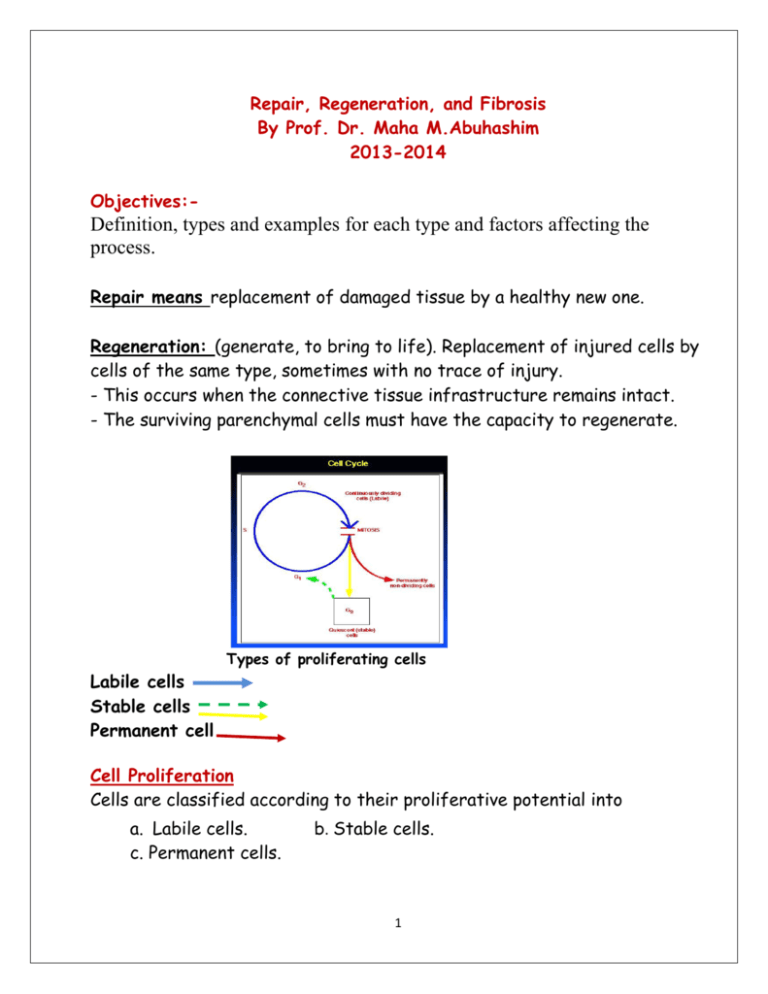
Repair, Regeneration, and Fibrosis By Prof. Dr. Maha M.Abuhashim 2013-2014 Objectives:- Definition, types and examples for each type and factors affecting the process. Repair means replacement of damaged tissue by a healthy new one. Regeneration: (generate, to bring to life). Replacement of injured cells by cells of the same type, sometimes with no trace of injury. - This occurs when the connective tissue infrastructure remains intact. - The surviving parenchymal cells must have the capacity to regenerate. Types of proliferating cells Labile cells Stable cells Permanent cell Cell Proliferation Cells are classified according to their proliferative potential into a. Labile cells. c. Permanent cells. b. Stable cells. 1 a. Labile cells: - These are continuously dividing cells which pass directly from M to G1 phase of the cell cycle. - They are of short life span. - Examples are epidermis of the skin, surface epithelium of gastrointestinal and genito-urinary system and hemopoietic cells of the bone marrow. - Injury of these cell is followed by complete regeneration providing that the supporting stroma is intact. b. Stable cells: - Normally, these cells undergo few postnatal divisions but are capable of division when activated or after injury ( pass from Go to G1). They include hepatocytes, renal tubular cells, glandular cells, and mesenchymal cells e.g smooth muscle, osteoblasts, cartilage cells, endothelium and connective tissue cells. - Injury of these cells is followed by complete regeneration if the supporting framework is preserved. Renal tubular epithelium as an example for repair of stable cells BM is essential for organized regeneration c. Permanent cells: These cells have left the cell cycle and CANNOT undergo mitotic division in postnatal life. - Permanent cells are found in the central nervous system and heart. 2 Once they are destroyed, they cannot regenerate. - Injury of labile or stable cells if accompanied by an intact supporting framework will completely regenerate. If the framework is destroyed, repair occurs by fibrosis. - Injury of permanent cells will always heal by fibrosis and if in the brain, it heals by gliosis. Repair by Fibrosis - It is replacement of the damaged tissue by fibrous tissue. - It occurs when there is destruction of the supporting framework or when the parenchymal cells are unable to divide (permanent cells). - Replacement of non viable tissue (e.g thrombus, hematoma, necrotic tissue ) by fibrous tissue is called Organization. Steps of the repair by fibrosis:1-Removal of debris… begins early, there is liquefaction and removal of dead cellular material and other particulate material. Macrophage is the main active cell in this process 2- Formation of granulation tissue which is a transitory, highly vascular newly formed connective tissue formed of Capillaries and Fibroblasts. - It fills the gap left by the liquefied material. Characters of granulation tissue… i . Red in color and granular. ii- It is wet as the newly formed blood vessels are more permeable. iii- It is resistant to infection, and insensitive to touch as the nerve endings take more time to regenerate. 3- Scarring a. Collagen is produced by fibroblasts, and as the amount of collagen increases; the tissue becomes gradually less vascular and less cellular. 3 *Scar = hypo vascular strong fibrous tissue. b. Remodeling = rearrangement of collagen fibers parallel to the surface to get the maximal strength of tissue. c. Progressive contraction of the wound resulting in deformity of the original structure. What is the difference between granulation tissue and granuloma? Granulation tissue A gaping wound filled with granulation tissue Granulation tissue…. facts you must know…. Definition - Characters Mechanism of formation - Structure - Fate - Skin scar, wound becomes smaller in size and under the microscope, collagen is increased and vascularity becomes less prominent. Healing of stable cells (Bone healing):Steps:1-Hematoma formation + mild acute inflammation. 2- Macrophages+ osteoclasts clean the area. 3- Area is invaded by osteogenic granulation tissue formed of capillaries and osteoblasts which lay down osteoid matrix 4 that become calcified forming a mass of woven bone called Callus. 4- External and internal callus are reabsorbed. 5- Woven bone is remodeled and change to lamellar bone. 1-6 Steps of healing of a bone fracture Cell- Cell Interactions • Cellular proliferation is mediated by a group of Growth factors each of which promotes the proliferation of certain cell types. Cytokines and Growth Factors 1- Epidermal growth factor (EGF) 2- Platelet-derived growth factor (PDGF) 5 3- Fibroblast growth factor (FGF) and vascular endothelial growth factor (VEGF) 4- Transforming growth factor-beta (TGF-b) and tumor necrosis factor (TNF) EGF &TGF-α PDGF bFGF TGF VEGF IL-1 -β &TNF Angiogenesis Proliferation - epith, endoth &fibroblasts Collagen synthesis Collagen degradation ++ + + + ++ _ _ + ++ _ + + + + + Wound Healing Primary intention: the usual case with a surgical wound, in which there is a clean, sterile wound with well-apposed edges, and minimal clot formation. Secondary intention: when wound edges cannot be opposed, usually infected with debris and blood clots (e.g., following crush injuries). A large scar usually results. I- Primary intention Clean surgical wound 6 Steps and dating of healing by primary intension II- Secondary intension Steps of healing :1- Inflammation subsides and macrophage begin to clean the area. 2- granulation tissue starts to form from the sides and depth of the wound to fill it. 3- Epithelialization starts when the gap of the wound if filled by granulation tissue. 7 4- A scar is formed and when it contracts it leads to deformity of the affected site. 5- This takes a longer duration than in primary intension. Healing by primary intension and secondary intension Factors that influence wound healing 1.Type, size, and location of the wound. 2.Vascular supply (diabetics heal poorly). 3.Infection - delays wound healing and leads to more granulation tissue and scarring. 4.Movement - wounds over joints do not heal well due to traction 5. Radiation - ionizing radiation is bad, UV is good 6. Overall nutrition: vitamin and protein deficiencies lead to poor wound healing, especially vitamin C, which is involved in collagen synthesis,also zink and calcium are important. 7. Age: younger is definitely better! 8. Hormones - corticosteroids drastically impair wound healing, because of their profound effect on inflammatory cells. 8 Complications of wound healing Defective formation of granulation tissue and fibrous tissue…. Wound dehiscence. Decreased cellular proliferation…..ulcer sinus and fistula. - Ulcer = discontinuity of the surface epithelium of the skin or mucous membrane. - Sinus = A blind ended tract opening on the surface. - Fistula = A tract joining two hollow organs. Weak scar….incisional hernia Wound dehiscence Keloid Excessive Formation of granulation tissue & scarring 1- Proud flesh Excessive formation of granulation tissue above the surface of the wound. - This prevents epithelialization of the surface. - Surgical removal or cautery is required for complete healing of the wound 2-Keloids (hypertrophy scars) - Are the result of over-exuberant production of scar tissue, which is primarily composed of type III collagen - The cause is thought to be due to genetic factors, perhaps due to lack of the proper collagenases to degrade type III collagen. - Surgical removal is usually followed by recurrence. What are the other complications that may result from fibrosis? 9
