HOMEWORK 1 SOME BASIC TERMS CNS / PNS
advertisement
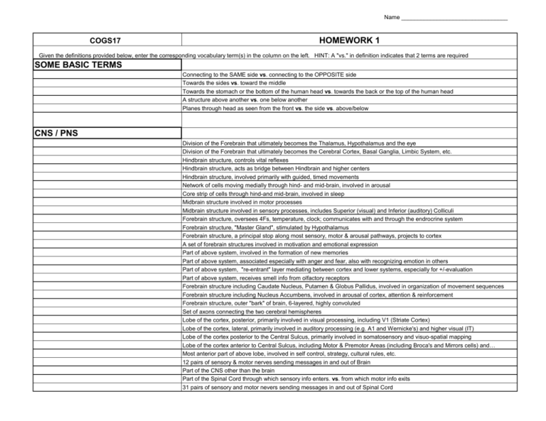
Name _________________________________ COGS17 HOMEWORK 1 Given the definitions provided below, enter the corresponding vocabulary term(s) in the column on the left. HINT: A "vs." in definition indicates that 2 terms are required SOME BASIC TERMS Connecting to the SAME side vs. connecting to the OPPOSITE side Towards the sides vs. toward the middle Towards the stomach or the bottom of the human head vs. towards the back or the top of the human head A structure above another vs. one below another Planes through head as seen from the front vs. the side vs. above/below CNS / PNS Division of the Forebrain that ultimately becomes the Thalamus, Hypothalamus and the eye Division of the Forebrain that ultimately becomes the Cerebral Cortex, Basal Ganglia, Limbic System, etc. Hindbrain structure, controls vital reflexes Hindbrain structure, acts as bridge between Hindbrain and higher centers Hindbrain structure, involved primarily with guided, timed movements Network of cells moving medially through hind- and mid-brain, involved in arousal Core strip of cells through hind-and mid-brain, involved in sleep Midbrain structure involved in motor processes Midbrain structure involved in sensory processes, includes Superior (visual) and Inferior (auditory) Colliculi Forebrain structure, oversees 4Fs, temperature, clock; communicates with and through the endrocrine system Forebrain structure, "Master Gland", stimulated by Hypothalamus Forebrain structure, a principal stop along most sensory, motor & arousal pathways, projects to cortex A set of forebrain structures involved in motivation and emotional expression Part of above system, involved in the formation of new memories Part of above system, associated especially with anger and fear, also with recognizing emotion in others Part of above system, "re-entrant" layer mediating between cortex and lower systems, especially for +/-evaluation Part of above system, receives smell info from olfactory receptors Forebrain structure including Caudate Nucleus, Putamen & Globus Pallidus, involved in organization of movement sequences Forebrain structure including Nucleus Accumbens, involved in arousal of cortex, attention & reinforcement Forebrain structure, outer "bark" of brain, 6-layered, highly convoluted Set of axons connecting the two cerebral hemispheres Lobe of the cortex, posterior, primarily involved in visual processing, including V1 (Striate Cortex) Lobe of the cortex, lateral, primarily involved in auditory processing (e.g. A1 and Wernicke's) and higher visual (IT) Lobe of the cortex posterior to the Central Sulcus, primarily involved in somatosensory and visuo-spatial mapping Lobe of the cortex anterior to Central Sulcus, including Motor & Premotor Areas (including Broca's and Mirrors cells) and… Most anterior part of above lobe, involved in self control, strategy, cultural rules, etc. 12 pairs of sensory & motor nerves sending messages in and out of Brain Part of the CNS other than the brain Part of the Spinal Cord through which sensory info enters. vs. from which motor info exits 31 pairs of sensory and motor nevers sending messages in and out of Spinal Cord Name _________________________________ "Law" governing above directions of information flow in Spinal Cord Areas of the Spinal Cord (as seen in cross-section) or Brian consisting of soma vs. of myelinated axons Tube through core of Spinal Cord containing fluid Four hollow chambers (plus aqueducts) in brain that produce the fluid that feeds, cleans and cushions brain Fluid, produced by ventricles, found within Spinal Cord and in covering surrounding CNS Three-layered (Dura-Mater, Fluid-filled Arachnoid-Space, and Pia-Mater) protective covering that surrounds CNS Semi-permeable barrier, controls what chemicals enter brain, via closed gaps between capillary cells & barrier of Astrocytes That part of the PNS that is responsible for the body's interaction with the environment That part of the PNS that is responsible for assessing and maintaining the body's internal environment That part of the ANS that produces the "fight or flight" response vs. that which facilitates relaxation and replenishment Extreme compensatory response of one system to extreme activation of the other - can lead to fainting, ulcers, voodoo death NEURAL FUNCTIONING Cells in the Nervous System responsible for information transmission Cells in the Nervous System responsible for support, feeding, recycling, development, etc Organelles in a cell that are the site of protein production, crucial to much neural functioning Organelles in a cell that are the source of energy (ATP) to power active (rather than passive) functions in cell Processes (branches) of a neuron that receive the incoming message vs. the one that releases the outgoing message Difference in the amount of a given chemical inside/outside a cell vs. a difference in charge inside/outside a cell Symbols for 4 key chemical elements in neural functioning - including 3 positive ions, 1 negative ion Name for and amount of difference in charge inside/outside cell, in millivolts (mV), in a polarized cell ready to fire Energy-requiring pump that helps restore membrane potential after cell fires A sequence of depolarization that moves along an axon, resulting in the all-or-nothing release of NT Section of axon where depolarization sequence begins A greater or lesser change in the polarity of a neuron that results in a greater or lesser release of NT Propagation of info down an axon by way of chemical gates opening/closing vs. by flow of electrons "Jumping" electrical conduction that occurs in myelinated axons Glia cells wrapping around sections of an axon to insulate it and speed its information transmission Gaps between myelin sheaths on an axon Disease that destroys myelin; no ion gates under sheath so neurons cannot fire Period following an Action Potential during which the cell cannot (or is more difficult to) fire The event in which one cell releases NT and that NT affects another cell The gap between cells across which NT passively floats The cell that releases the NT vs. the cell that receives the NT The end of the axon from which NT is released, also called "button" or "end bulb" Packets of NT released by a neuron The release of NT into cleft via its packet opening at a Fusion Pore in the cell's membrane Area, usually on a dendrite, that is specialized for the attachment of NT An increase vs. a decrease in a cell's likelihood of releasing neurotransmitter Less polarized, less difference between inside of cell and outside of cell vs. more difference Cumulative effect of the activity of multiple Presynaptic cells; Can be temporal or spatial When NT has direct effect on ion channels in Postsynaptic cell vs. indirect effects via internal metabolic processes Name _________________________________ Chemical in Postsynaptic cell involved in energy-requiring processes (including altering ion channels) triggered by NT Chemicals released by Presynaptic cells that directly affect local Postsynaptic cells vs. ones that widely influence neural activity Chemical (endogenous or man-made) that acts to facilitate vs. to reduce the effects of specific NTs Process by which NTs or their components re-enter the Presynaptic cell for re-use. Enzyme in cleft that breaks down Acetylcholine Site on Presynaptic terminal that reacts to that cell's own NT, usually acting to turn off/down that cell's further NT release Synapses at a Presynaptic terminal that reacts to NT from another cell, excitatory or inhibitory PLUS: List six important NTs (with their abbreviations) and three important hormones NTs Hormones DEVELOPMENT In the new embryo, the outermost layer of cells - becomes the nervous system and skin In the growing (wormlike) embryo, the surface along the back that thickens and hardens A pair of ridges all along the above that begin to curl towards each other The long hollow chamber that is formed when the above meet and fuse; Inner surface becomes the CNS Outer surface of the above ridges that separate off and become the PNS A pathological condition involving a failure of the edges above to completely fuse, leading to birth defects or death Hollow core of developing embryo, source of cells of nervous system The original type of cells in this area that undergo division to populate the nervous system General term for the production of new cells Cell division that produces two identical offspring vs. produces one identical and one new (neuron or glial) cell The movement of cells from their place of origin to their later position An early type of glial cell that extends its processes out like wheel spokes for the developing neurons to move along The process by which neurons form new connections The specialized tip of a growing axon that detects the chemicals that guide its path Glia cells that are positioned to direct growing axons towards their target cells Chemicals that attract/repel Axon growth, help prevent cell death, and/or promote Axonal branching One type of the above, from muscles & organs, that promotes survival and growth of axons in the brain and Sympathetic NS Cell Death as determined by "suicide genes" that cause developing neurons to package their contents & destroy themselves Newly formed axonal branches that replaces another (that has died off) at a synapse New outgrowths on, or subdividing of, the processes that receive NT, in response to an enriched enviornment, learning, etc. A mnemonic for the rule that co-activated cells tend to be strengthened in their connectivity and out-compete neighboring cells Name _________________________________ BRAIN STUDY TECHNIQUES Name 3 types of neuronal stain that are injected live, but then examined in brain tissue slices Creating or exploiting brain damage to determine if that area is necessary to a certain function Method used to generate, for example, the "Penfield Map" of somatosensory cortex in live patients Do all three of the above get good spatial or temporal resolution? Which of the above yield information on brain FUNCTION? Record activity using a micro-electrode probe in an active subject Using a "electrode cap", technique detects the electrical dipoles generated by changing electrical potentials Does the above record localized changes in electrical activity or summation of changes over thousands of neurons? The time-locked average of many EEG trials to factor out other brain activity & focus on a particular response Detection of naturally occurring changes in magnetic fields created by brain activity (complementary to EEG) Devise used to measure extremely weak magnetic fields, such as those produced by brain activity Of the above four techniques, which requires confining the subject in a large apparatus? Of the above four techniques, which is the only one with poor temporal resolution? Of the above four techniques, which has the best spatial resolution? Of the above four techniques, which is the most expensive? Aspect of MRI that involves using pulse of radio waves to make hydrogen protons gyrate in body's fluid Aspect of MRI that involves aligning the magnetic fields of those gyrating protons Aspect of MRI that involves the release of energy when the protons are allowed to return to 'natural' alignment Example of a neurological disease revealed by MRI's capacity to distinguish white from grey matter Technique that makes use of the diff in how oxygenated vs deoxygenated hemoglobin in blood respond to magnetic fields Is deoxygenated hemoglobin more likely to be found at Active or Non-active sites in the brain? What does the "f" in "fMRI" stand for? Patient is injected w/radioactive fluid that is absorbed w/glucose into active cells & detected as gamma emissions Technique using 2-D x-rays of tissues that vary in how x-rays penetrate, to build up 3-D image Order of above four scanning techniques, best to worst, for detail resolution Order of above four scanning techniques, lowest to highest, for cost

