Treating greenstick fractures
advertisement
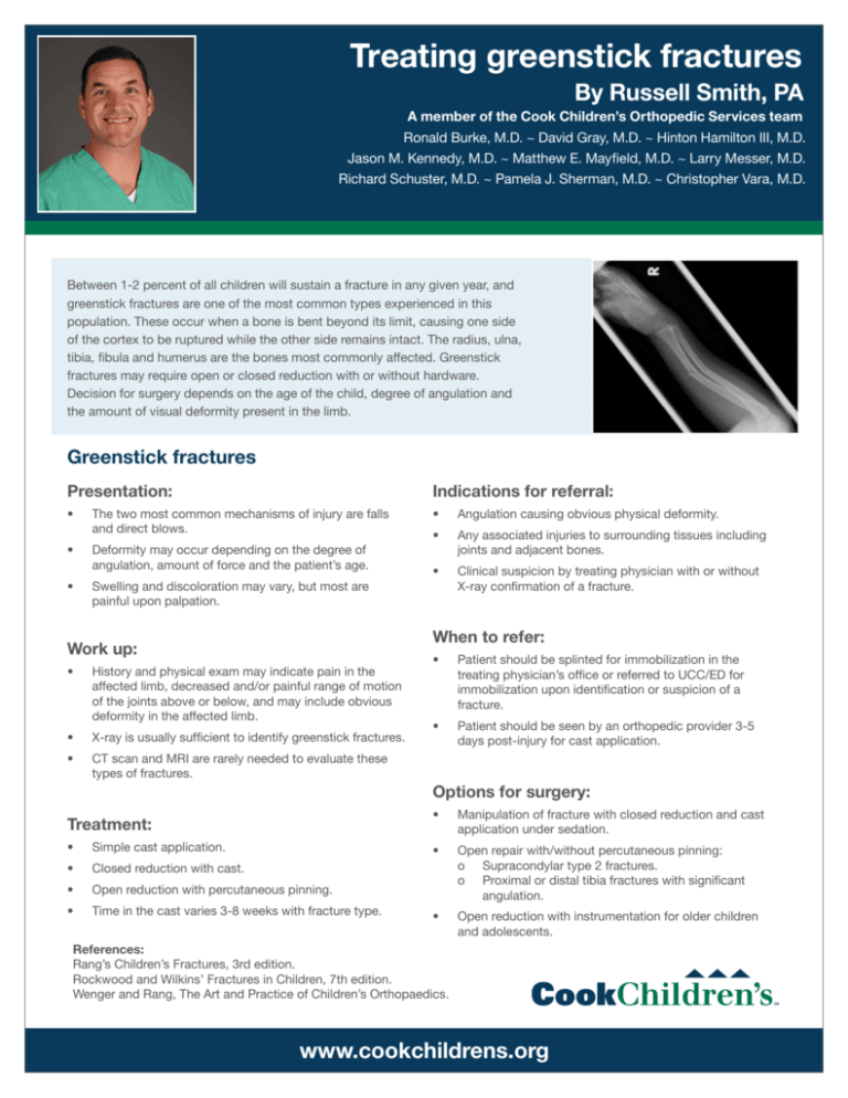
Treating greenstick fractures By Russell Smith, PA A member of the Cook Children’s Orthopedic Services team Ronald Burke, M.D. ~ David Gray, M.D. ~ Hinton Hamilton III, M.D. Jason M. Kennedy, M.D. ~ Matthew E. Mayfield, M.D. ~ Larry Messer, M.D. Richard Schuster, M.D. ~ Pamela J. Sherman, M.D. ~ Christopher Vara, M.D. Between 1-2 percent of all children will sustain a fracture in any given year, and greenstick fractures are one of the most common types experienced in this population. These occur when a bone is bent beyond its limit, causing one side of the cortex to be ruptured while the other side remains intact. The radius, ulna, tibia, fibula and humerus are the bones most commonly affected. Greenstick fractures may require open or closed reduction with or without hardware. Decision for surgery depends on the age of the child, degree of angulation and the amount of visual deformity present in the limb. Greenstick fractures Presentation: Indications for referral: • The two most common mechanisms of injury are falls and direct blows. • Angulation causing obvious physical deformity. • • Deformity may occur depending on the degree of angulation, amount of force and the patient’s age. Any associated injuries to surrounding tissues including joints and adjacent bones. • • Swelling and discoloration may vary, but most are painful upon palpation. Clinical suspicion by treating physician with or without X-ray confirmation of a fracture. When to refer: Work up: • History and physical exam may indicate pain in the affected limb, decreased and/or painful range of motion of the joints above or below, and may include obvious deformity in the affected limb. • X-ray is usually sufficient to identify greenstick fractures. • CT scan and MRI are rarely needed to evaluate these types of fractures. • Patient should be splinted for immobilization in the treating physician’s office or referred to UCC/ED for immobilization upon identification or suspicion of a fracture. • Patient should be seen by an orthopedic provider 3-5 days post-injury for cast application. Options for surgery: Treatment: • Simple cast application. • Closed reduction with cast. • Open reduction with percutaneous pinning. • Time in the cast varies 3-8 weeks with fracture type. • Manipulation of fracture with closed reduction and cast application under sedation. • Open repair with/without percutaneous pinning: o Supracondylar type 2 fractures. o Proximal or distal tibia fractures with significant angulation. • Open reduction with instrumentation for older children and adolescents. References: Rang’s Children’s Fractures, 3rd edition. Rockwood and Wilkins’ Fractures in Children, 7th edition. Wenger and Rang, The Art and Practice of Children’s Orthopaedics. www.cookchildrens.org For referrals and consultations: Cook Children’s Orthopedic Services 682-885-4405 phone 377 Rhome Highland Village 114 Keller N North Richland Hills Office locations Lewisville Dodson Specialty Clinics Grapevine 1500 Cooper Street Fort Worth, TX 76104 Southlake Southwest Multi Specialty Cook Children’s Northeast Hospital 6210 John Ryan Dr., Ste. 107 Fort Worth, TX 76132 121 360 Cook Children’s Medical Center 30 Dodson Surgery Center Fort Worth Dallas 750 Mid Cities Blvd., Ste. 100 Hurst, TX 76054 Grand Prairie Arlington Cook Children’s Urgent Care and Pediatric Specialties 20 Benbrook Mansfield Desoto 1187 Surgery locations Cook Children’s Medical Center 801 Seventh Ave. Fort Worth, TX 76104 Dodson Surgery Center 1500 Cooper St. Fort Worth, TX 76104 Hurst Multi Specialty Cook Children’s Northeast Hospital 6316 Precinct Line Rd. Hurst, TX 76054 www.cookchildrens.org 2727 E. Southlake Blvd. Southlake, TX 76092


