The diencephalon gives rise to the: •Thalamus •Epithalamus (pineal
advertisement
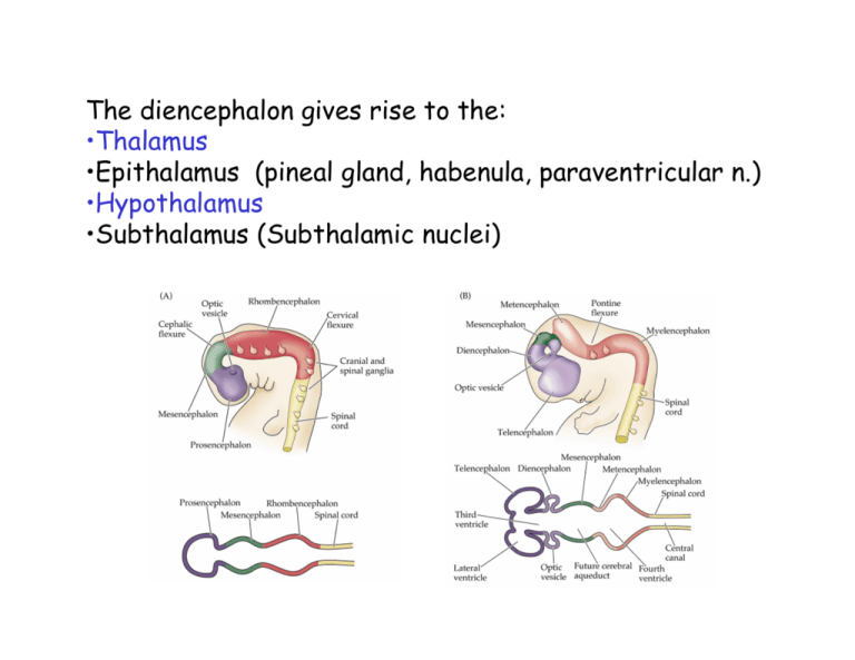
diencephalon The diencephalon gives rise to the: •Thalamus •Epithalamus (pineal gland, habenula, paraventricular n.) •Hypothalamus •Subthalamus (Subthalamic nuclei) For the most part when we refer to the ‘thalamus’ we really mean the dorsal thalamus. Most of the following material refers to the dorsal thalamus but you should be aware of the ventral thalamus that consists of the: •thalamic reticular nucleus •ventral lateral geniculate nuc. •zona incerta . The dorsal thalamus is divided into a number of nuclei. A basic definition of a thalamic nucleus is “a circumscribed region of cytoarchitecture receiving a particular set of afferent connections and projecting within the borders of a particular cortical field or fields.” The rat thalamus Nissl stain Myelin stain Thalamic nuclei of the rat drawn by Le Gros Clark (1932) Sir Wilfred Le Gros Clark The human thalamus Retinal afferents to the LGn Cortical afferent to thalamic cells p5 Lemniscal afferents to the VPL Dorsal Thalamic Nuclei •Project to the cerebral cortex (and some to basal ganglia) •Receive projections from the cerebral cortex •No descending projections •All sub-cortical information (except olfaction) passes through the thalamus to get to the cortex •All parts of cortex receives projections from thalamus Retinotopy (LGn) Somatotopy (VPL) LGn Y-cell LGn X-cell LGn local circuit neuron Thalamic nuclei contain two basic cell types: Projection neurons and interneurons (local circuit neurons) Some nuclei like the LGn have more than one class of projection neuron p5 p9 Afferents to thalamic projection neurons have a characteristic distribution to dendritic trees p10 GABA in the rat thalamus Human thalamus Extraglomerular neuropil Synaptic architecture the glomerulus is a distinctive feature of many thalamic nuclei •D thalamocortical neuron dendrite •T1 principal afferent (Glu) •T2 local circuit neuron presynaptic dendrite (GABA) •G glial cell The glomerulus p7 Attenuation of injected current in a relay cell (A, B) and in two local circuit neurons (C, D) p7 Tonic and Burst Response Modes: Determined by voltage- and time-dependent state of IT If cell is relatively depolarized by ≥5 mV for ≥50-100 msec, IT is inactivated and response is tonic mode: sustained firing of unitary action potentials with no role for IT 40 mV 100 msec 300 200 p8 -47 mV 100 tonic (linear) burst (nonlinear) 0 0 -70 mV -83 mV -59 mV -77 mV -59 mV Response (spikes/sec) If cell is relatively hyperpolarized by ≥5 mV ≥50-100 msec, IT is de-inactivated and response is burst mode: IT is activated, leading to all-or-none Ca2+ spike and burst of action potentials 800 1600 2400 Current Injection (pA) 3200 Luminance p9 Tonic Burst linear nonlinear detectability: poor detectability: good cortical activation: poor cortical activation: good Hypothesis: Bursts as a “Wake-up Call” tonic firing is better for stimulus analysis and bursting is better for detecting changes or novelty in a relatively unattended scene; bursts act as a “wake-up call”. indirect evidence: more bursting during inattention or drowsiness and a tendency for novel stimuli to elicit bursts. but there is still much about burst and tonic firing in behaving animals to be explained. p3 thalamic nuclei can be categorized on their location within the thalamus Name Cortical target Lateral geniculate (LGd) Retina Striate Cortex area 17 Ventroposterior lateral (VPL) Medial lemniscus (Dorsal columns) Spinothalamic tract SI and SII Ventroposterior medial (VPM) Trigeminal nuclei SI and SII Ventrolateral (VLp) Deep cerebellar nuclei Vestibular nuclei Area 4 (Primary motor area) (VLa) Central lateral (CL) p4 Afferents Globus pallidus Spinothalamic tract Striatum Area 4, SI, parietal Thalamo-cortical projections Specific and non-specific: Thalamocortical relationships in the cat based on degeneration studies by von Monakow (1895) Constantine von Monakow Corticothalamic afferents terminate in: •Layer 4, spill into 3 and 5 •Layer 6 •Layer 1 spill into 2 Individual nuclei have projections to combinations of layers, partly depending on cells size. p6 Generalized scheme of thalamic circuitry including interneurons and the thalamic reticular nucleus p6 layer 4 layer 6 Visual Cortex Glu GABA TRN ACh excitatory inhibitory relay cells Input to beRetina Relayed interneurons Thalamic LGN Relay p10 PBR midbrain Scheme of thalamocortical projection in the cat (Macchi, 1983) 1. Project densely onto a single cortical area 2. Project densely to one area and diffusely to another 3. Project diffusely on several areas with concentration in one 4. Project diffusely to many regions Conventional Alternate based on Parvalbumin and calbindin (Jones) Retinal afferents cortical afferent Afferents revisited: There is strong evidence that cortical afferents form a heterogeneous population. Some have a morphology similar to sub-cortical afferent and arise from layer 5 cortical cells. Together with the sub-cortical afferents these can be seen as ‘drivers’ and deliver the message to be processed by the projection neuron. The small cortical afferents from layer 6 are ‘modulators’ and together with other non-driver afferents alter the responsiveness of the cell. p11 First and higher order thalamic nuclei The searchlight hypothesis Reciprocal connections Segregation of input into TRN p11 All information that goes to cortex passes through thalamus Thalamus can act as a gate. •TRN opens and closes the gate. •TRN might mediate selective attention/lateral inhibition •Synchronize input Local circuit neurons •Filter inputs •Lateral inhibition •Synchronize input p12 Low threshold calcium conductance underlies the ‘tonic’ and ‘burst’ modes of thalamic projection cells Sleep: Two stages of sleep. •Slow wave sleep is characterized by low frequency, high amplitude oscillations •Desynchronized (REM) sleep high-frequency, low amplitude oscillations. During slow wave sleep cholinergic input is reduced and cells enter burst mode •The bursting in cells is synchronized •Depends on TRN relay cell interactions •Note bursting cells are not silent During REM sleep cholinergic input increases, relay cells enter tonic mode Epilepsy Reverberatory circuits between TRN, relay cells and cortex Thalamic pain syndrome Dejerine-Roussy syndrome
